|
|
|
|


|
Dr. Chris Doumen |
|
|
|
|
|
|
Dr. Chris Doumen |
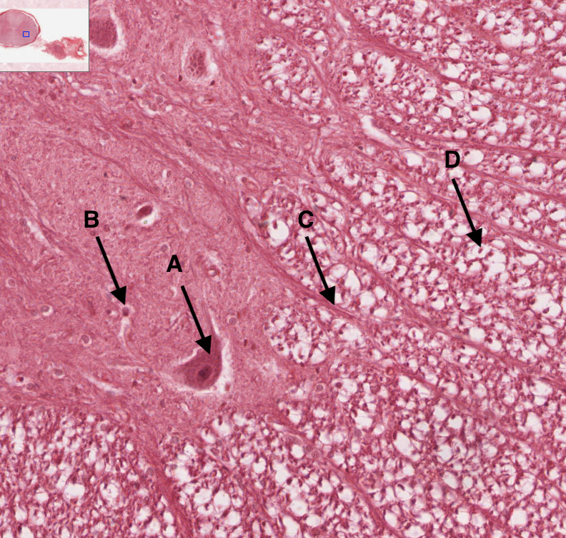 |
What you see is an enlarged area of the little blue box within the upper left image. From what specific region of the Spinal Cord is this ? The ventral horn If you observe this histology section closely, you can see that the left side is different than the right side. Why ? The left side is gray matter, the right side is white matter Identify the following structures now. A : Somatic motor nerve cell body B : Neuroglial cell C : Axons from motor neurons going towards the ventral root D : Individual axon (the black little cicrcle) with myelination Name two possible neuroglial cells visible in this section . B could be an astrocyte or microglial cell - the white area around D is myelination from an oligodendorcyte |