Biology 2404 A&P Basics Lab Exercise 4 Cells Dr. Weis
| Objectives | Background | Medical Terms | Activities | Applications | Careers | WWW | Review Questions |
Students should be able to
* name the parts and function of a eukaryotic cell
* name the transport processes that occur at the cell membrane
* name the stages of the cell cycle
* name the parts and function of a light microscope
Read related information in textbook
Cytology and Cells
The cell, by definition, is the smallest functional unit capable of carrying on all life processes. The Latin word part associated with this structure is cyt/o, which means cell. Cytology is the study of cells and their function.
Types of Cells
There are two major types of cell : prokaryotic and eukaryotic.
The prokaryotic cells were first in existence approximately 3.5 billion years ago and their name means "before the nucleus". Examples of prokaryotic cells are bacteria and cyanobacteria (blue green algae). The true nucleated cells, called Eukaryotes, appeared about 1.5 billion years ago. Eukaryotes can be unicellular, such as yeast, or multicellular like plants and animals.
Similarities between the two major types of cells exist.
Both Prokaryotic and Eukaryotic cells have:
* basic metabolic functions
* ribosomes
* externally bound membranes
The major difference is that Eukaryotes have membrane bound organelles,
including, the nucleus which contains the genetic material, DNA.
Other differences include:
* Size : Eukaryotes are approximately 10x larger
* DNA : Eukaryotes have more extensive DNA
* Cell Wall : Prokaryotes have a cell wall composed of peptidoglycan,
a large single polymer of Amino acids.
NOTE : Not all Eukaryotes have cell walls.
Those that do, such as plants, do not have the peptidoglycan structure.
Peptidoglycan structure of Prokaryocytes such as bacteria are important in staining for identification such as the Gram stain and in the use of antibiotics.
Around 1665, an English Scientist by the name of Robert Hooke used an early light microscope to describe the tiny empty chambers he saw while viewing a dried cork. He named these empty chambers cells. Other scientists continued observing the fluid filled spaces in living structures such as found in plants. From this early research the cell theory was derived and a few of its concepts were discussed in their earlier definition.
The concept of homeostasis or balance that is maintained by the cell reflects the equilibrium maintained by a continued hierarchy of organization that anatomists use to study living systems. This level of organization was discussed earlier and is as follows:
* Chemical / molecular
* Cellular
* Tissues
* Organs
* Systems
* Organism
* Population
* Communities
* Ecosystems
Cytology of Eukaryotic Cells
Cytology is the study of cells and how they function.
Cells are between 75-90% water and the remaining percentage (10-25%) consists mainly of proteins. Other molecules such as carbohydrates, lipids, and nucleic acids each make up about 10-15% of the remaining half of the solids or solutes.
Total composition by element is
60% Hydrogen (H),
25% Oxygen (O)
12% Carbon (C)
5% Nitrogen (N).
Other notable elements that form biological molecules are Phosphorus (P), Sulphur (S), Sodium (Na),
Magnesium (Mg), Chlorine (Cl), Potassium (K), Calcium (Ca), and Iron (Fe).
Every Eukaryotic cell has three main parts :
1. Cytoplasm = Cytosol + organelles
2. Cell or plasma membrane
3. Nucleus which contains the genetic material, DNA
The cytosol is the liquid form of the cell. It's major components are water, ions, proteins, wastes and insoluble materials known as inclusions.
The cell membrane separates the cytosol from the surrounding fluid and encloses some organelles.
In the medical field, the cytosol of animal cells is known as Intracellular Fluid (ICF) and the surrounding fluid is called Extracellular Fluid (ECF). ECF has three subdivisions : plasma, lymph, and interstitial fluid.
In Latin, organelles mean small or little organs and they are structures that perform dedicated functions within the cell. Two ways to classify organelles are those that are bound by a membrane (membranous) like the cell membrane and those that have direct contact with the cytosol (nonmembranous).
Membranous Organelles include the nucleus, mitochondria, chloroplasts, endoplasmic reticulum, the Golgi apparatus, lysosomes and peroxisomes.
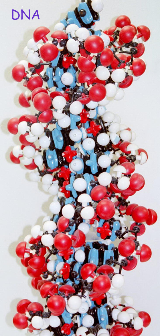
1. Nucleus : The nucleus is a membranous organelle by definition, but because of its unique function, it is also considered as a major part of the cell. In Eukaryotic cells, the genetic material known as DNA is kept here. Remember that in Prokaryotic cells the DNA is free floating. The nucleus has a double membrane that is fused together to form pores through which certain substances can pass. Because of the nuclear membrane pore size, the DNA does not leave the nucleus, but other nucleic acids called RNA are used to write (transcribe) and read (translate) the genetic code.
DNA and RNA structure, specific function, and processes are discussed in your textbook.
2. Mitochondria are double bound membranous organelles found in all portions of the cytoplasm. The number of mitochondria will vary per cell depending on the energy demand. They also vary in size and shape and contain their own DNA so they can self replicate. Their double membrane consists of one outer smooth membrane that surrounds the organelle and an inner membrane that is folded called the cristae. The cristae folds come in contact with the matrix fluid contents and enzymes run the chemical reactions that oxidize nutrients to carbon dioxide and water. These reactions will release Energy that is used to synthesize ATP in a process called aerobic respiration. Biologists now theorize that mitochondria are descendants of bacteria due to the double membrane and the fact that they have their own DNA and ribosomes. The mitochondrial ribosomes were found to be more similar to bacterial ribosomes.
3. Endoplasmic Reticulum (E.R.) is a tubular structure that may be elongated. These organelles are present in cells that secrete large amounts of proteins or spherical (in cells that secrete steroid hormones). A cross sectional view of E.R. reveals a space in the center called cisterns and a fluid medium called the matrix. The function of E.R. involves storage, transport, and synthesis. Its membranes are continuous with the nucleus. E.R. may be classified two ways : rough and smooth. When ribosomes are associated with the surface of the E.R., it is referred to as rough E.R. and will function in protein synthesis. If there are no ribosomes, the E.R. is smooth and functions involve lipid metabolism, cholesterol synthesis, glycogen synthesis/storage, the storage/retrieval of Ca++, and detoxification of drugs.
4. Golgi Apparatus is closely associated with the E.R. and will appear as flattened stacked discs called saccules. There are usually between 4 -6 saccules in a typical Golgi complex. Functions include packaging secretions for the cell, packaging secretions for export (exocytosis), and remodeling the cell membrane. Packaging involves placing the needed secretions, enzymes, lipids in vesicles for transport.
5. Lysosomes are vesicles formed at the Golgi Apparatus and are found throughout the cell. They contain hydrolytic enzymes called hydrolases that cleanup and recycle substances to remove unwanted or damaged structures or remove foreign substances. When its membrane is unstable in certain disease states, the rupture of lysosomes can result in cell self-digestion known as autolysis.
6. Peroxisomes are thought to originate from the R.E.R. and contain different enzymes known as peroxidases. Peroxisomes neutralize toxins and protect cells from the damaging affects of free radicals. It has been theorized that the production of free radicals contributes to the aging process.
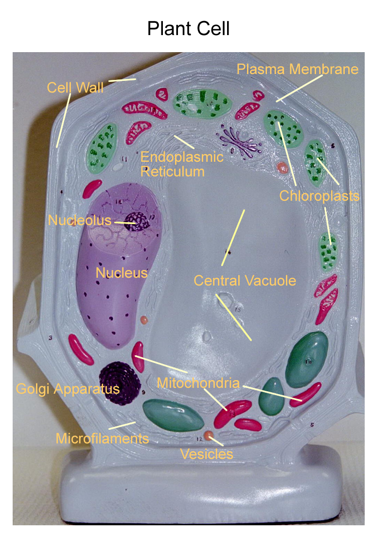
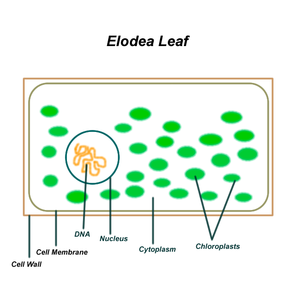
7. Chloroplasts are double bounded membrane organelles found in the leaves of plant cells. Inside the chloroplasts there are photosynthetic membranes called thylakoids. The thylakoids are stacked on top of one another and these stacks are called granum (singular). Grana (plural) are held together by lamellae and the interior of the entire chloroplast is filled with a semiliquid fluid called stroma. Chloroplasts will be discussed again in the photosynthesis laboratory unit.
Non membranous organelles include the nucleolus, ribosomes, filaments, and microtubules. Filaments will come in various sizes : microfilaments, actin filaments, intermediate filaments, and thick filaments. Various filaments will be involved with the cytoskeleton and microvilli. Microtubules will create other structures such as the cytoskeleton, cilia, flagella, and centrioles.
1. Nucleoli are found in the nucleus and function to synthesize ribosomal RNA, rRNA. They also assemble ribosomal subunits for transport out of the nucleus through the nuclear pores. Nucleoli will appear as 1-4 dark staining areas inside the nucleus. They will be most prominent in cells that manufacture large amounts of proteins, since ribosomes are needed in greater number. Changes in the nucleolus numbers and staining characteristics are used to help identify cancer cells.
2. Ribosomes are organelles that manufacture proteins. They can be found free in the cytoplasm or associated with endoplasmic reticulum, known as R.E.R. Ribosomes are made of RNA and protein and work together as two subunits. The smaller subunit, the light ribosomal subunit will pair with the larger subunit called the heavy ribosomal subunit in order to read the mRNA strand as part of the process of protein synthesis. The free ribosomes manufacture proteins for the cell. Fixed ribosomes, those attached to E.R., manufacture proteins for packaging and export by the Golgi apparatus.
3. Filaments
a). Microfilaments are slender protein strands that help form the internal framework of the cell known as the cytoskeleton. They also help support the finger-like folds of the cell membrane known as microvilli.
b). Actin filaments are a specific type of microfilament that will interact with other protein filaments to help form the consistency of the cytoplasm. In certain specialized cells, actin interacts with another protein, myosin, to achieve movement or to change the shape of the cell.
c). Intermediate filaments are larger than microfilaments and function to stabilize cell shape, organelle position, and the cells position with other cells.
d). Thick filaments are massive bundles of the protein myosin. These filaments appear only in muscle cells and will interact with actin to provide for muscle shortening for a contraction.
4. Microtubules are hollow tubes built from the globular protein, tubulin. They form part of the cytoskeleton and have the same function as the intermediate filaments. They also form the structural components of cilia, flagella, and centrioles.
a). Cilia contain 9 pairs of microtubules around a central pair, known as the 9+2 array. The beating action of cilia allows for movement of fluids or secretions across a cell surface.
b). Flagella resemble cilia, but are much longer. The movement of the flagella propels the cell through its fluid environment.
c). Centrioles contain 9 pairs of microtubules with no central pair, known as the 9+0 array. Centrioles are involved with cells that undergo division by forming the spindle fibers that attach and move the chromosomes during this process.
Cell Membrane
The cell membrane or plasma membrane forms the boundary between intracellular (ICF) and extracellular (ECF) fluid. The structure is a phospholipid bilayer with associated proteins, carbohydrates, and cholesterol.
This membrane structure creates certain properties of the cell which include these functions are:
1. Barrier : separation of fluid compartments
2. Semipermeable : regulation of exchange with the environment
3. Transmembrane Potential : electrically charged membrane
4. Structure : connections and support between two membranes or the cell membrane and its environment
5. Sensitivity: recognizes and responds to changes in its environment
Prokaryotes
Prokaryotes do not contain membrane bound organelles, including the nucleus. The organelles are organized macromolecules that have specific function within the fluid called the cytoplasm.
The cytoplasm of Prokaryotes is enclosed in a cellular membraneand is supported by a cell wall covered by a gelatinous capsule. The largest Prokaryotes are cyanobacteria (blue-green algae) and contain chlorophyll and other pigments necessary for photosynthesis.
Bacteria have a cell membrane that lies flat against the cell wall.
In the cytoplasm, the ribosomes and fine threads of DNA can be seen.
Cell Poster (bacteria)
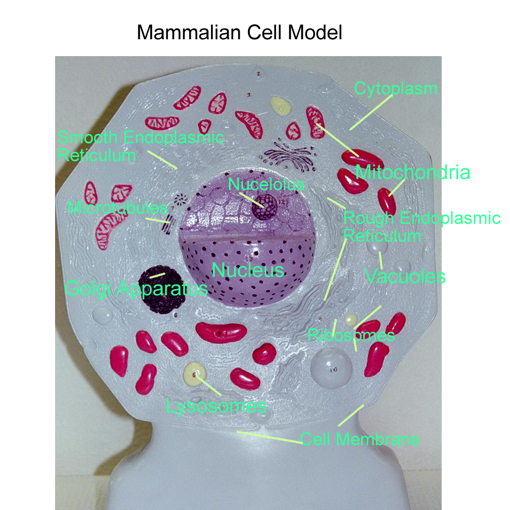
Cell models
Eukaryotic Cells
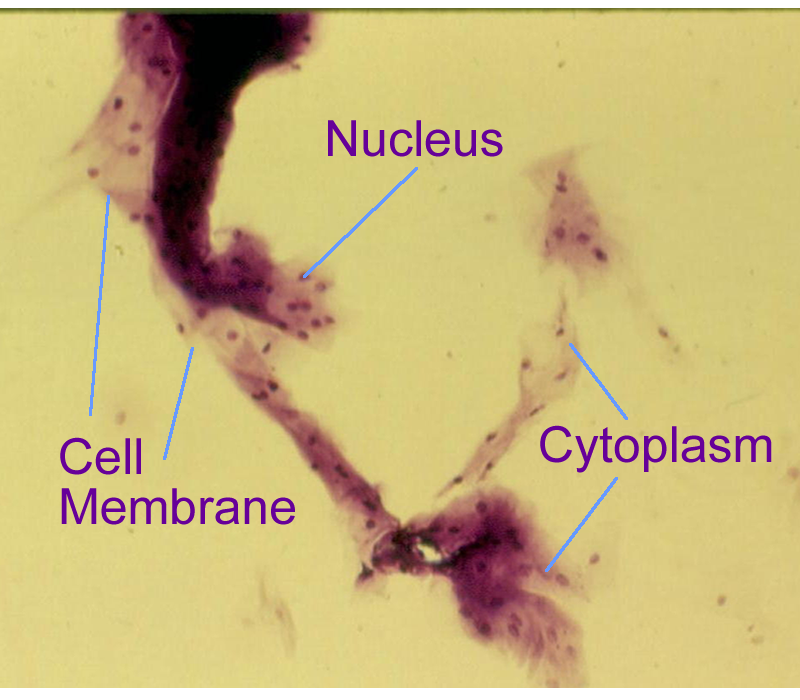
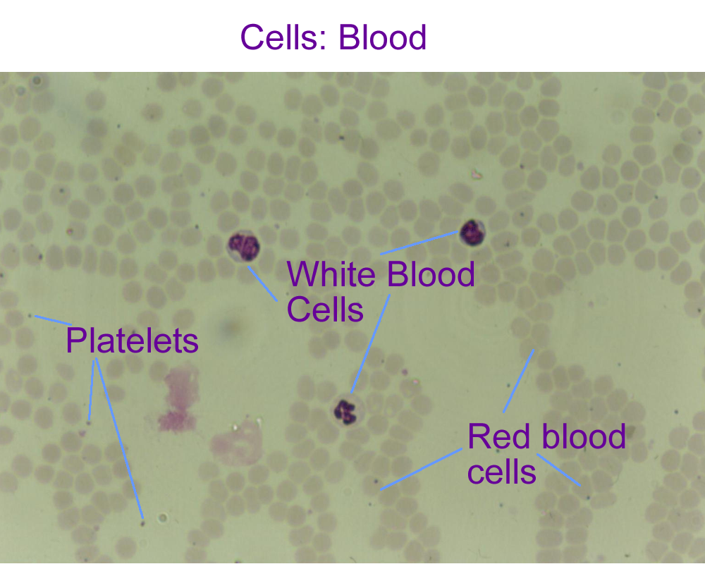
Mammalian : cheek cell, blood cell
Prokaryotic Cell Drawing
Bacteria : Cocci,
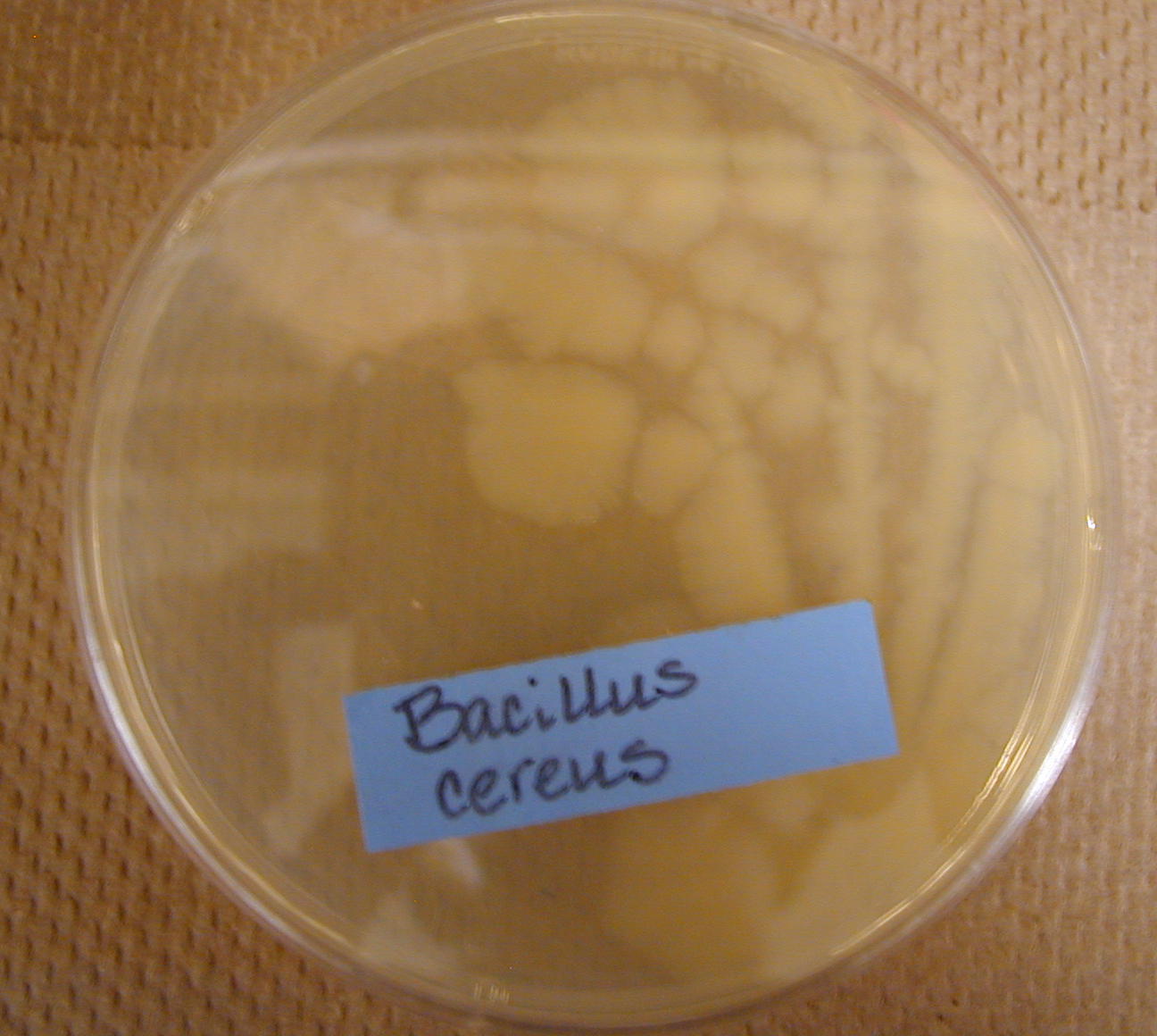
Rods (Bacillus)
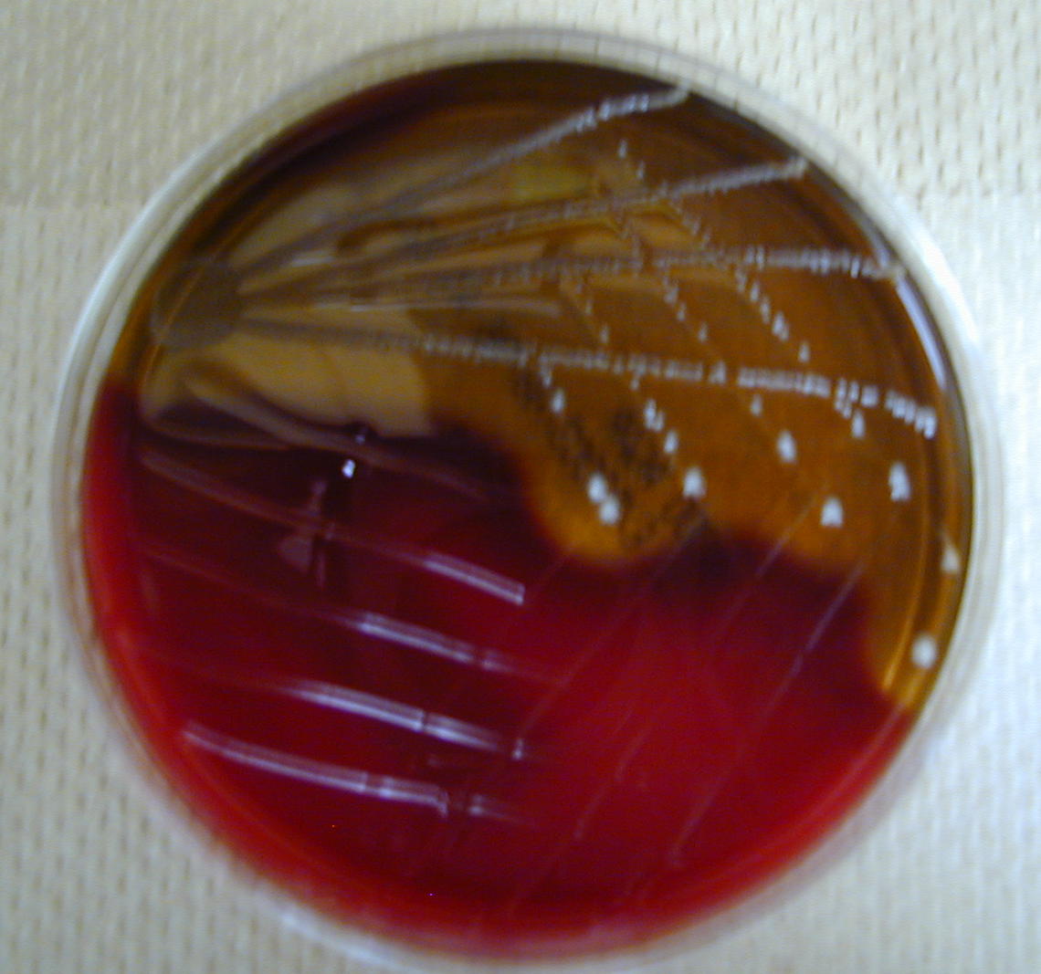
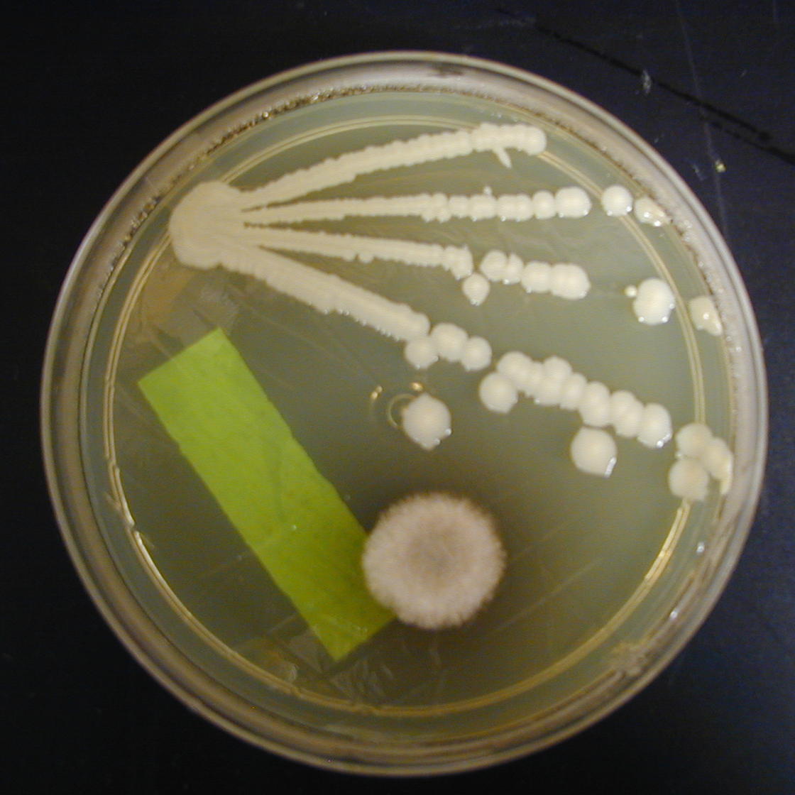
Bacterial Agar Plate Cultures
Cell Cycle
The DNA of the cell controls cell division and replication. Hormones help in the communication and regulation of this cycle. The DNA also directs the daily function of the cells and their internal chemical processes. For most cells, their normal daily functions form a part of the cell cycle known as Interphase. It can also be represented by the term G-0, which is growth phase 0. Depending on the DNA and the response to the communication signals, the division and replication phases can then begin.
The nucleus is the first to complete its division, followed by the cytoplasm.
Nuclear divisions create exact copies with the same number of chromosomes. This process is called mitosis. Prior to these events, the DNA, cytoplasm, and organelles need to be duplicated. These phases of cell duplication are termed G-1, S, and G-2. G-1 is growth phase one and the cytosol and organelles are duplicated. In the S phase, the DNA is duplicated by semi-conservative replication. The final phase before nuclear division is growth phase two (G-2) and any structures needed for nuclear division are made.
Examples of such structures are centrioles and spindle fibers.
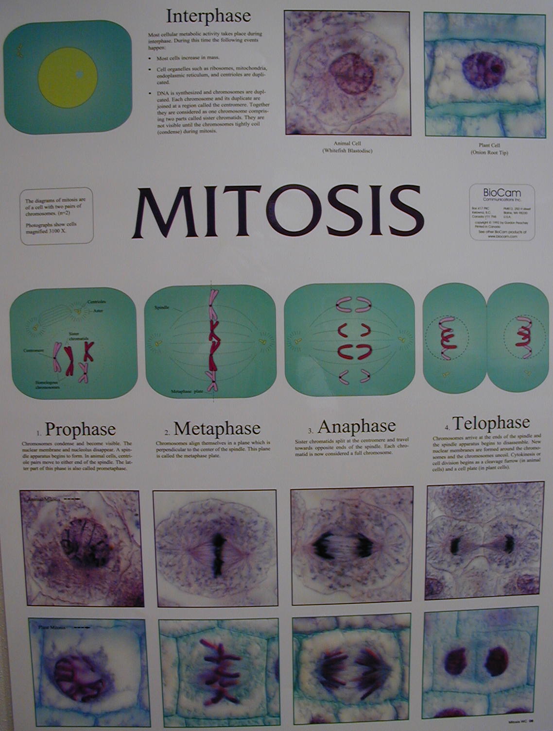
Mitosis then begins and happens in ordered stages.
At the end of mitosis, there will be two daughter nuclei containing the diploid (2n) number of chromosomes.
Mitotic phases in order are:
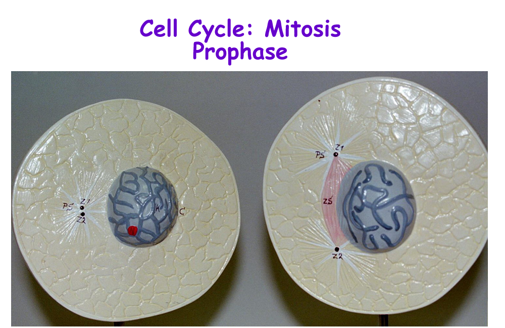
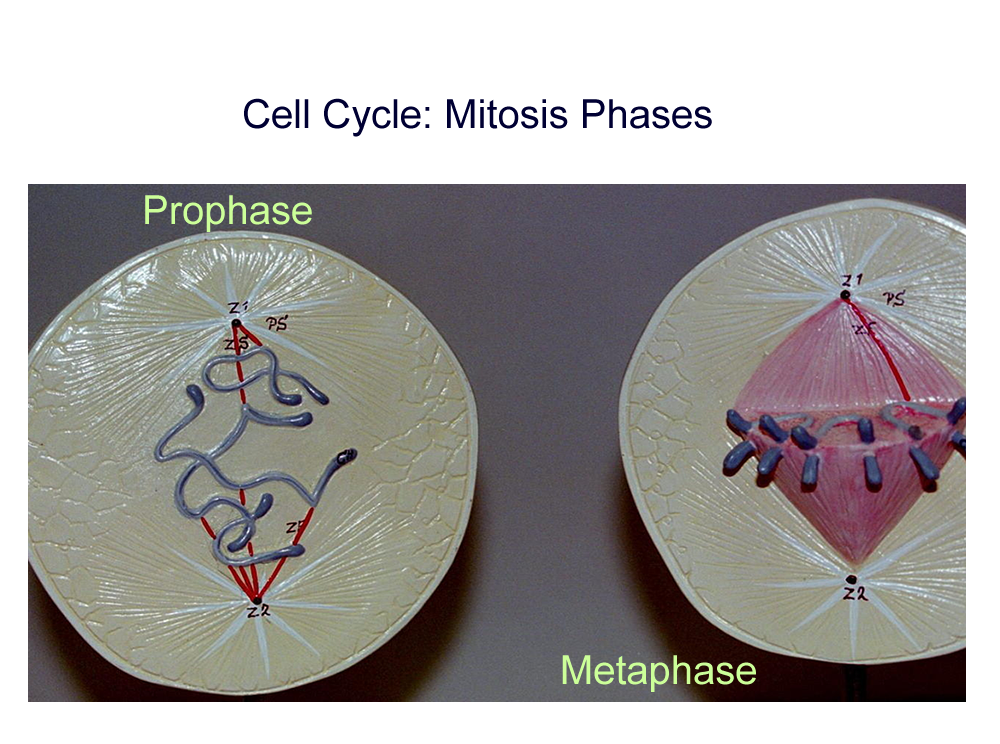
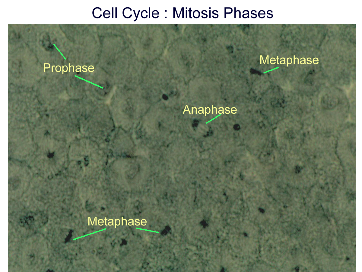
Prophase Nuclear envelope dissolves, chromatin condenses, spindles attach to centrioles
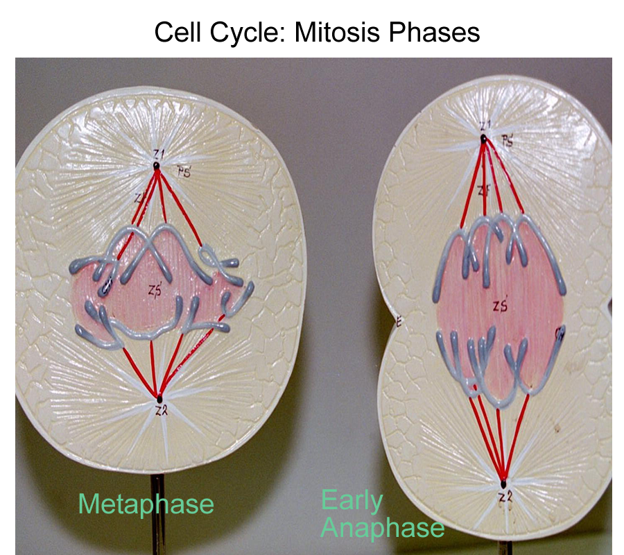
Metaphase chromosomes line up at midline or central equator of the spindle
Anaphase chromosomes pull apart and go to opposite ends of the cell directed by spindle fibers
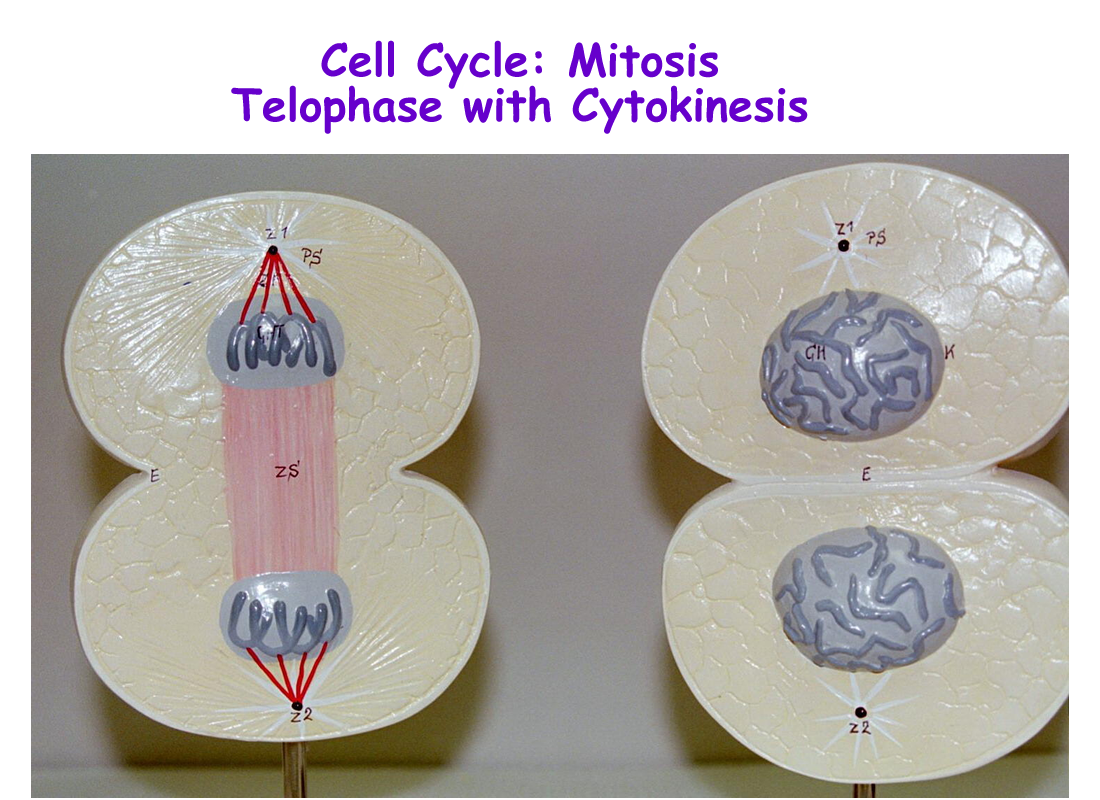
Telophase end phase of nuclear division. Spindle fibers dissolve, chromosomes return to normal form, nuclear envelope reforms.
Nuclear division is now complete. All that remains is to complete the cell division by dividing up the cytoplasm (cytosol and organelles). This final phase of cell division is called Cytokinesis and begins in animal cells with the formation of a cleavage furrow down the midline or equator.
Eventually the furrow pinches the cell into two parts, thus ending this cell division.
The two new daughter cells then begin G-0, which consists of normal cell processes and function.
There is another type of cell division called meiosis that occurs in the reproductive organs that produce gametes or sex cells. Meiosis has some similarities as well as differences when compared to mitosis. The biggest difference is that the resulting meiotic division creates specialized cells that only have half (haploid or 1n) the number of chromosomes.
Meiosis will be looked at in greater detail in the reproductive systems.
The importance of the mitotic phases is in identifying pre-cancerous (hyperplasia) verses cancerous cells. Cells that contain a high amount of mitotic figures (phases), double nuclei, prominent nucleoli, different sized nuclei, and a 1:1 nuclear to cytoplasmic ratios are all warning signs of a malignancy.
The importance of the cell cycle is in repair and replacement of cells that form tissues and organs.
Mitosis Models : Prophase, Metaphase, Anaphase, Telophase
White Fish Blastula Mitosis
Prophase and Metaphase and Late Anaphase
Early Anaphase, Telophase, and Cytokinesis
Microscope
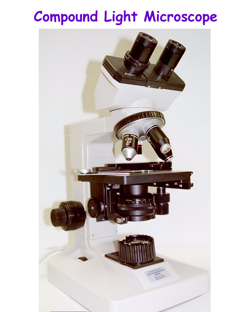
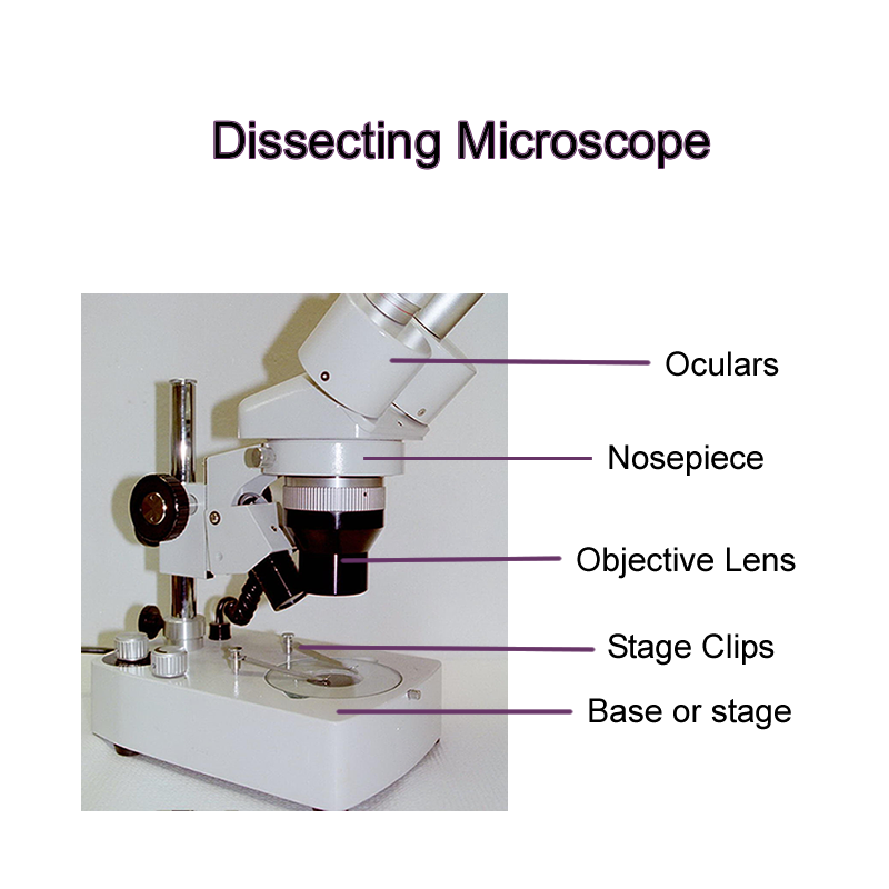
In order to view cells and tissues, an instrument called the microscope is used to magnify the size and structures seen. Most all laboratories have at least a compound light microscope to view cytological samples, wet mounts, smears, or thin prepared tissue specimens. Other microscopes that may be used in a clinic, hospital, or research setting include dissecting microscopes and scanning electron microscopes.
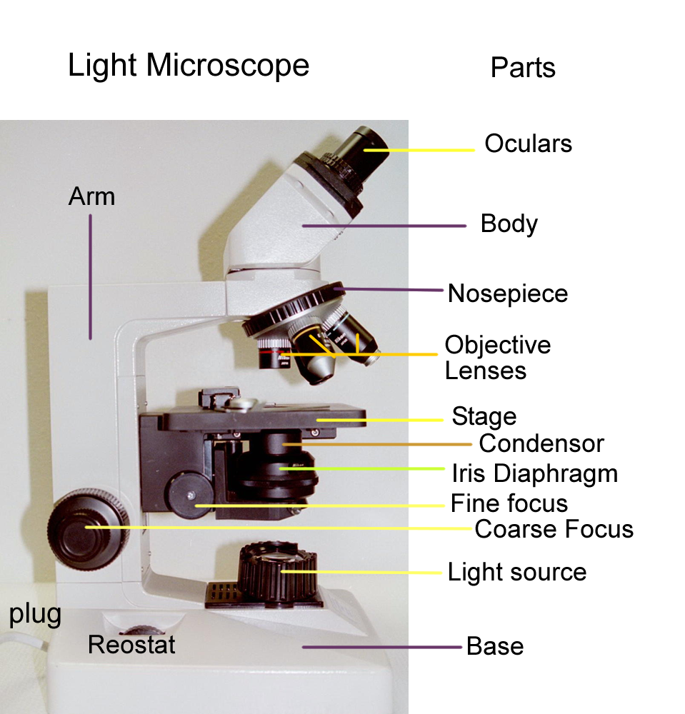
Light microscopes consist of a two lens system: the ocular lens or eyepiece and the objective lens that magnifies the object. The objective lens forms a real image which is then magnified again by the eyepiece to create a virtual image. The light source comes from the base and travels through the condenser’s iris diaphragm to help focus light through the specimen on the stage and then into the lens system. The objective lens is mounted to a rotating nosepiece and usually come in four different magnification powers.
Possible objective lenses include:
4X lens scanning objective lens used to survey the sample
10X lens low power objective used to view wet mounts
40X lens high dry objective used to view stained cells and urinalysis sediments
100X lens oil immersion objective used to view smears, bacterial stains,
and other cytological preps for cancerous changes.
The ocular or eyepiece lens is usually at 10X magnification and is supported by the arm.
When viewing a specimen or reporting specimen size, it is good to keep in mind
that the total magnification of the specimen is defined as the eyepiece multiplied
by the objective, that is Ocular X Objective = Total Magnification. An example
for total magnification would be 10X x 40X = 400X total magnification. At time
you may see histology slides magnification is labeled based on the objective
magnification, such as 10x or 40x. Remember that this is not the total magnification.
In order to help bring the object into focus there are two focus knobs: course and fine focus that allow the stage to be moved up and down. Mechanical stage knobs allow the movement of the specimen in two other directions, right and left.
Care and proper use of the microscope are important to the longevity of the instrument and are discussed in detail before the instrument is used by students.
Lenses are kept clean with proper cleaner and lens paper
Oil is only used with the oil immersion lens and any oil that is spilled or comes in contact with other objectives must be cleaned immediately.
Scanning power is used first to locate the object of interest, then the low power is used. If needed the high dry objective is then rotated into position for a closer view of the specimen or area of interest. Oil immersion is used in special cases and this objectivs requires the use of oil to concentrate the light through the specimen.
Principles of Microscopy
A. Magnification is the factor by which a specimen is enlarged and is calculated by using the total magnification formula.
B. Parfocal is the ability of the microscope image to remain in focus when switching form one objective to the next.
C. Diameter of Field is also called the field of view or area seen when viewing a slide through the lens system.
D. Depth of Focus is also known as depth of field and relates to the thickness of an object and the ability to keep it in focus in a vertical (up and down) plane.
E. Image Orientation is due to the optics of the lenses involved, the image seen will be real, inverted, and magnified by the objective and ocular lenses.
F. Resolving Power is the degree at which two points on a specimen are seen as separate detailed images.
G. Contrast is how well the details of a specimen
stands out against a background.
Cell Membrane Transport and Physiology
The plasma membrane allows for separation of ICF and ECF and the control of fluid (solvent) and solid (solute) movement. Due to the chemical nature of the plasma or cell membrane it is considered to be selectively permeable. The structure is a phospholipid bilayer with associated proteins, carbohydrates, and cholesterol.
This membrane structure produces certain properties of the cell, which include the following:
1. Barrier: separation of fluid compartments
2. Semipermeable: regulation of exchange with the environment
3. Transmembrane potential: electrically charged membrane
4. Structure: connections and support between membranes of two cells or between the cell membrane and its environment
5. Sensitivity: recognizes and responds to changes in its environment
All of these cell membrane properties allows for homeostatic or steady state balanced control for proper function of each cell that makes up their respective tissues and organs.
A selective permeable membrane allows for inward movement of solutes and solvents that are necessary for survival, and the outward movement of solutes and solvents that form waste products. The phospholipid bilayer allows for fat soluble molecules and compounds to freely pass, while water soluble molecules and compounds require different transport mechanisms involving the cell membrane proteins.
Some transport mechanisms do not require energy and are passive; others require energy and are therefore active. The terms used to discuss membrane transport mechanisms are therefore: Passive Transport and Active Transport.
Passive Transport
Two types of passive transports that are important to cell membrane transport mechanisms are diffusion and filtration.
Diffusion is the movement of molecules from an area of high concentration to an area of lower concentration until equilibrium is achieved. Diffusion requires no driving energy and is based on the differences in concentration gradients. Molecules have their own kinetic energy or energy of motion which is the driving force for passive processes.
There are three types of diffusion.
1) Simple diffusion
2) Facilitated diffusion
3) Osmosis
Simple diffusion involves fat soluble molecules that move across the fatty phospholipid membrane.
Facilitated diffusion involves small water soluble molecules that need a protein carrier to get through the fatty phospholipid membrane. Proteins can act as carriers for molecules such as glucose and amino acids, or as open channels for ions such as calcium ion, potassium ion, sodium ion, and chloride ion.
Osmosis involves the movement of water through the membrane down its concentration gradient across a Semipermeable membrane from an area of high water (dilute solution) to an area of low water (concentrated solution).
In order to understand the differences between these types of passive transports, the components of a solution are important. A solution is made up of solids called solutes suspended in a liquid called the solvent. In biological processes, examples of solutes can be ions, glucose, and amino acids. The solvent for biological processes is water. Solutions can also be classified in relationship to the cells they come in contact with thus resulting in water movement (osmosis) between the two fluid areas.
Isotonic solutions have the same solute concentration as cells. Isotonic solutions for the body are 0.9% NaCl. No net movement of water occurs.
Hypertonic solutions have a higher solute concentration than the inside of cells. An example of a hypteronic solution would be 10% NaCl. Since the outside of the cells is more concentrated, the water within the cells moves out and the cell shrinks or crenates.
Hypotonic solutions have a lower solute concentration than compared with the inside of cells. An example of a hypotonic solution would be distilled water. Since the inside of the cell is more concentrated than the outside fluid environment, water moves into the cell and the cell swells and can eventually burst or lyse.
Even though diffusion processes are passive, they are not static and can change.
Diffusion rates can be affected by temperature, size of the molecule, and the kinetic energy of the molecule. Diffusion rates increase with increases in temperature and decreases in molecular weight of the substance. Diffusion rates decrease with decreases in temperature and increases in the molecular weight of the substance.
Filtration
The driving force for molecules during the filtration process is a hydraulic (fluid in motion) pressure that is generated by the heart muscle pump. Filtration pressures happen across the plasma membrane capillary beds and allows for plasma substances to be “pushed” or filtered through capillaries into the interstitial spaces that bathe the tissues.
Filtration also happens at the kidney, once again due to blood pressure. It is nonselective and depends on the pressure gradients, the integrity of the membrane, and size of the capillary cell membrane pores.
Active Transport
Active transports involve use of proteins that use cellular energy, such as ATP and moves substances against a concentration gradient from low to high.
Examples of active transport at a membrane are:
Solute Pump
Gated Ion channels
Bulk Transport
a) Phagocytosis
b) Pinocytosis
c) Receptor mediated endocytosis
dia- through ex- out, from
ferr- iron hydro- water
-ia state lyso- dissolve
meta- after, beyond micro- small
morpho- form -oid form
pino- drink phago- eat
-plasty mold, form pro- before
pyro- heat, fire therm/o- heat
Water properties
Three water states, time, and temperature
Measure out three separate cups of water. Place one cup of water in a freezer safe container. Place another cup of water in a saucepan and the third cup of water in a glass. Note the time and place the water in the freezer safe container in the freezer and start the water in the saucepan on "High" to boil. How long did it take to freeze the water? How long did it take to boil the water? Let the boiled water cool and note the time until its temperature reaches that of the room temperature water in the glass. Remove the frozen water from the freezer and note the time that it takes to reach the same temperature as that of the water in the glass. What are the chemical reasons for the different states of water and the time it takes to change from one state to another?
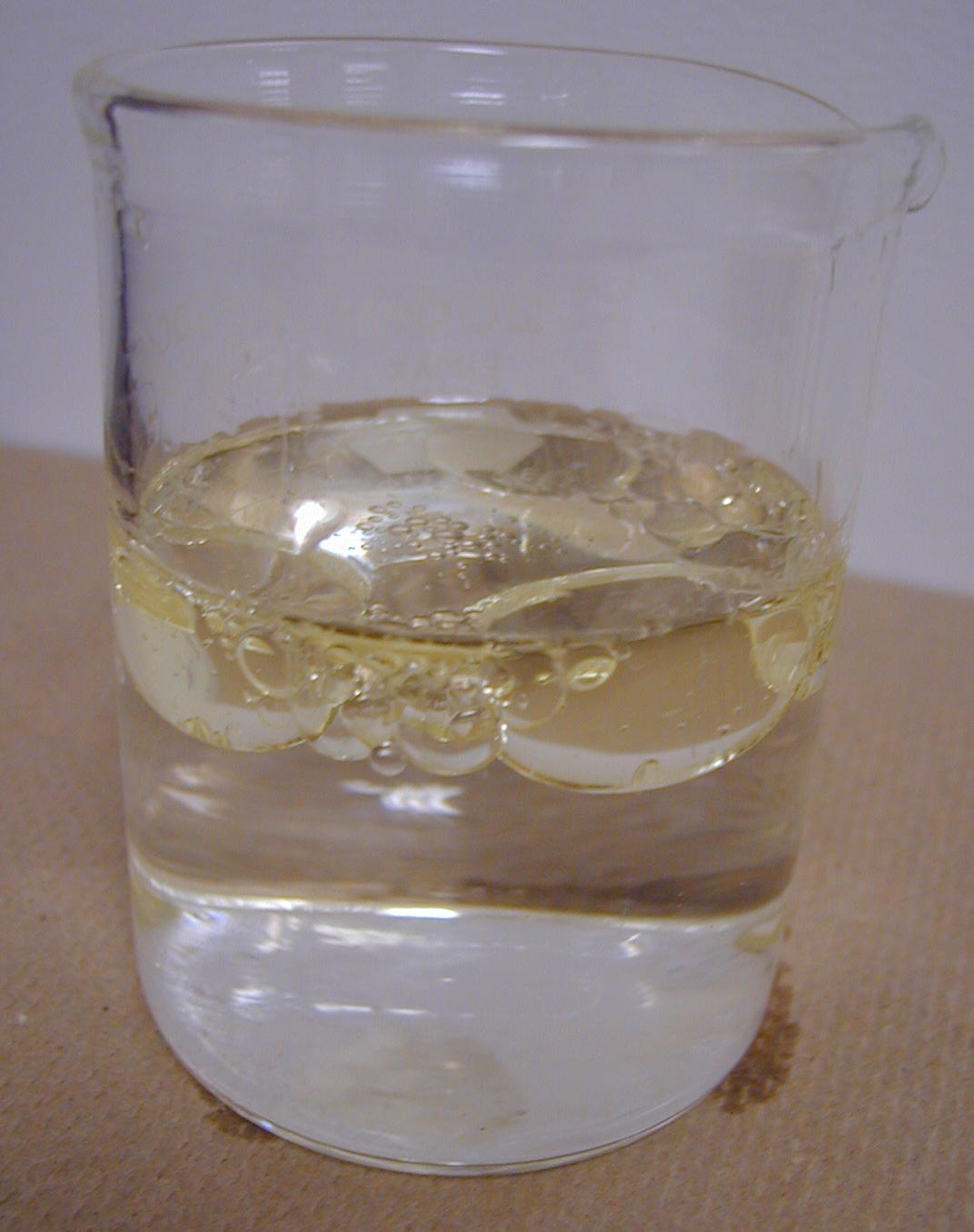
Fat and water interactions (Oil and vinegar)
Place an equal amount of vinegar and oil into a clear glass or measuring cup.
Note the interaction of the two.
Color 3 tablespoons of water with a green or blue food coloring. Add the colored water to the oil/vinegar mixture. Where does the dyed water go (into the oil or the vinegar)?
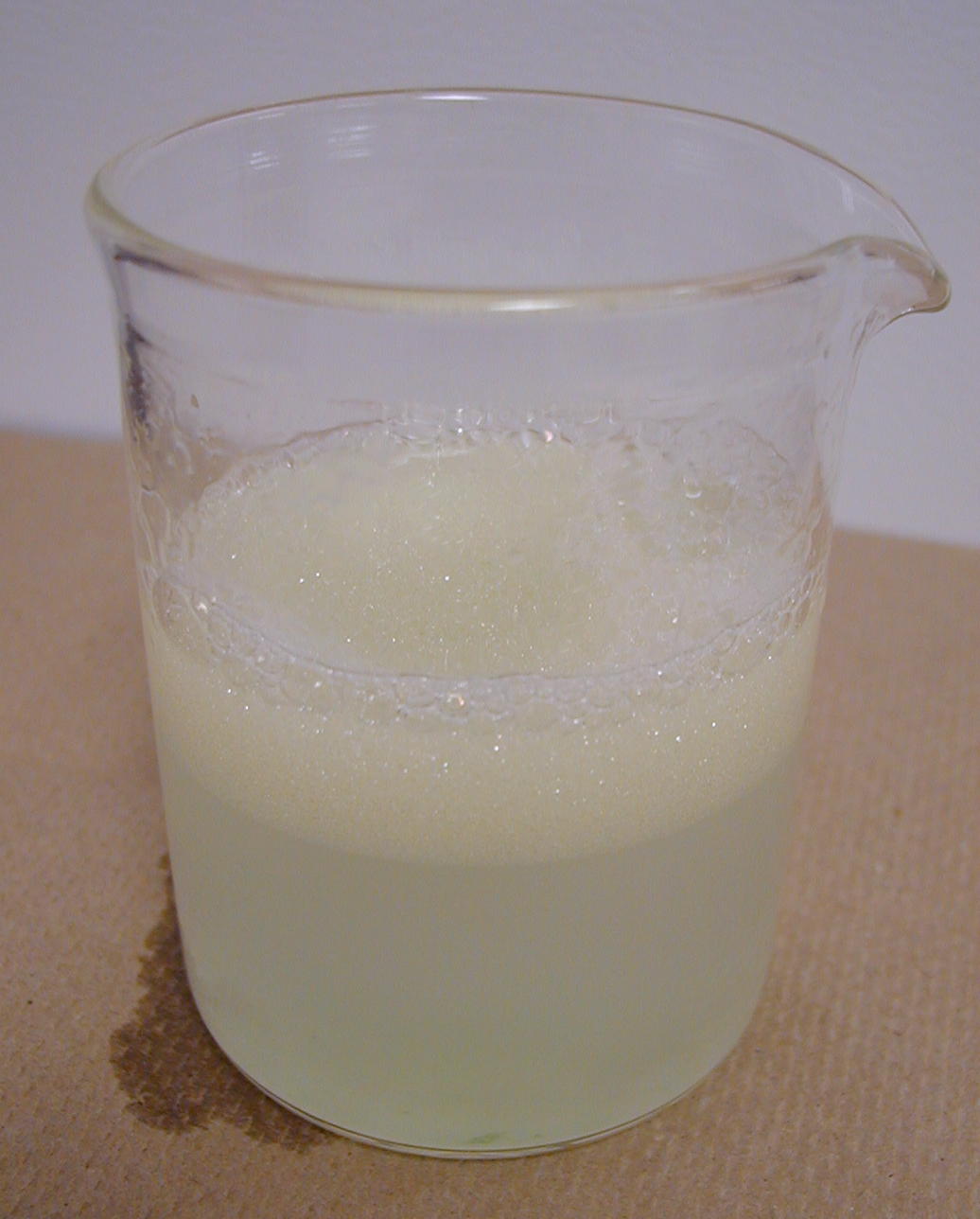
Now add one tablespoon of liquid detergent. Mix well. Note the interaction of the oil and vinegar.
What does the detergent do to the oil and vinegar mixture?
Look at the picture of the emulsion with the red dye. Why is the red dye found throughout the beaker ?Water, Oil, and Liquid Detergent in Beaker
Water, Oil, Liquid Detergent, Fat Soluble Dye
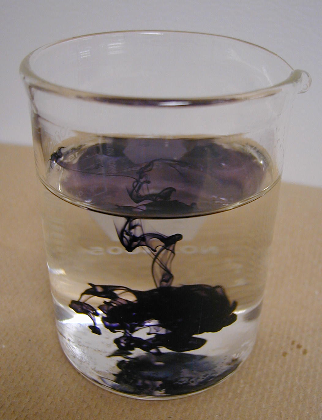
Diffusion
Ink in Water: Place a drop of ink in water and see how long it takes to dispurse. Look at the two photographs and explain why the dye is not as well diffused as the india ink. Assume that the times are the same.
Food Coloring: Place a drop of food coloring in water and see how long it takes to dispurse
Now compare the two times. What does water solubility have anything to do with the times of the dye, india ink, and food coloring.
Agar plates / temp : The plate on the left was kept at room temperature (25 degrees celsius) for 24 hours. The plate on the right was kept in 42 degrees celsius for 24 hours. How does temperature affect the rate of diffusion ? What if a third plate was kept in 5 degree celsius area for 24 hours?
Cell Membrane Permeability Experiment
Recall that in the chemistry exerise the chart showing the results for testing
of specific macromolecules.
Click the links to see the solution, reagent, and results for reference.
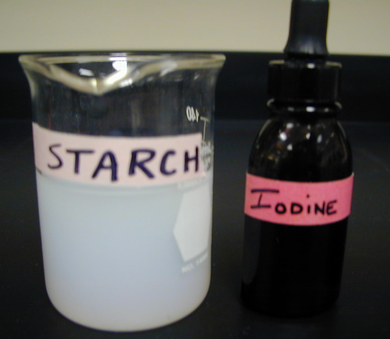
| Solution | Reagent | Positive Result | Negative Result |
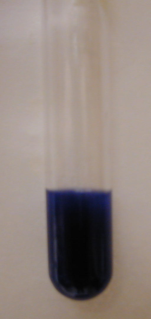
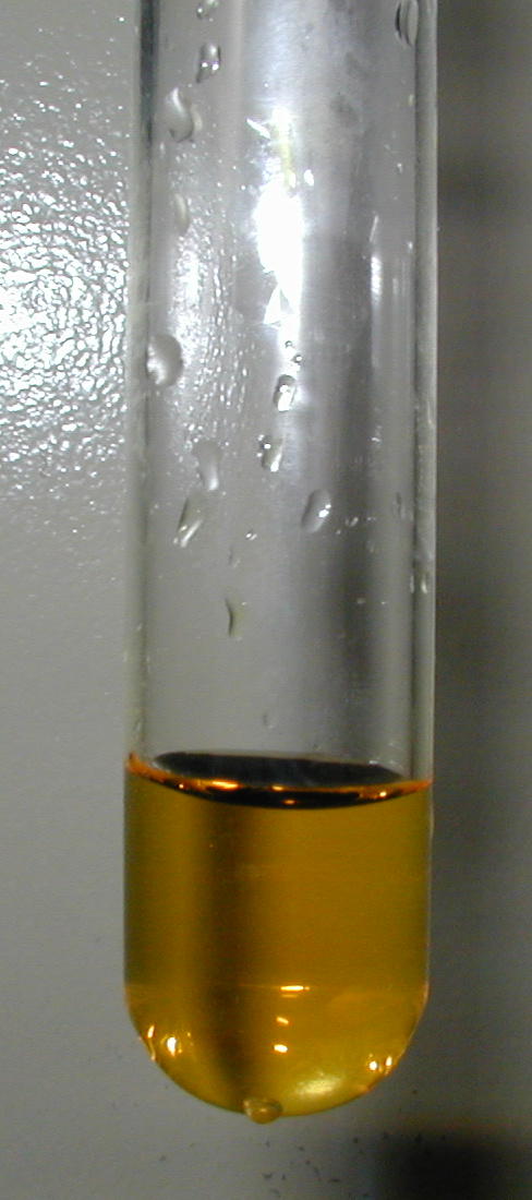
| Starch | Iodine | black | no color change |
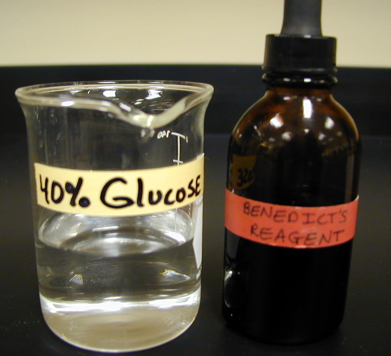
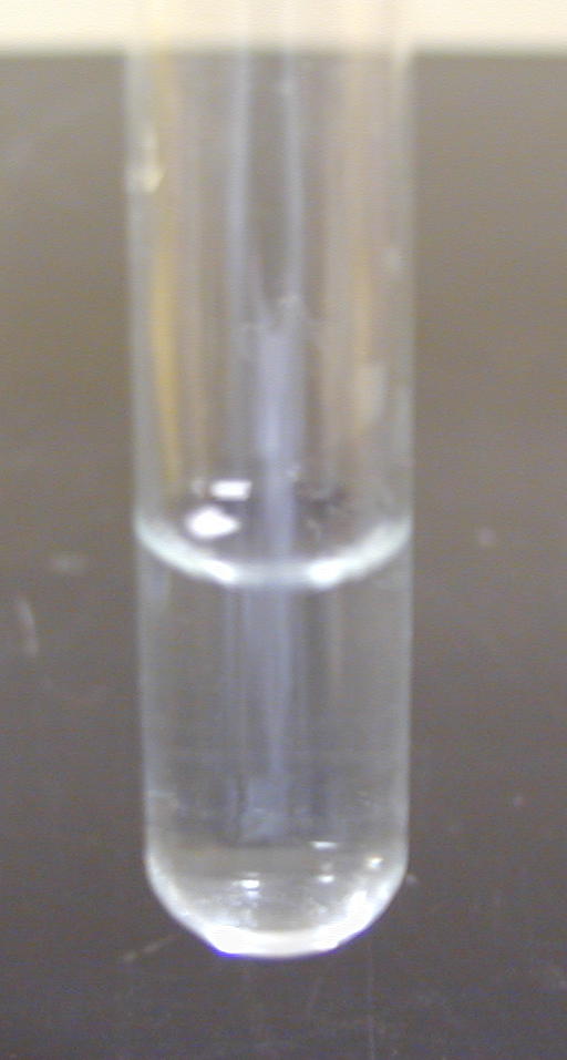
| Dextrose | Benedicts | green-orange-red | original blue color of reagent |
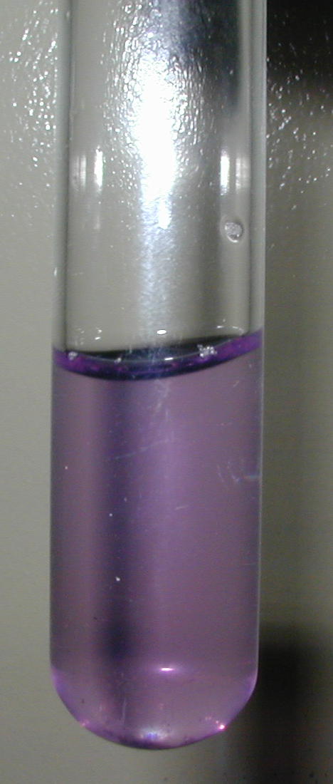
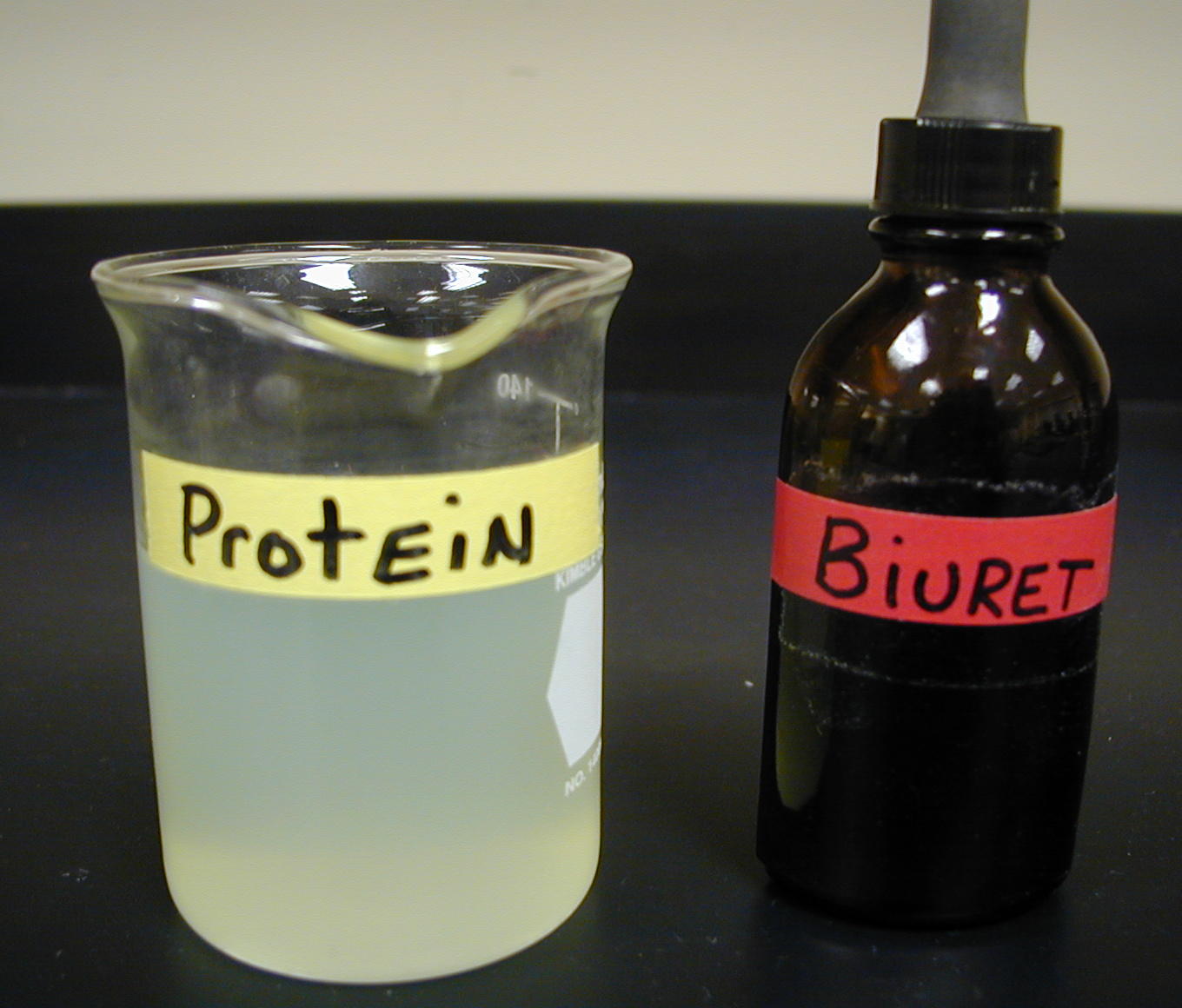
| Protein | Biuret | blue - purple | no color change |
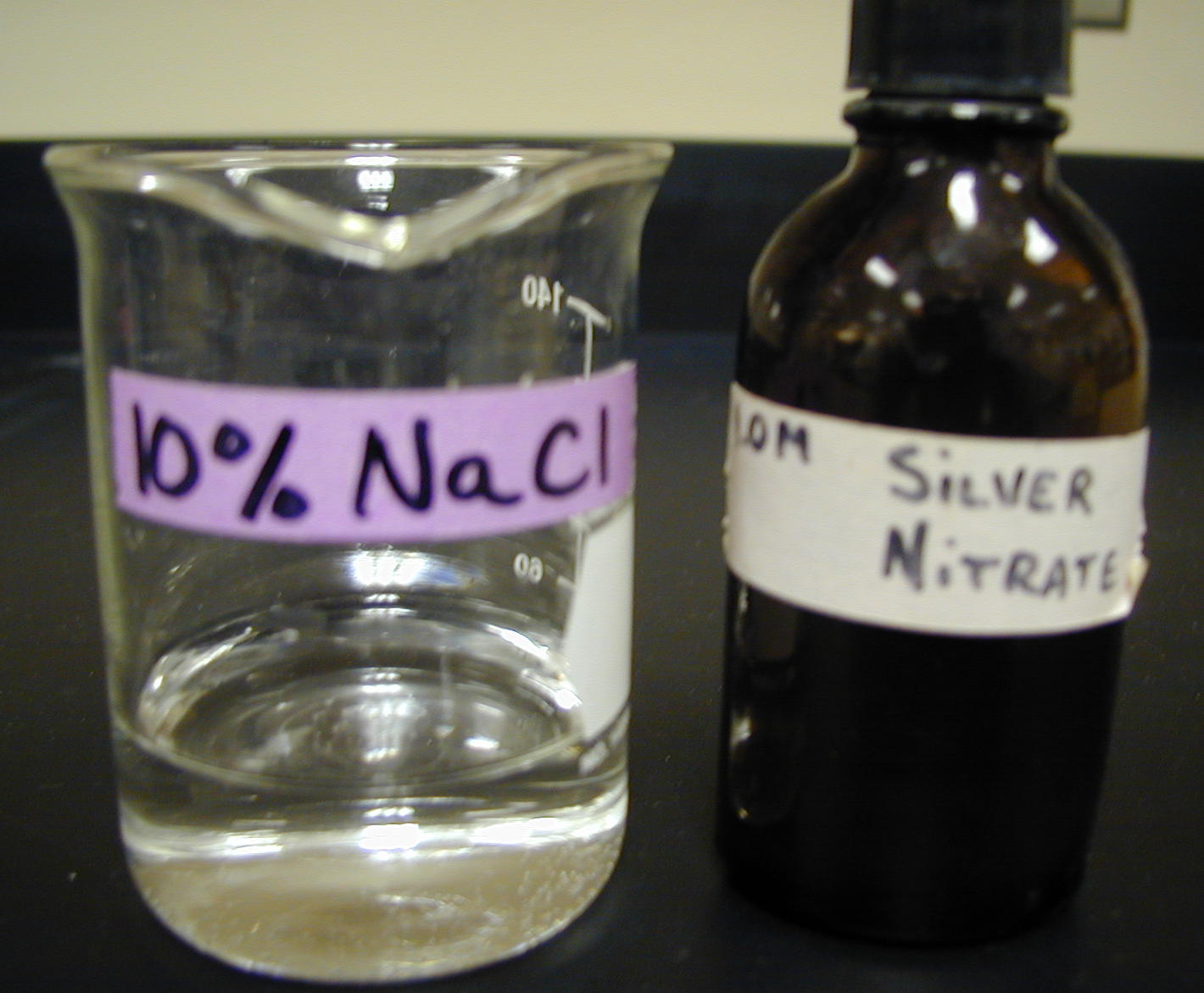
| Salt | Silver Nitrate | white precipitate | no color change |
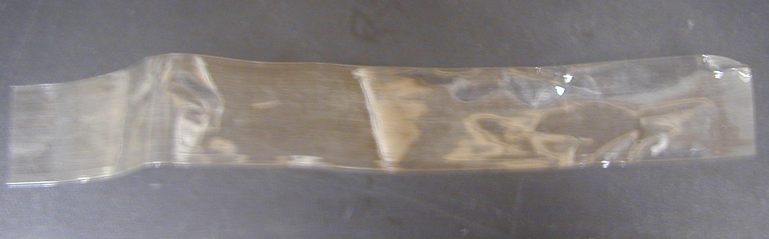
In order to study and understand cell membrane selective permeability, a dialysis
bag is used to
represent the cell membrane
.
It is filled with a mixture
of solutions and tied off. The mixture of solutions represents the cytosol
or ICF.
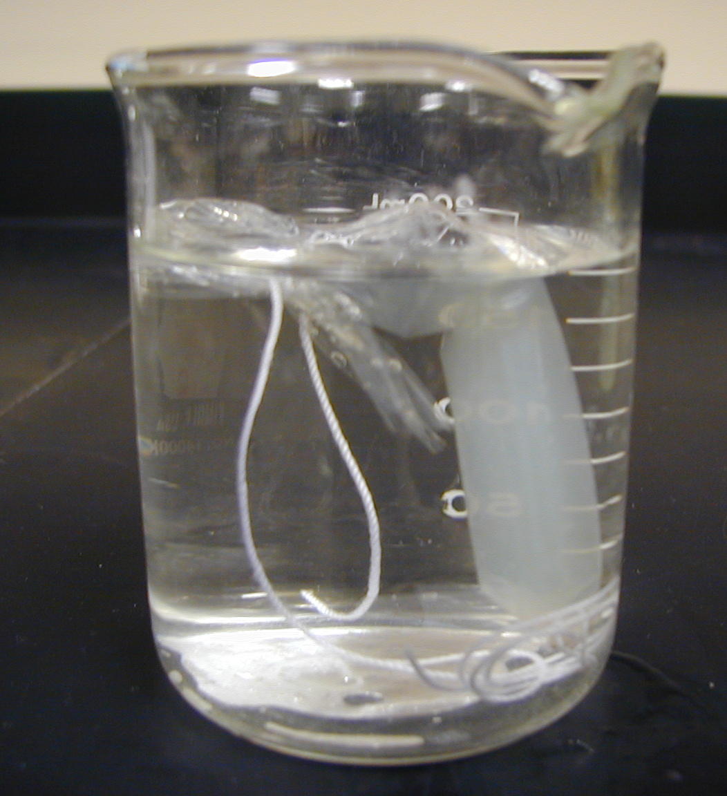
It
is then placed in a beaker
of distilled water which represents the fluid outside the cell,or ECF.
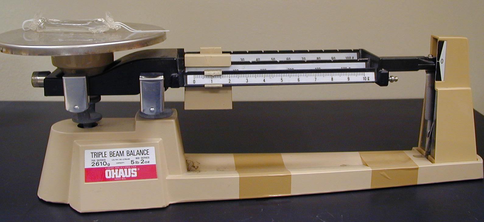
Throught the lab experiment, the bag
is weighed periodically in 20 minute intervals.
Once the 60 minute time limit has been reached, the bag is removed and weighed
for the last time.
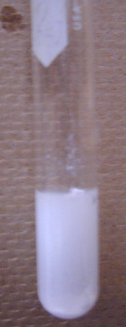
Next, the contents of the beaker are analyzed to see which of the solutions
has diffused through
the dialysis bag membrane by using the reagents shown in the chart above.
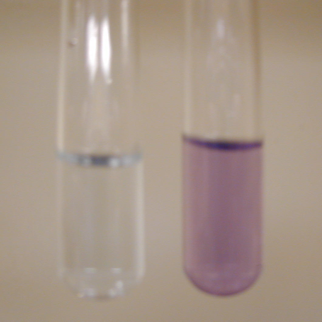
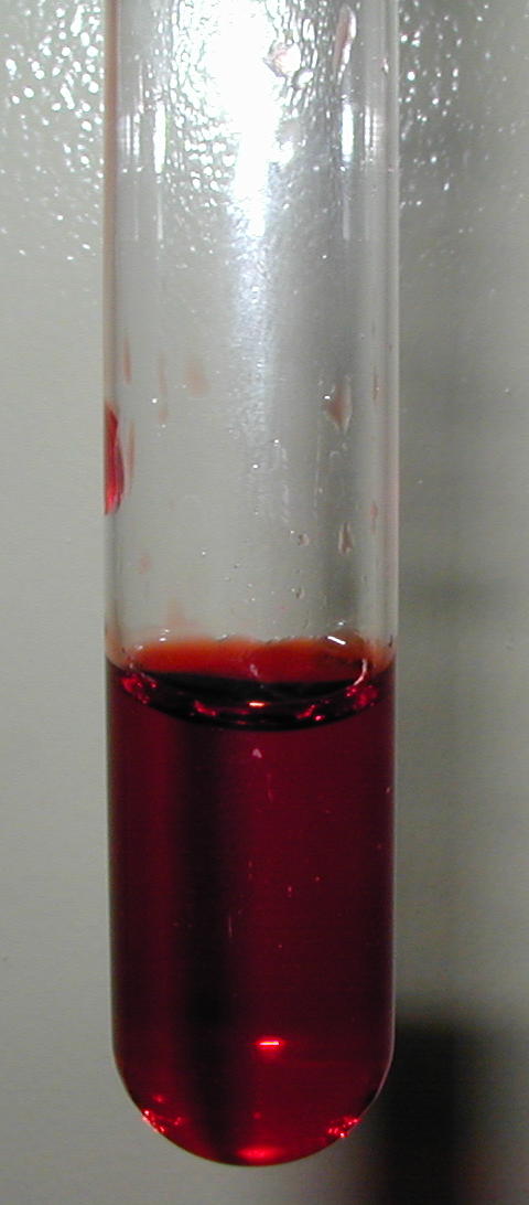
Observe the following results for the beaker solution tests performed and
record your answers :
unknown test tube results link : protein
test ; starch
test; salt
test ; glucose test
Bases on the above unkown results, did some substances "diffuse" through
and other substances did not ?
If so, why ? If not, why ?
Filtration
Coffee filter, machine, and coffee.
Using a coffee machine, fill the machine using water to make 1-2 cups of coffee and place the appropriate filter in the dispensor and add coffee granules. Allow machine to percolate and make coffee in the normal manner. Do any of the granules come through using a filter ? Repeat the experiment without the filter. Do any granules come through? When would filtration be a necessary process in the body? What happens if the body's filter is damaged or missing?
Osmosis
Vegetable baggies, molasses, salt, food coloring, chunky soup, containers, water
Purchase the special baggies for vegetables (the ones with small holes). Obtain four clear containers and fill them with one cup room temperature water. Into one of the baggies place two tablespoons of molasses (honey, kayro syrup) and place this baggie into one of the water baths. Record the time that you start. Check the water solution every 10 seconds and record any changes in the taste of the water.
Repeat this experiment using two teaspoons of salt in the baggie and immersing in a fresh water bath. Record the start time and check the water solution every 10 seconds for any change. For the food coloring, use 20 drops in two tablespoons of water. Place the solution in the baggie and record the start time and if / when you notice a color change to the water bath. With the last baggie, use a chunky style soup (your choice of flavor). Place three tablespoons of soup into the baggie and immerse in the fourth water bath. Record your start time and check the water every 10 seconds for any changes in color or taste.
Since these baggies have small holes, how might this demonstrate the process of osmosis? What if the holes were missing, how might that change the process? What if the holes were larger? How might changing pore (hole) size benefit the body?
Tonicity: Potatoes and Water Experiment. Include results in your LAR (if selected)
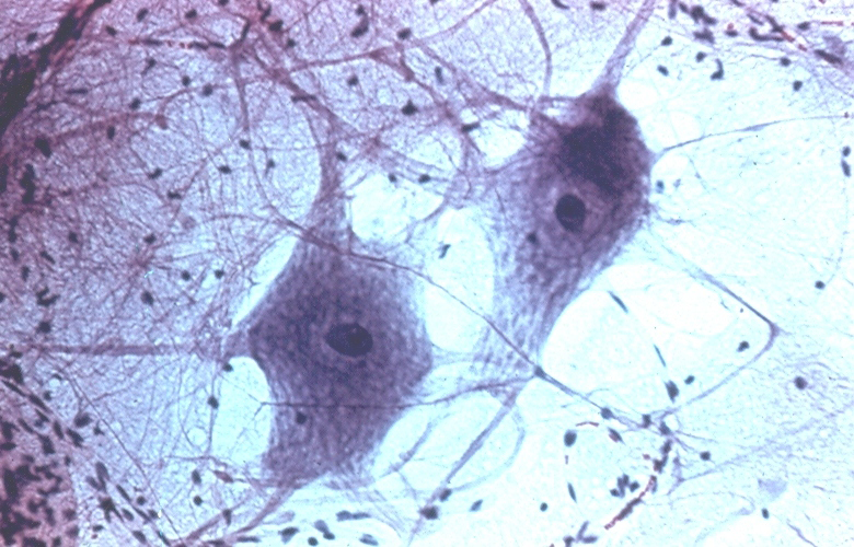
Cell Compare and Contrast: Draw and label a neuron and a simple columnar epithelial cell. Compare and contrast with regards to location, shape, function, and life span. Why do cells need to differentiate?
Concept
Map: Make a concept map about the structure and function
of the major cell parts (nucleus, PM, cytosol) and all the eukaryotic
organelles and their function. Insert a copy of this map into your report
as a document insert or send a PDF scan of the map as an email attachment
when you send your LAR report (if this exercise was selected as your activity
write up).
Cell Game
Go to the cell game website and play the game of cell division supervisor
Make a cell
Make an edible cell for your family and friends. Items needed : One 8" round or square baking pan, 2 boxes of jello mix (any flavor) and edible items such as candies, fruit cocktail, marshmallows, etc. Mix the jello as directed and allow to cool to room temperature. Poor the jello in a saran wrap lined pan. Place the edible items in the jello noting what each item represents for the cell (organelles). Place pan in refrigerator and let cool for several hours or overnight. Completely cover the jello with saran wrap. After setting up, remove the jello mold from the pan and keep the saran wrap on.
Take a picture or send a description of your cell (ingredients used). Identify each ingredient as to what it represents and give its function as its legend. Send picture/description and the legend to your instructor. What does the jello represent ? What does the saran wrap represent ? What do your edible items represent?
Then share, eat, and enjoy your jello cell.
This project will count 5 extra points on the cell lab quiz if done and turned in before the tissue lab quiz.
TV program : C.S.I.
Watch the program Crime Scene Investigators (Las Vegas, New York, or Miami shows) and make note of descriptive references to cells and tissues.
Send your findings to your instructor via email for 3 points extra credit on the cell lab quiz. Must be turned in before the next lab quiz over tissues.
Cytology
DNA analysis
Receptor or Channel Blocking drugs
IV fluids
Cancer
Infectious Diseases (and causes)
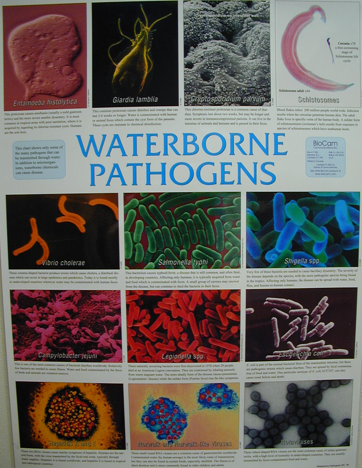
Bacterial pathogens, Water pathogens
Common Colds (viruses, Mycoplasma)
Influenza (Orthomyxoviridae) ~ virrus
Chronic Fatigue Syndrome (Retrovirus)
Acquired Immunodeficiency Syndrome (Human Immunodeficiency Virus)
Lyme disease (Spirochete)
Measles (Rubeola virus)
Rubella (Rubella virus)
Mumps (virus)
Chickenpox (varicella-zoster virus)
Diptheria (Corynebacterium diphtheriae) ~ bacteria
Whooping cough (Bordetella pertussis) ~ bacteria
Lockjaw (Closteridium tetani) ~ bacteria
Forensic Scientist
Microbiologist
Biotechnologist
Clinical Pathologist
Toxicologist
Medical Technologist
http://www.science.ubc.ca/~biomania/tutorial/osmosis/outln01.htm
http://www.cancerindex.org/medterm/index.htm
http://www.cancernet.nci.nih.gov/
http://www.cancer.org (Americal Cancer Society)
http://www.nigms.nih.gov/news/science_ed/life.html inside the cell
http://gslc.genetics.utah.edu/index.html
Study Guide and Review Questions
1. Define cell and give the major parts and their functions.
2. Define mitosis, list the phases in order.
3. Name and give the function of 4 membranous organelles.
4. Name and give the function of 2 non-membranous organelles
5. Define ion and give 4 examples
6. Define magnification and give the formula
7. Name the passive cell membrane transports and define each.
8. Give two examples of active transport mechanisms and the substance affected.
9. Define ICF and ECF.
10. Give the importance of understanding the entire cell cycle.
11. Define pH and explain its importance in body fluids
12. What is the purpose of cholesterol in the cell membrane?