Biology 2404 A&P Basics Lab Exercise 6a Nervous System: CNS Dr. Weis
| Objectives | Background | Medical Terms | Activities | Applications | Careers | WWW | Review Questions |
Students should be able to:
* Name the anatomical and functional divisions of the nervous system
* Name the three major neuron types and their function
* Name the neuroglial cells and their function
* Describe the basic neurophysiology events
* Define synapse and give its structure
* Name the major areas of the brain and their functions
* Give the structure and function of the spinal cord
* Name four neurotransmitters and their function
Read related material in the textbook
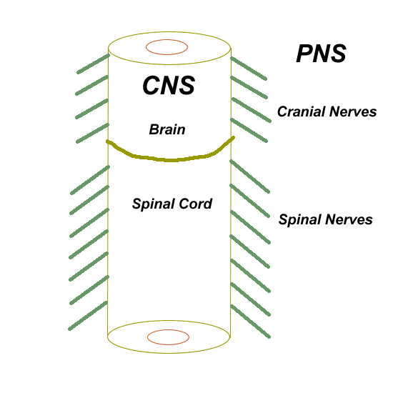
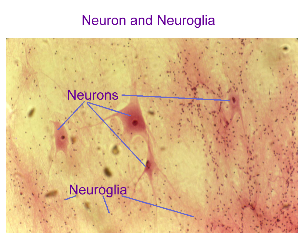
The nervous system comes from embryonic neural ectoderm. This flat sheet of neural ectoderm cells forms a tube that will become the central nervous system, the CNS. Outgrowths and extensions from the CNS tube will become part of the peripheral nervous system, the PNS. The CNS and PNS divisions of the nervous system are formed by two types of neural tissue: neurons and neuroglia.
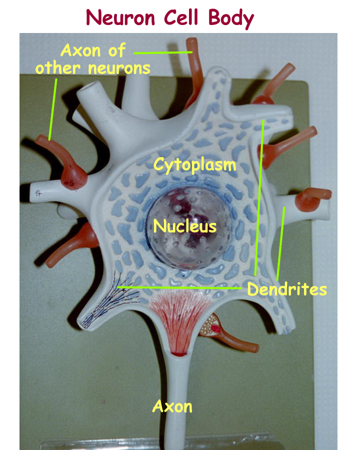
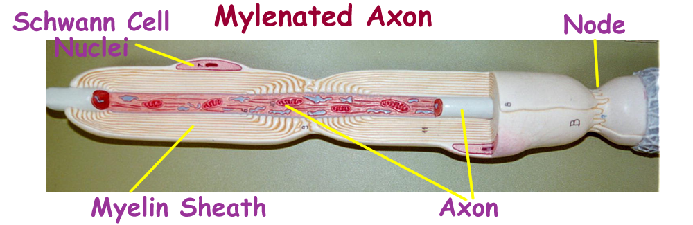
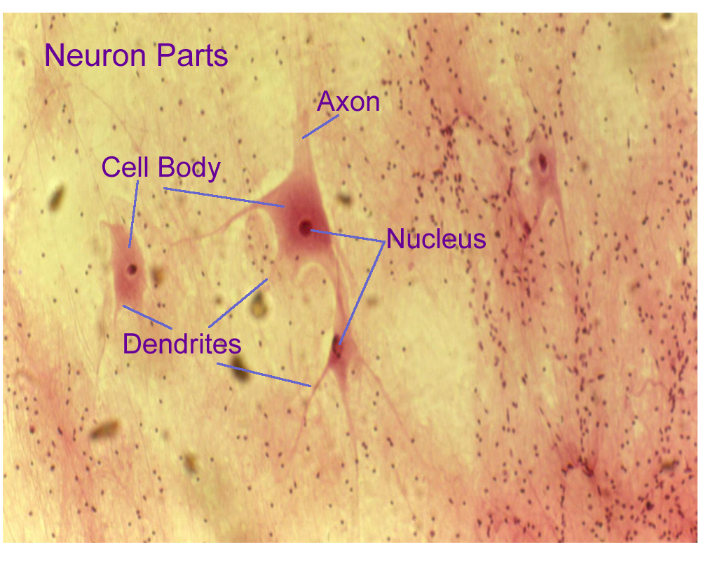
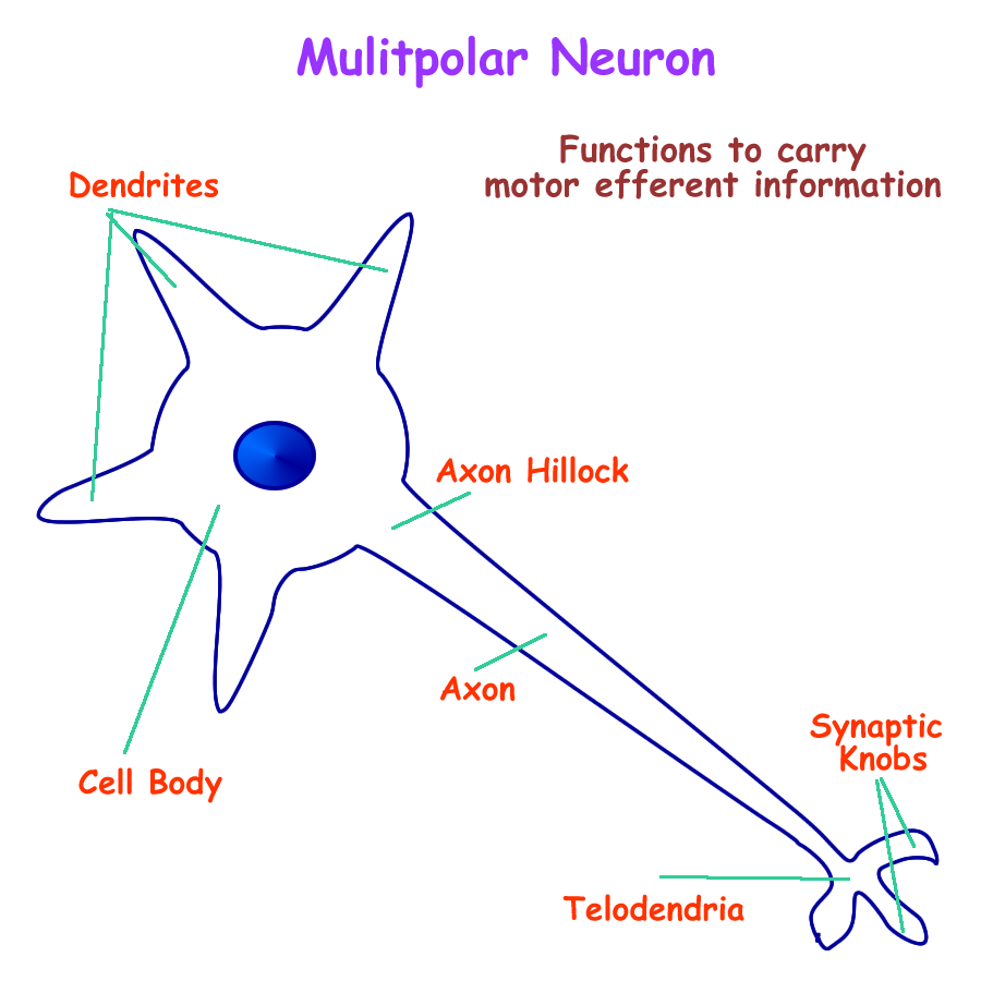
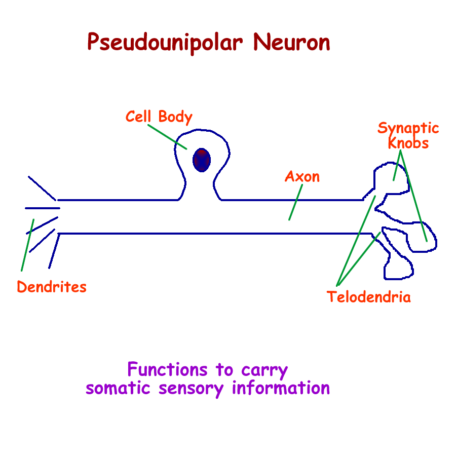
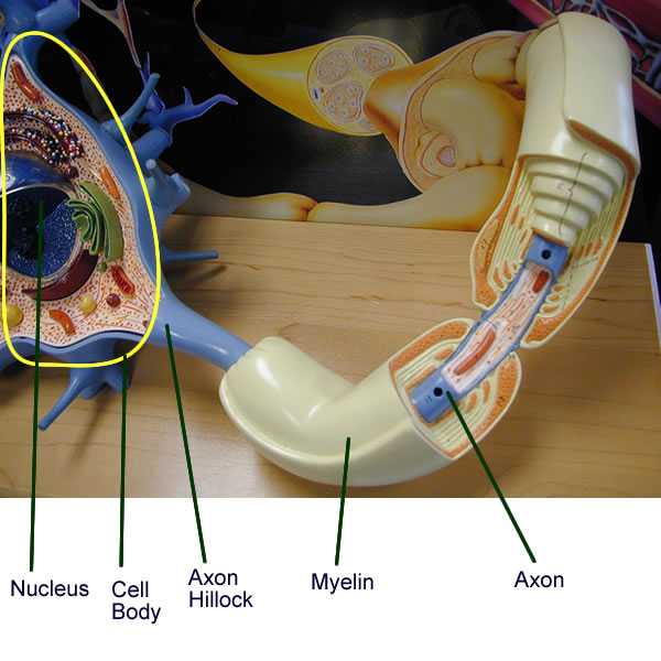
Neurons are specialized cells for conducting electrical impulses. Neurons have a cell body that contains the nucleus and most organelles, short extensions called dendrites receive signals, and a single longer extension called the axon is used for sending impulses. The terminal portion of the axon forms finger like processes called telodendria that end in terminal buttons also called synaptic knobs.
The nucleus of the neuron directs the production of the neurotransmitter, the chemical signal that is stored and released at the synaptic knob. Neurons can be classified according to structure, function, and even neurotransmitter produced.
Functionally, neurons are divided into cell body with dendrites and axons.
The three major types of neurons seen are:
Multipolar CNS sensory afferent
CNS motor efferent
PNS motor efferent
Pseudounipolar PNS somatic sensory afferent
Bipolar PNS special sensory afferent
Structural classification is based on number of processes, primarily dendrites as most neurons only have one axon. Afferent and efferent are the directional terms for the signal and their names are referenced according to the CNS. Afferent means to carry the information to the CNS and efferent means to carry the information away from the CNS to the PNS for motor response.
Somatic sensory information is detected by the skin sensory receptors from the body and deals with pain, temperature, touch, pressure and position. Sensory information can be received from the special senses of hearing, vision, taste, and smell.
Motor information deals with signaling movement in the form of a muscle contraction by smooth, cardiac, or skeletal muscle or with movement in the form of a glandular secretion.
Neuron Model : whole, cell body, axon
SYNAPSES
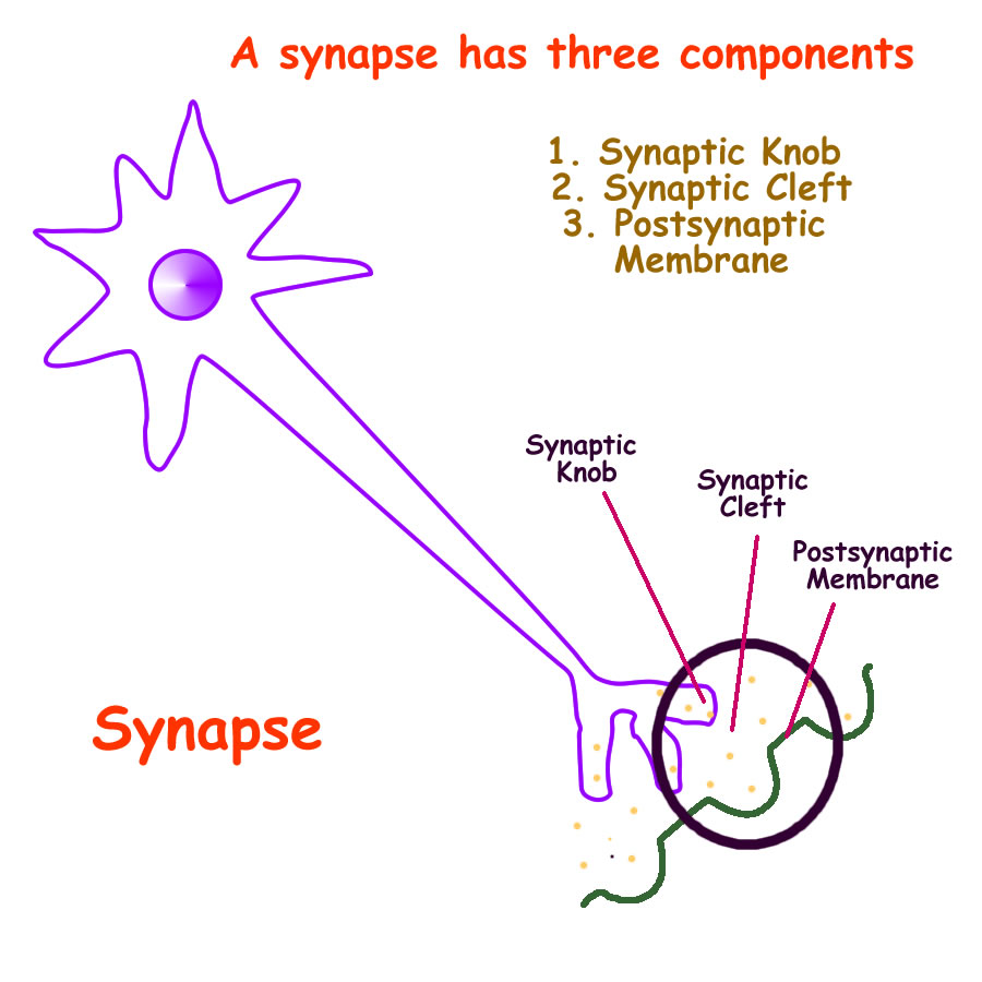
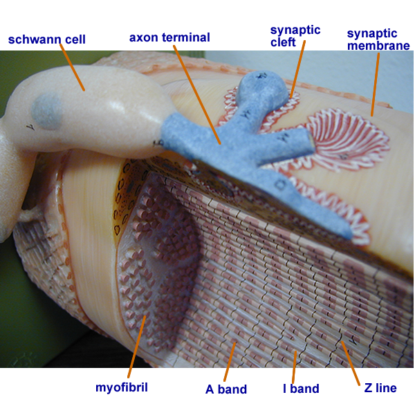
Nerves to nerve junctions for transmission of impulses are termed a synapse. Nerves that transmit signals to other tissues such as muscles and glands are termed junctions, such as the neuromuscular junctions and neuroglandular junctions. A synapse and junction have similar structures:
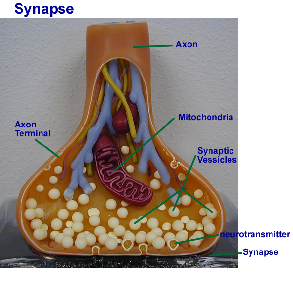
Synaptic Knob store and release neurotransmitters
Synaptic Cleft small fluid filled space between communicating cells
Post synaptic Membrane contain the receptors for the neurotransmitters
Synapse Drawing : Neuron to Neuron, Neuromuscular Junction, Neuroglandular Junction
Neurotransmitters
Neurotransmitters are the general term for chemicals made in the synaptic knob of neurons and released into the synaptic cleft of the synapse or junction. Chemically, neurotransmitters can be classified according to their structure and function. Major neurotransmitters used in the CNS are acetylcholine, serotonin, dopamine, norepinephrine, GABA, and endorphins.
Major neurotransmitters used in the PNS are acetylcholine and norepinephrine. Structural and functional classifications are based on chemical class and how the neurotransmitter acts at its receptor. Most neurotransmitters are based on amino acids. Direct acting means that the neurotransmitter opens the ion channels linked to the receptor and indirect acting means that the neurotransmitter signals other chemicals to open or close ion channels linked to the receptor. Excitatory means that the neurotransmitter depolarizes the neuron to reach threshold, while inhibitory means that the neurotransmitter hyperpolarizes the neuron to prevent it from sending a signal. Neurophysiology terms are discussed in the next section.
1. Acetylcholine Ach CNS, PNS direct acting, excitatory
2. Amines
Dopamine CNS indirect acting *
Serotonin CNS indirect acting inhibitory
Norepinephrine CNS, PNS indirect acting *
* excitatory and inhibitory, depending on receptor
3. Amino Acids
GABA CNS direct acting, inhibitory
4. Peptides
Endorphins CNS indirect acting, inhibitory
NEUROPHYSIOLOGY
In order to signal other neurons, muscles, or glands, an electrical impulse must be generated in response to a chemical, electrical, or mechanical signal. Neurons are excitable tissue capable of changing ion concentrations across their membrane.
When ions move, electrical impulses are created causing other ion channels to open or close. Neurophysiology terms used are:
| Resting Membrane Potential | ion concentrations established by the Na+/K+ pump in the cell membrane Na+ concentration is slightly higher outside K+ concentration is slightly higher inside |
| Depolarization | Opening of Na+ gated ion channels to allow influx of sodium and lower the Resting membrane potential |
| Threshold | When the movement of Na+ = K+. The trigger point for an impulse to be fully generated at the axon hillock |
| Action Potential | Continuation of Na+ influx to create the nerve impulse along the axon |
| Repolarization | Opening of K+ gated channels to allow Outflux of K+ to help re-establish theionic charges of the resting membrane potential |
| Hyperpolarization | Opening of K+ or Cl- gated channels when the neuron is at rest. Prevents the neuron from reaching threshold and creating an impulse. |
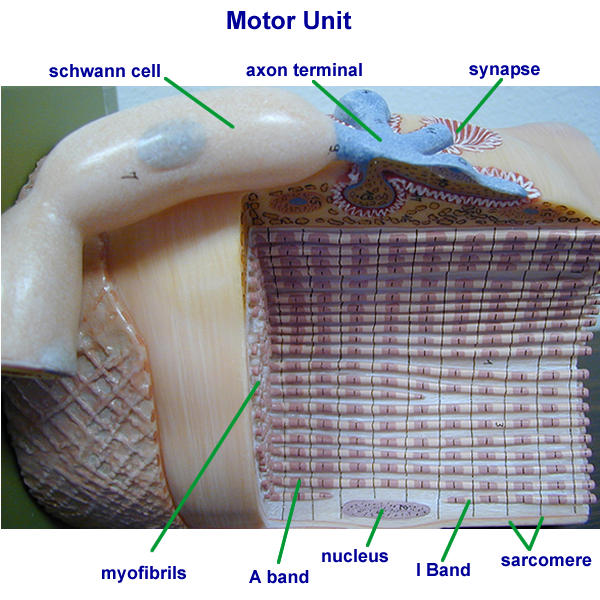
Neuromuscular junction and motor unit
Nervous System review of action potentials : click on appropriate site link for this review
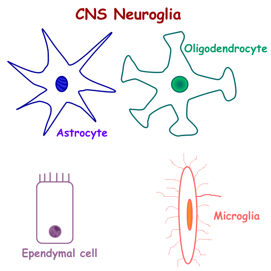
Neuroglial cells are the support cells for the neuron’s two functional parts : cell body with dendrites and axons.
In the CNS, the neuroglial cells are:
Astrocytes support the cell body and dendrites
Regulate the ionic environment of this region
Helps form the blood brain barrier
Oligodendrocytes support the axon
Insulates with a single wrapping of its cell membrane
Myelinates with multiple wrappings of its cell membrane
Ependymal Cells lines the inside of the CNS tube
Helps form and circulate Cerebral Spinal Fluid (CSF)
Microglial Cells WBC macrophages that provide immune defense
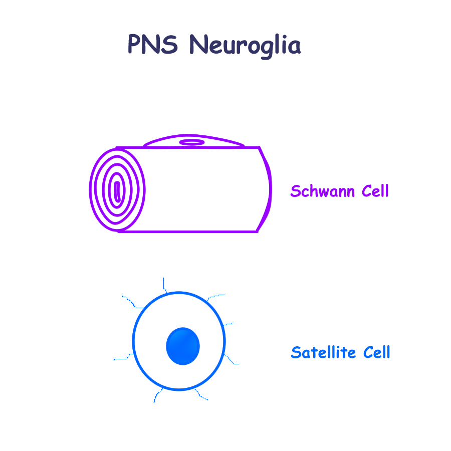
In the PNS, the neuroglial cells are:
Satellite cells support the PNS cell body and dendrite regions
Schwann Cells supports the PNS axon
Insulates with a single wrapping of its cell membrane
Myelinates with multiple wrappings of its cell membrane
Neuron Drawing : Multipolar, Bipolar, Pseudounipolar
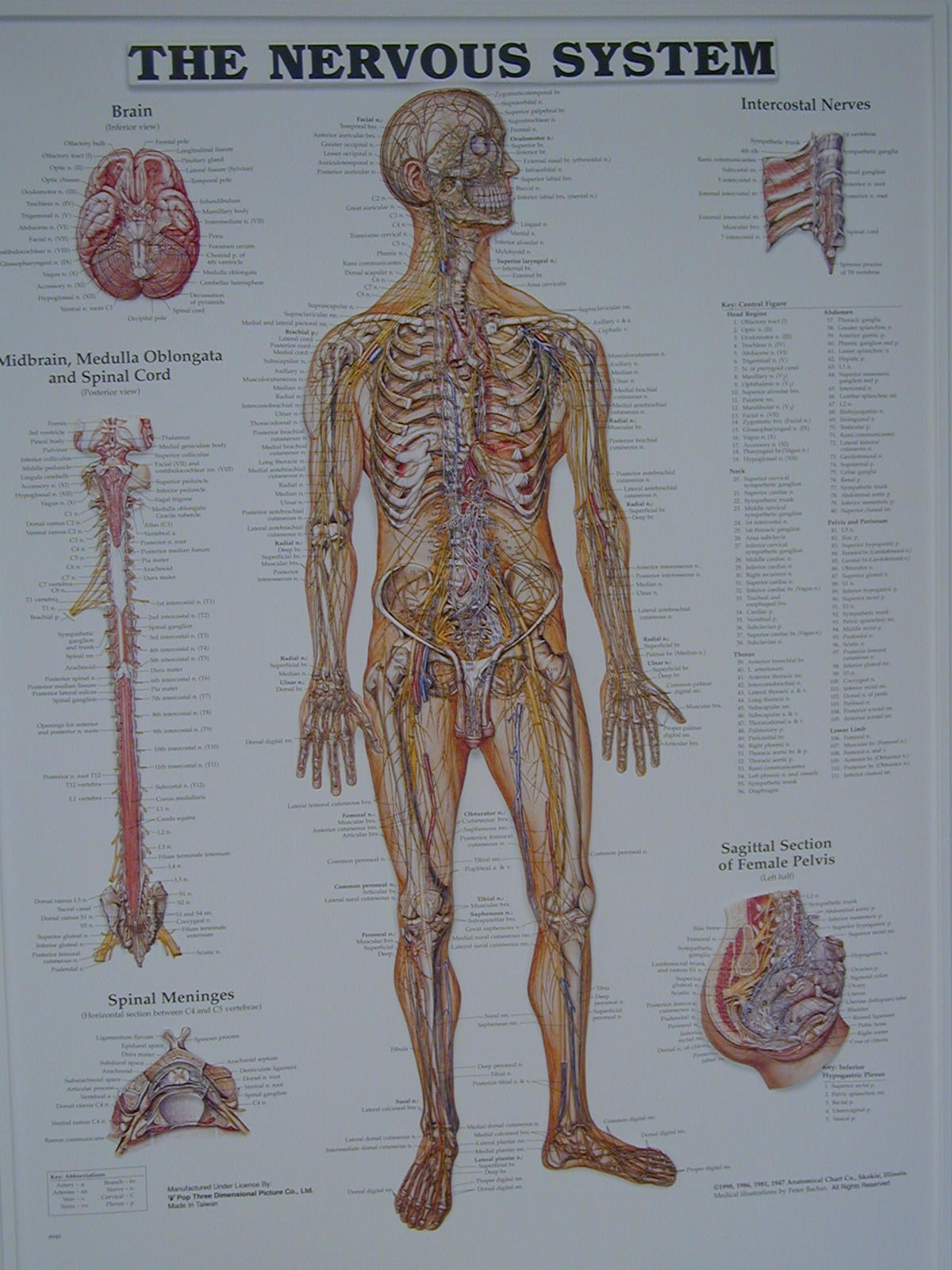
Central Nervous System (CNS)
The central nervous system consists of the brain and spinal cord. The brain develops at the proximal end of the neural tube. Vesicles develop to form adult structures:
Telencephalon Cerebrum
Diencephalon Thalamus, Hypothalamus, Epithalamus
Mesencephalon Midbrain
Metencephalon Pons, cerebellum
Myelencephalon Medulla oblongata
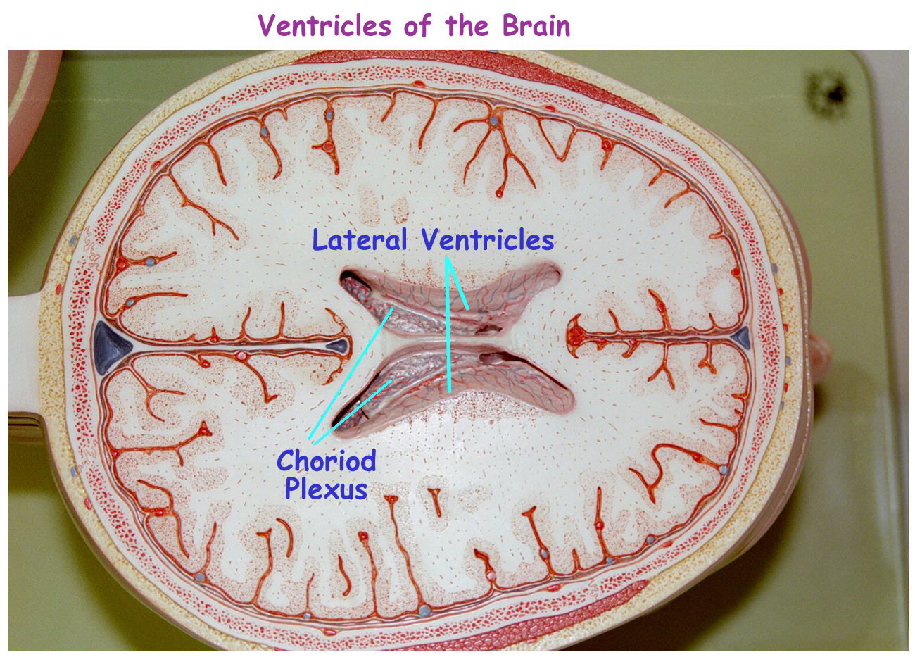
The CNS tube stays hollow throughout the length and these hollow areas will form the ventricles of the brain and the central canal of the spinal cord. The ventricles and central canal are both lined with the ependymal cells that produce CSF in the ventricles and these cells help to circulate this fluid in and around the CNS.
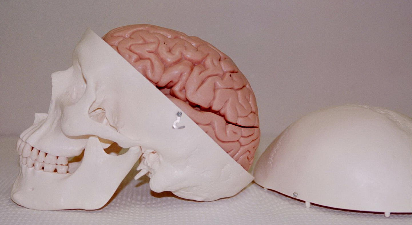
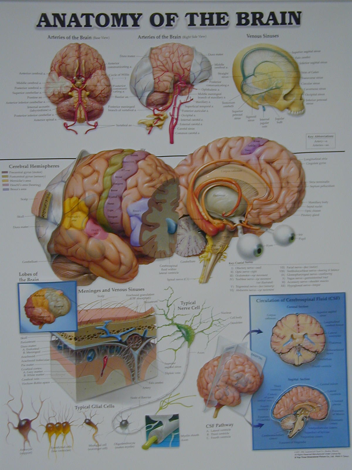
Protection for the CNS consists of many bones of the axial skeleton, CSF, and the connective tissue meninges. The brain has microglia and the blood brain barrier for extra protection. The spinal cord relies on local tissue defenses for its extra protection.
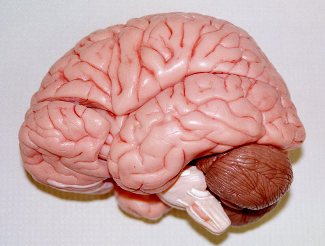
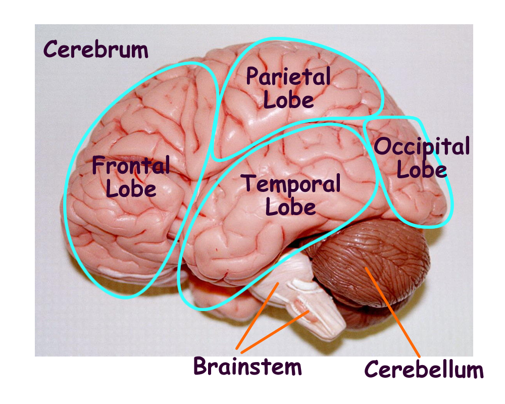
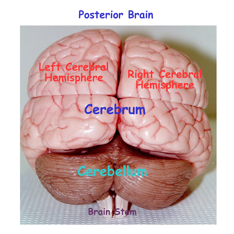
Cerebrum
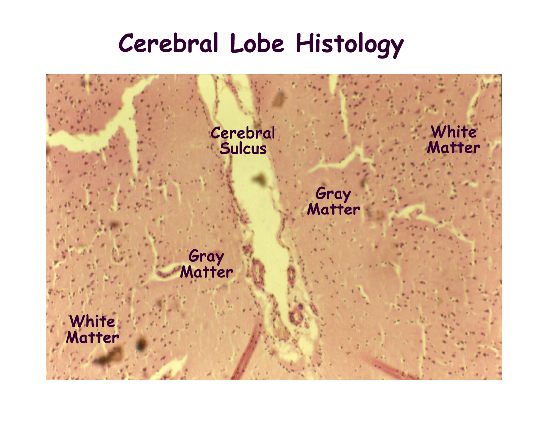
The cerebrum is the largest part of the brain and is divided into right and left hemispheres by the longitudinal fissure. Grossly it has outward folds called gyri and inward depressions called sulci which help to increase surface area.
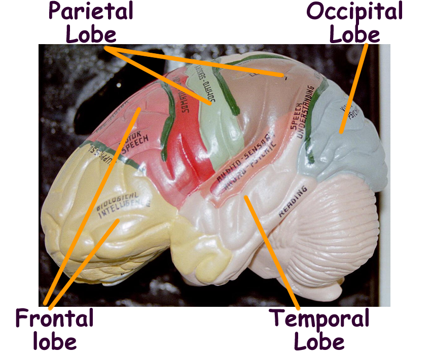
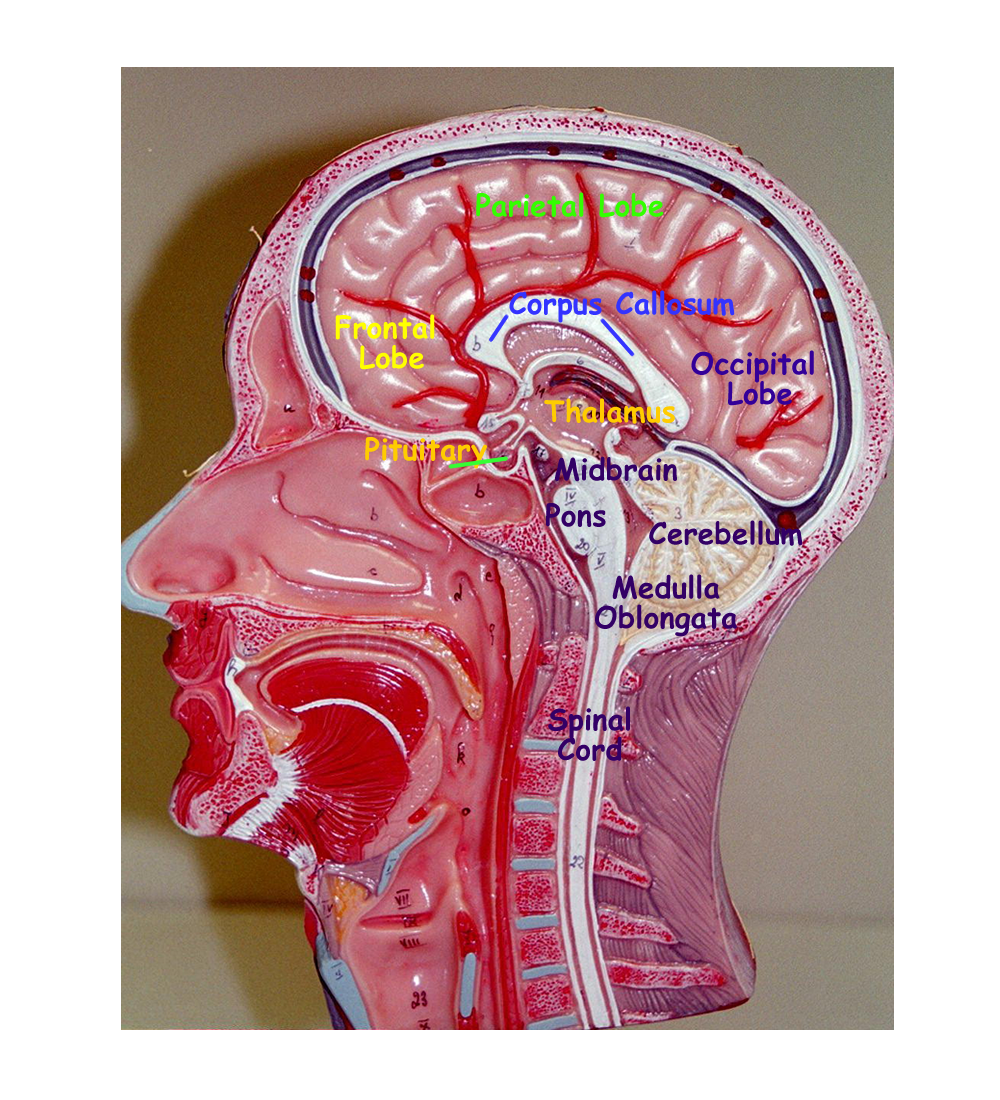
The cerebrum is divided into functional lobes protected and referenced by the cranial bones. The cerebral lobes and their functions are:
Frontal Lobe primary motor cortex, learned motor skills, eye reflexes, Speech
Pareital Lobes primary somatosensory cortex for body senses
Temporal Lobes primary auditory lobe
Occipital Lobe primary visual lobe
Within each lobe there are association areas where information is stored for retrieval to compare old and new information.
Prefrontal association area in frontal lobe planning, moods, motivation
Parietal association area in Parietal lobe pain, temperature, touch, pressure
Temporal association area in Temporal lobe auditory sounds
Occipital association area in Occipital lobe visual images
A fifth internal lobe, the insula, exists for autonomic information, storage, and control
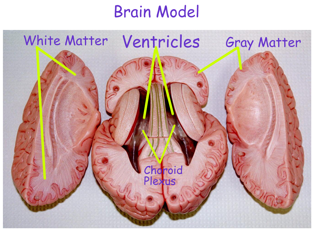
Histologically, the cerebrum has an outer grey matter and an inner white matter. The grey matter which forms the cerebral cortex is the nonmyelinated portions of neurons, primarily the cell body and dendrites. The white matter which forms the myelinated portions of the neuron, primarily axons whose fiber connections to and within the brain creates the association fiber tracts within each lobe, the commissural fibers such as the corpus callosum that connects right and left hemispheres of the cerebrum, and the projection fibers that connect the cerebrum to the rest of the brain structures. White matter also forms basal nuclei, most of which deal with motor information to the spinal cord for the skeletal muscles of the body.
The lateral (1st, 2nd) ventricles are located within the cerebrum and produce CSF for cushioning, support, and nutrition of the CNS neurons.
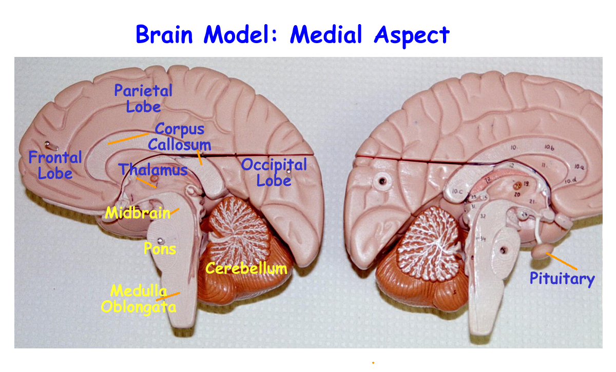
Brain models : Lateral, Posterior, Medial , Superior
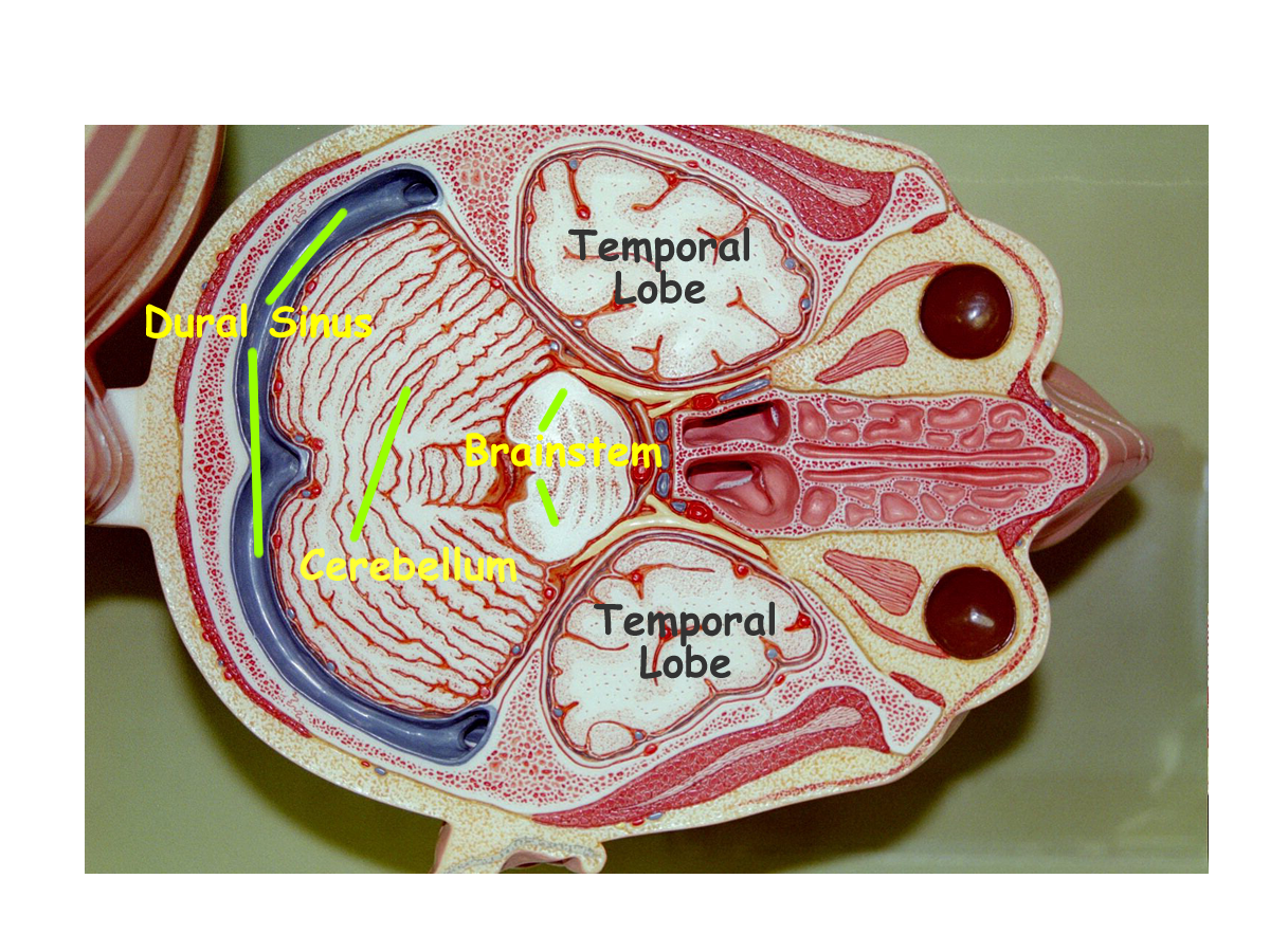
Transverse section head with ventricles
Brain with skull: Dorsal, Lateral
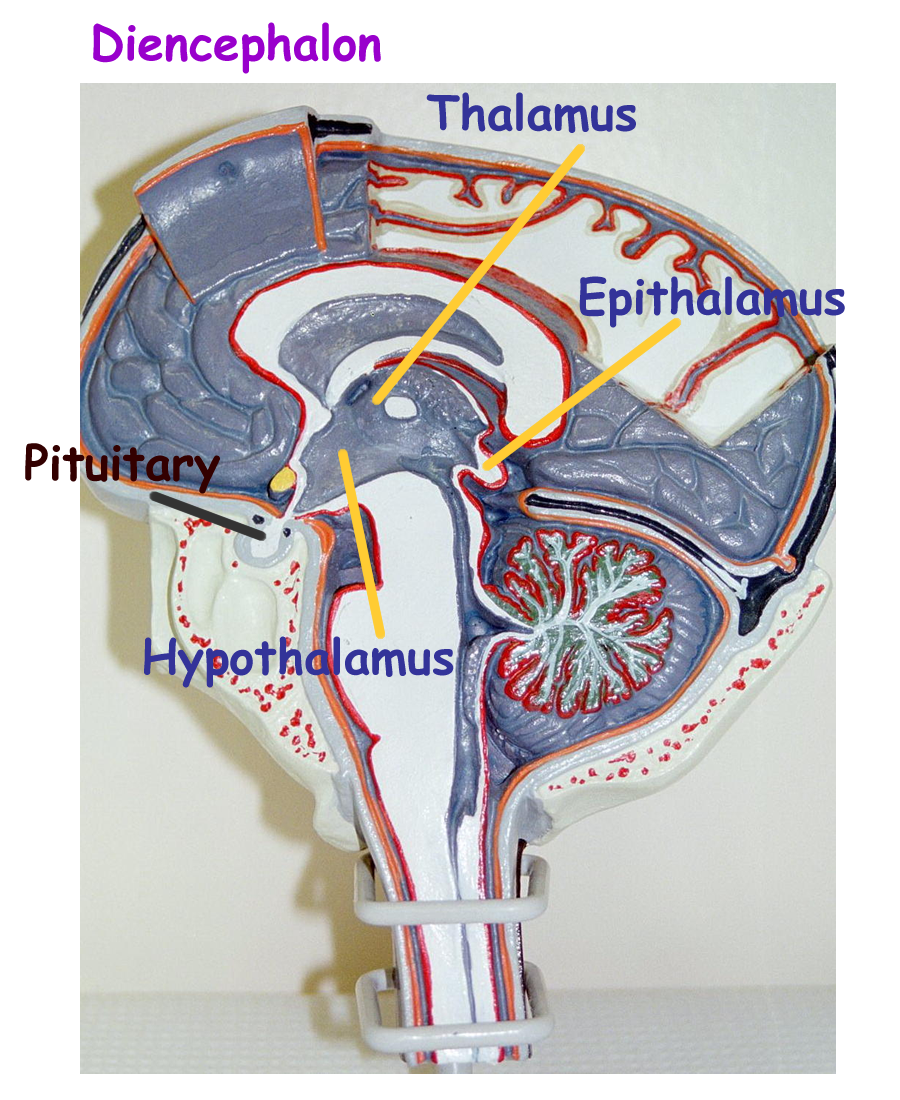
Diencephalon
The diencephalon is the major functional part of the brain and is found encircling the third ventricle. It is divided into three regions: the thalamus, the hypothalamus, and the epithalamus
The epithalamus is superior and caudal to the thalamus and functions to house the pineal gland, and endocrine structure that secretes melatonin from the amino acid tryptophan to help regulate sleep/wake cycles.
The thalamus is the paired middle portion of the
diencephalon and functions as a direct relay for all sensory information except
smell.
Smell will eventually be routed through the thalamus after it is processed
in the limbic system. The sensory
relays direct new incoming sensory information to the proper lobe for processing,
storage, and analysis. The thalamus is also involved with memory and learning.
The hypothalamus forms the lateral and ventral walls of the third ventricle and its many functions are:
1) Autonomic nervous system control
2) Endocrine function and control of the pituitary gland (both Anterior and Posterior)
3) Temperature regulation
4) Sex drive
5) Feeding and thrist reflexes
6) Emotions
7) Sleep wake cycles
8) Memory and learning
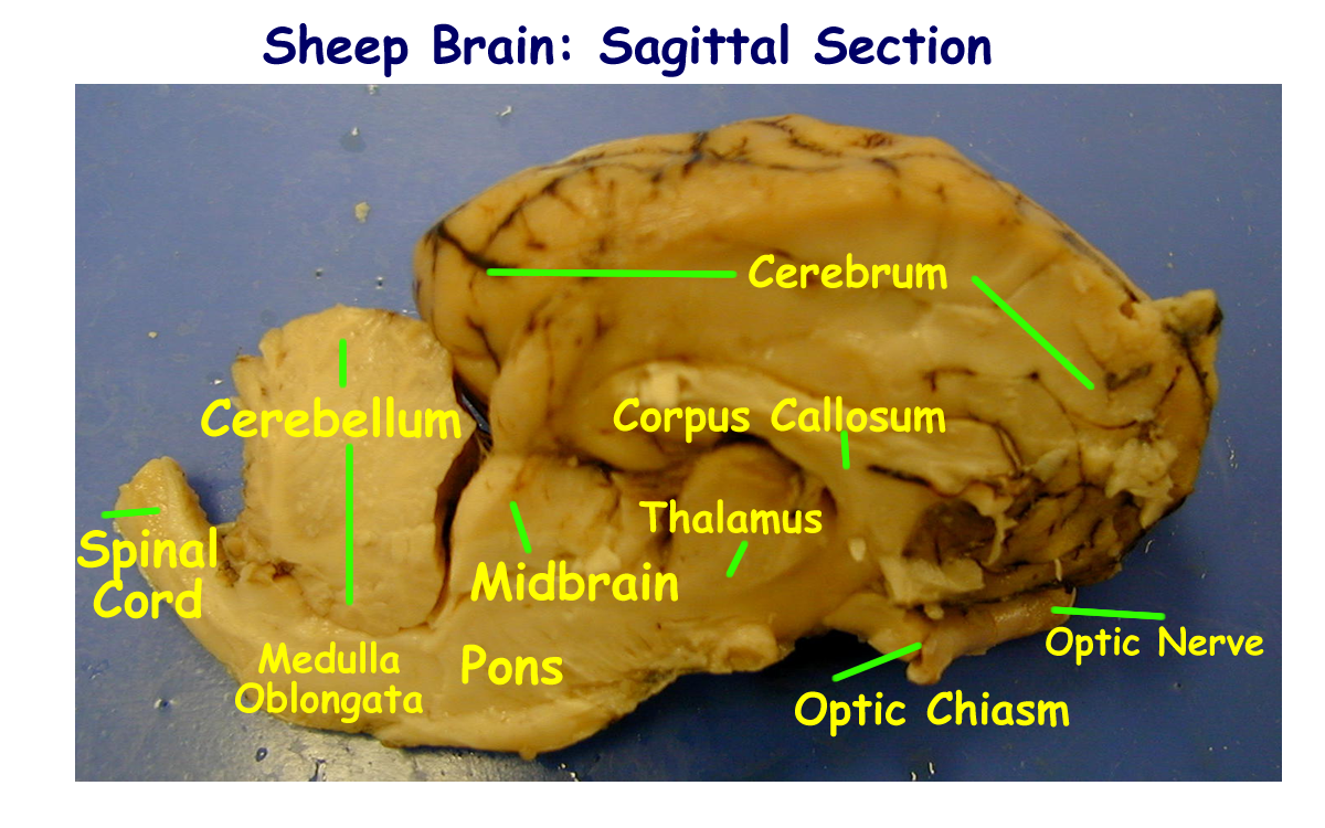
Sagittal cut brain Latex model
Internal Brain: Limbic System / Diencephalon
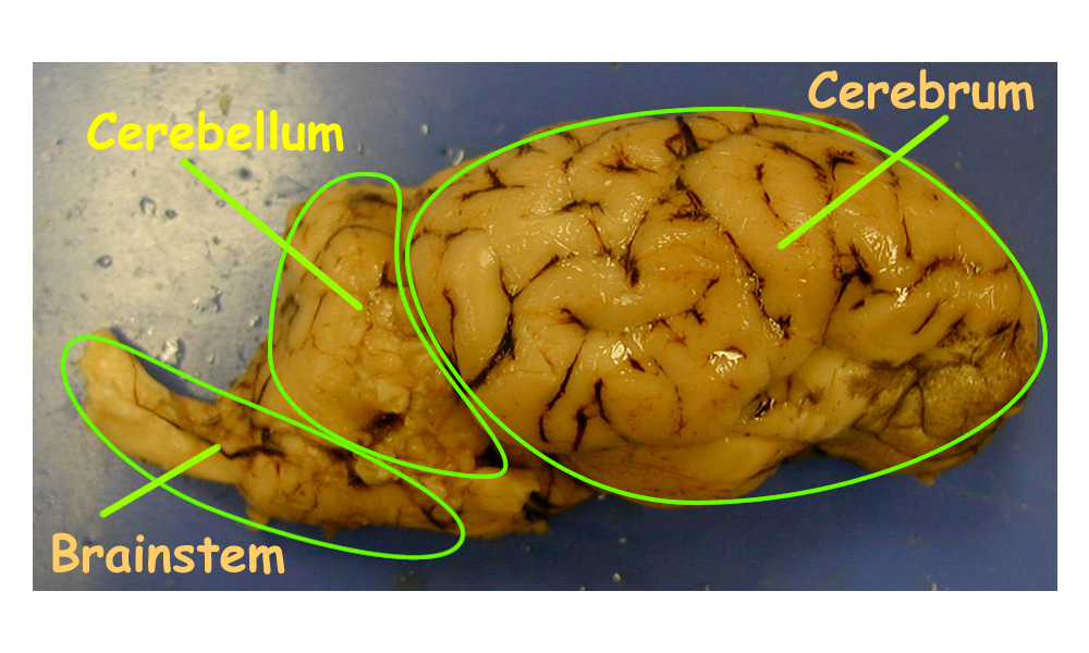
Brainstem
The brainstem consists of the midbrain, pons, and medulla oblongata. All regions of the brainstem allow for fibers to transmit sensory ascending information to and motor descending information from the spinal cord, cerebellum, diencephalons, and cerebrum. The brainstem also contains cell bodies of most of the 12 pairs of cranial nerves.
Midbrain
The midbrain or mesencephalon contains the mesencephalic aquaduct for CSF circulation and is also responsible for auditory and visual motor reflexes.
Pons
The pons of Varolii contains the fourth ventricle for CSF production and circulation. Additional functions include respiratory centers to change ventilation by increasing or decreasing normal breathing rates.
Medulla Oblongata
The medulla oblongata also contains the fourth ventricle for CSF productio and circulation. Additional functions include setting the normal heart and respiratory rates, vomiting reflexes, sneezing reflexes, and coughing reflexes. This is the area where the major axon fiber tracts carrying motor information cross to the opposite side of the body for skeletal muscles on the contralateral side.
Cerebellum
The cerebellum forms the second largest structure of the brain and is located ventrally to the occipital lobes of the cerebrum, separated from the cerebrum by the transverse fissure. The cerebellum has right and left hemispheres, but a thickened band of tissue known as the vermis separates the two sides. The gray matter of the cerebellum forms the outer leaf like folia of the cerebellar cortex while the white matter axon fiber tracts form the inner arbor vitae, the tree of life. The base of the arbor vitae forms cerebellar peduncles that connect the cerebellum to the cerebrum by way of the brainstem for sensory and motor exchange.
Functions of the cerebellum include coordinating and modifying motor movement and directions of the frontal lobe of the cerebrum by changing and applying signals for the appropriate rate, range, and force adjustments. Other functions of the cerebellum are the regulation of muscle tone, and the maintenance of body position and equilibrium based on input from the vestibular portion of the inner ear.
Sagittal Cut brain Latex Model
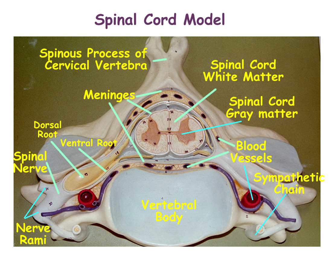
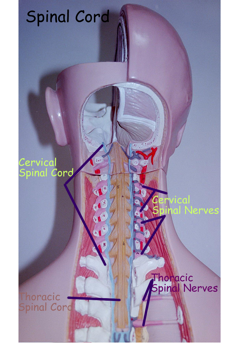
Spinal Cord
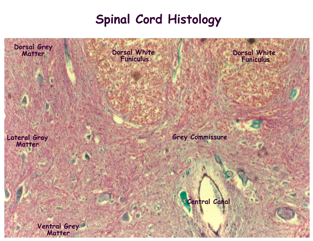
The spinal cord is the distal continuation of the CNS neural tube. Grossly it begins at the end of the medulla oblongata and exits at the foramen magnum of the occipital bone of the skull. The length of the spinal cord in humans extends to the first or second lumbar vertebrae of the spinal column and tapers to a point called the conus medullaris. The hollow center of this tube is called the central canal and is lined with ependymal cells that help circulate CSF produced in the ventricles of the brain.
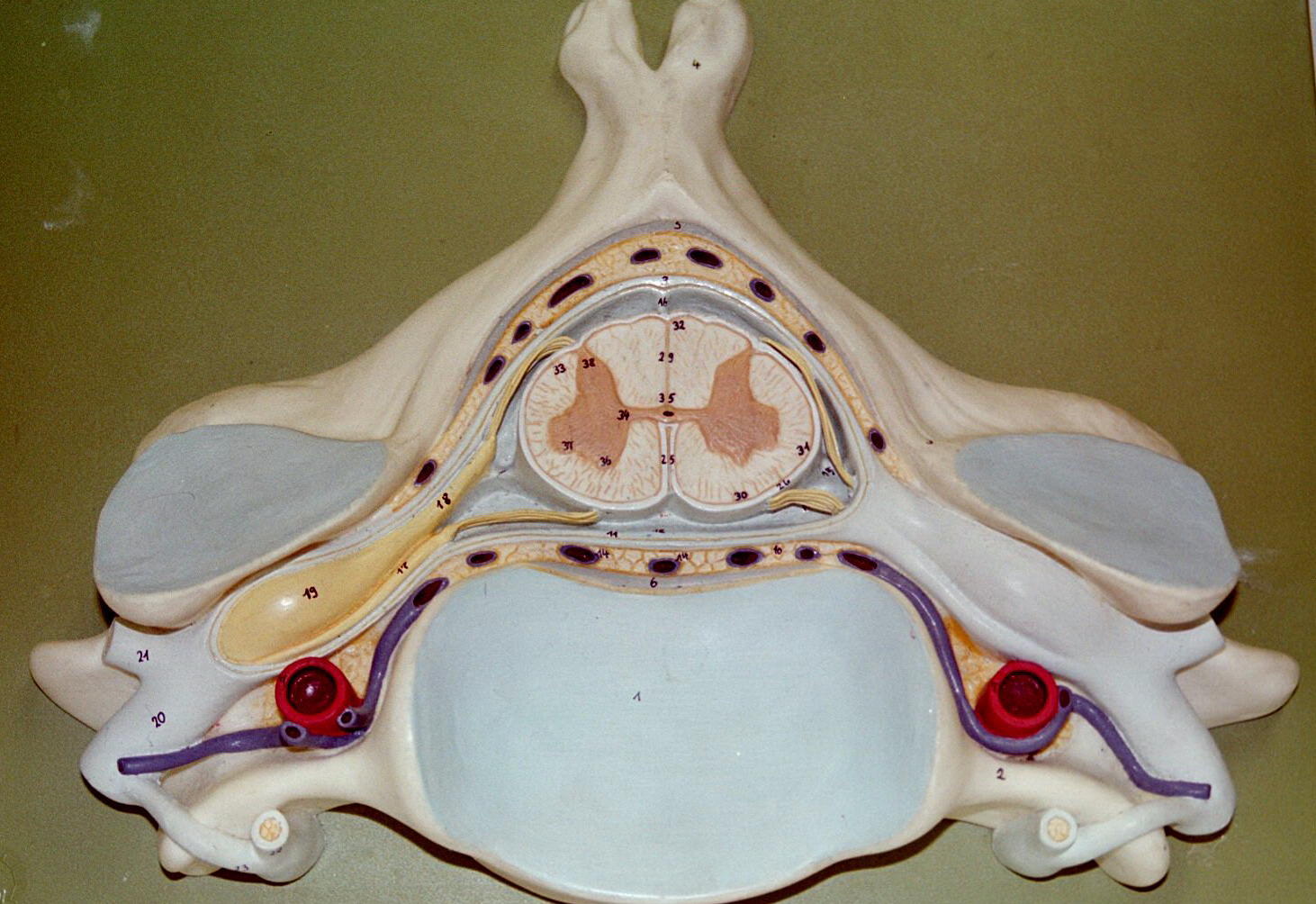
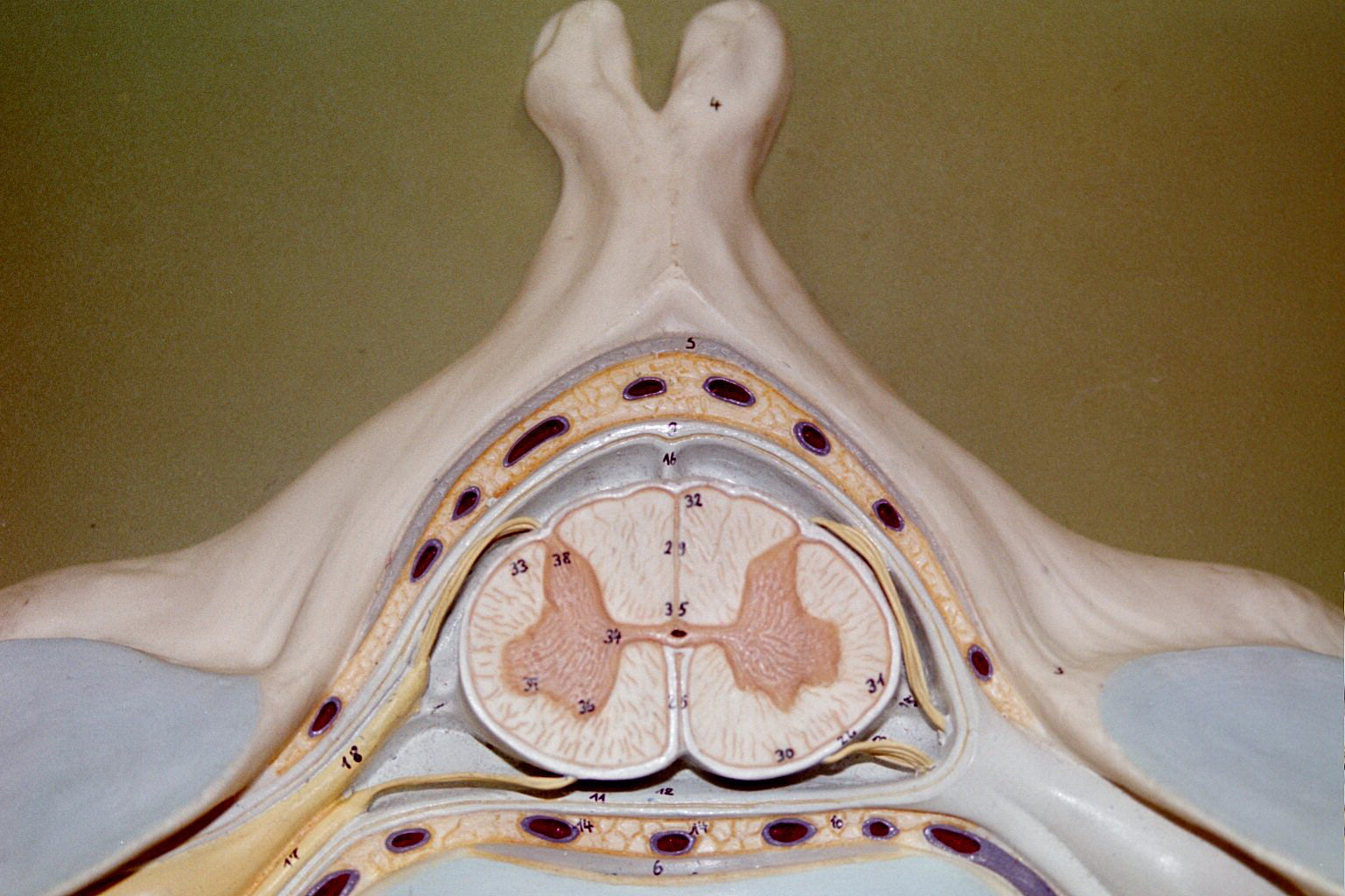
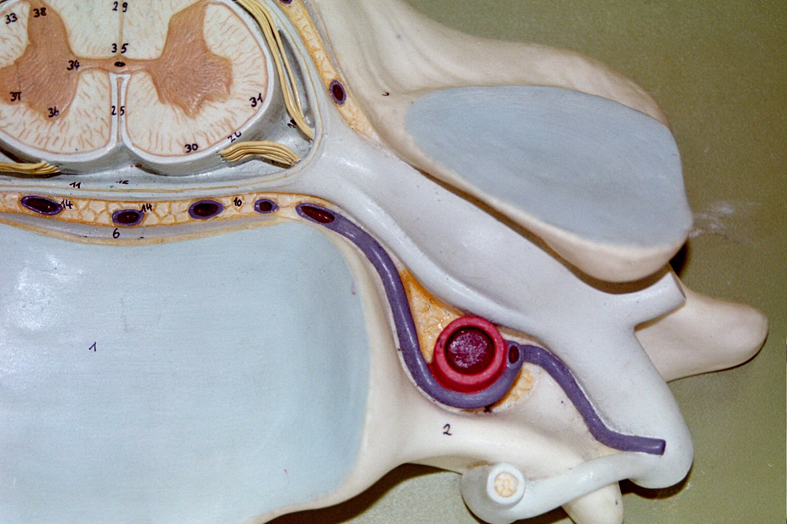
Histologically, the spinal cord consists of an internal gray matter and external white matter. Grey matter is the nonmyelinated portions of neurons, the cell body and dendrites and forms an H around the central canal. The white matter is usually myelinated portions of neurons, the axons that create ascending and descending tracts.
The function of the spinal cord is to carry sensory information from the periphery to the brain along the ascending sensory fiber tracts and to carry motor information from the brain to the peripheral tissues along the descending motor fiber tracts. Most sensory and motor information fiber tracts cross at some point, either in the spinal cord or the brainstem. The ascending and descending tracts have specific names and locations within the cord and several tests are used to evaluate the integrity of the system. An example of a test is the spinal cord reflex. Reflexes will be discussed in the PNS section of this A&P review.
Body Torso Model of Spinal Cord: Cervical & Thoracic Regions, Lumbosacral Regions
Spinal Cord Models : view one, view two, view three, view four
arachn- spider
Astro- star cephal/o- head
Cerebr/o- brain demi-, semi half
encephal/o- brain dura- hard
Lept/o- small, soft mening/o- meninge meninges
phobe, -phobia fear myelo- spinal cord
pseud/o- false psych/o-, ment/o- mind
som, somat/o- body telo- end
-fuge flee -glia glue
gyr/o- ring hypn/o-, somni- sleep
lep- take, seize malac/o- soft
narc/o- numbness necr/o- corpse
odyn- pain pont- bridge
schis- split crani/o- skull
thalam/o- thalamus ventricul/o- ventricle
-phasia speech brady- slow
splanch- viscera junct- yoke
Identify the types of neurons:
Identify the brain regions
Spinal Cord: Identify and give the function of the labeled structures.
VARK : Take this test to find out how best you learn. What were your results?
Brain
Works : Take this test to find out which hemisphere dominates and whether
you are an auditory or visual learner.
What were your results? Do you agree or disagree?
Humanmetrics: Take this test to see what personality type you are. What were your results? Do you agree or disagree?
Concept Map: Make a concept map of the CNS using its structures (gross and histological) anaotmy, location, and physological functions. Include this map in your LAR lab report (if selected) as a document insert or as an additonal emailed document by PDF scan.
Brain neurotransmitters:
Pick one neurotransmitter to research. Submit your findings to the instructor
for extra credit (+5 points) on CNS lab quiz. Findings should include class
of chemical, where secreted, type [excitatory or inhibitory or both], effects,
and elimination [how is the neurotransmitter removed from the synapse].
Cerebrospinal Fluid (CSF) tap results: Normal values have 0-3 cells per microliter of fluid with lymphocytes predominating, 50-100 mg /dL of glucose and 20-45 mg /dL of protein. Explain what might happen to the CSF results when the body is exposed to a viral infection, such as West Nile Virus.
Mental Diseases:
Depression, Bipolar, OCD, Schizophrenia, Phobias, Posttraumatic Stress Disorder,
Hypochondriac, Munchausen Syndrome
Meningitis: Cerebral, Spinal
Cerebral Vascular Accident (Stroke)
Transient Ischemic Attacks (TIAs)
Concussion
Contusion
Epidural and Subdural Hematoma
Encephalitis
Seizure, Epilepsy
Tumors of the Brain: Gliomas, Astrocytoma
Cerebellar Disease
Hydrocephalus
Headache, Migraine
Insomnia
Amnesia
Huntington’s Disease
Parkinson’s Disease
Alzheimer’s Disease
Multiple Sclerosis (MS)
Amyotrophic Lateral Sclerosis (ALS)
Gender Identity Disorder
Drug Abuse
Neurologist
Neurosurgeon
Psychologist
Counselor
Psychiatrist
http://www.nlm.nih.gov/medlineplus/healthtopics.html
http://www.lumen.luc.edu/lumen/meded/histo/frames/histo_frames.html
http://www.kcmetro.cc.mo.us/maplewoods/Biology/Bio110/Labs.htm
http://www.medem.com/MedLB/article_detaillb.cfm?article_ID=ZZZB2KOBGJC&sub_cat=185
http://www.neurosurgery.org/health/patient/answers.asp?DisorderID=51
http://www.neurosurgery.org/health/patient/answers.asp?DisorderID=44
http://www.medem.com/MedLB/article_detaillb.cfm?article_ID=ZZZR9WH46JC&sub_cat=509
http://www.medem.com/MedLB/article_detaillb.cfm?article_ID=ZZZYUAM46JC&sub_cat=509 brain side view
http://www.medem.com/MedLB/article_detaillb.cfm?article_ID=ZZZCLJKBGJC&sub_cat=185
http://www.nlm.nih.gov/medlineplus/brainandnervoussystem.html
http://www9.biostr.washington.edu/da.html
http://www.vh.org/Providers/Textbooks/BrainAnatomy/TOC.html
http://www.vh.org/Providers/Textbooks/BrainAnatomy/BrainAnatomy.html
http://www.med.harvard.edu/AANLIB/home.html
http://www.uofs.edu/sheep/ieframerow.html
http://www.quia.com/fc/37303.html
http://www.leeds.ac.uk/chb/humbmods.html
http://www.stemnet.nf.ca/CITE/body.htm
http://www.neurologychannel.com
http://dbsalliance.org (Depression and Bioplar support)
1. Name the two major divisions of the nervous system
2. Name the three types of neurons and their function
3. Name two CNS and one PNS neuroglial cell and their function
4. Define sensory afferent and motor efferent
5. Name two neurotransmitters and give their function
6. Define synapse and describe its anatomy and function
7. Define neurophysiology:
resting membrane potential
depolarization
threshold
repolarization
8. Name the lobes of the cerebrum and their function
9. Name the divisions of the brainstem and their function
10. Name the divisions of the diencephalons and their function
11. Name the two functions of the cerebellum
12. Define white matter and gray matter of the CNS
13. Describe how the CNS is protected
14. Give the source and function of cerebral spinal fluid
15. What is the purpose of myelinating an axon fiber?