Biology 2404 A&P Basics Lab Exercise 6b Nervous
System:
| Objectives | Background | Medical Terms | Activities | Applications | Careers | WWW | Review Questions |
Students should be able to
* Name the divisions of the PNS
* Describe the anatomy of the nervous tissue seen in the PNS
* Name the 12 pair of cranial nerves and their function
* Name a test that can be used to detect proper function for each of the 12 CN
* Give the Human spinal nerve formula
* Define dermatome and give its use
* Define spinal nerve plexus, their locations, and a nerve that comes from this region
* Define reflex by naming the components
* Give examples of the major types of reflexes
* Name the divisions of the Autonomic Nervous System and their effects
Read related material in textbook
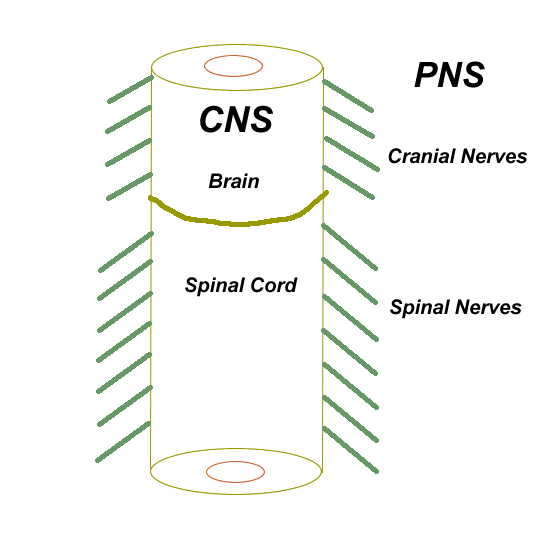
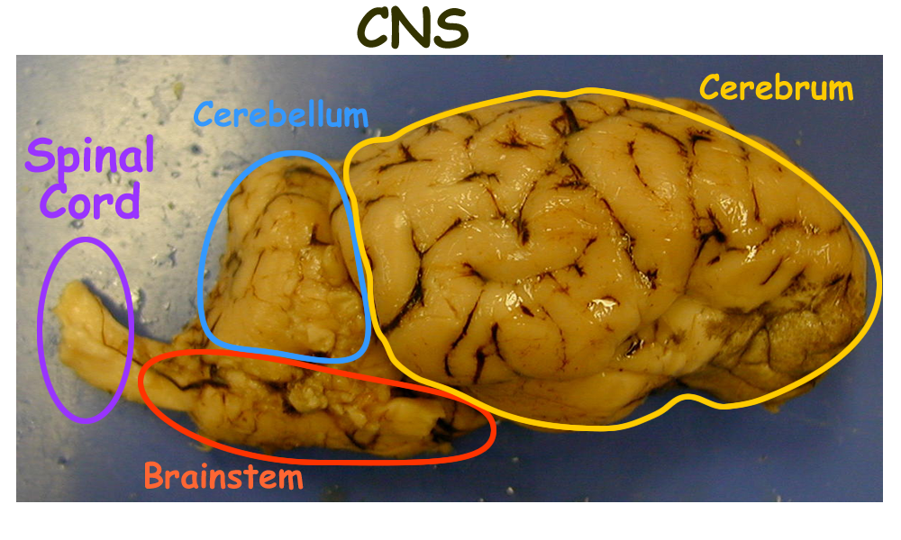
Embryologically, the nervous extensions from the central nervous system (CNS) tube are part of the peripheral nervous system or PNS. The PNS extensions from the brain are the cranial nerves and the PNS extensions from the spinal cord are the spinal nerves.
PNS major divisions are somatic and autonomic and each PNS division has sensory and motor components:
Somatic Sensory Skin receptors for afferent signals
Special Sensory Special sensory receptors for afferent signals
Autonomic Sensory Visceral pain sensations that follow somatic sensory
Somatic Motor Efferent signals to skeletal muscle
Autonomic Motor Efferent signals to smooth muscle, cardiac muscle, and epithelial glands
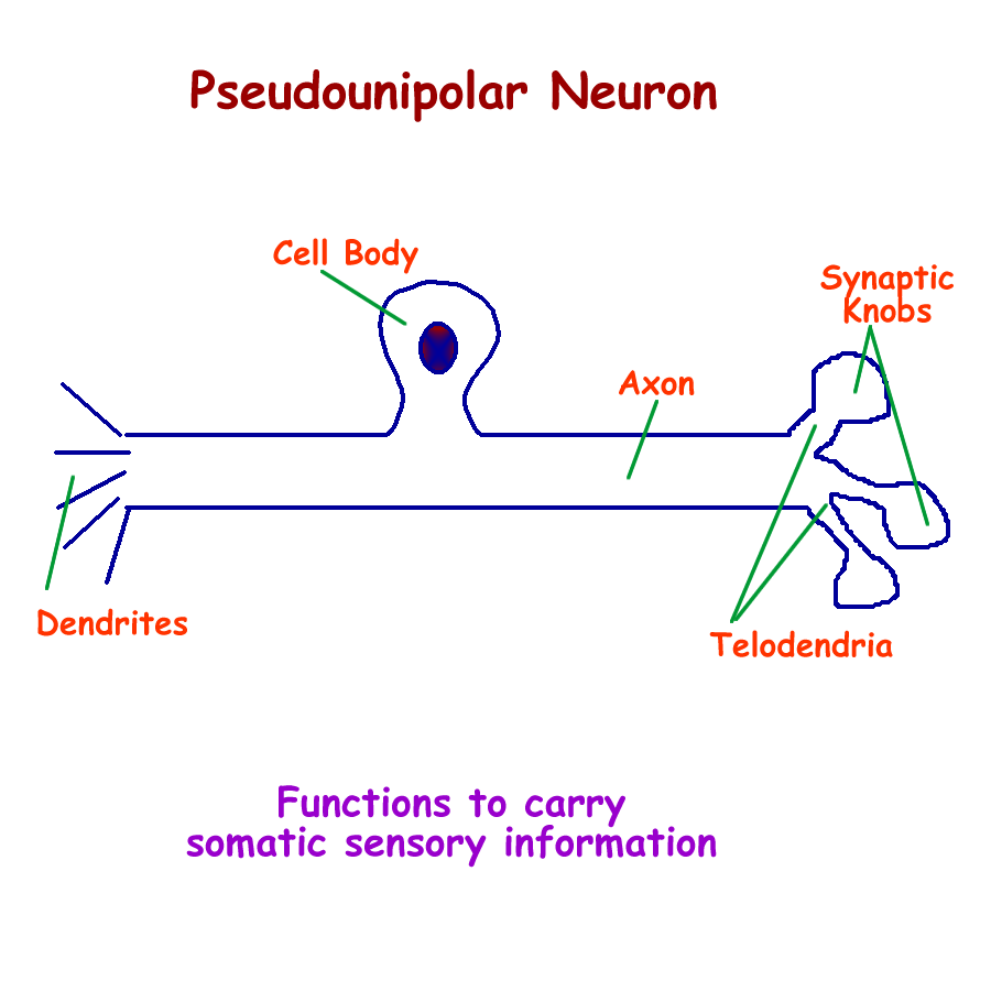
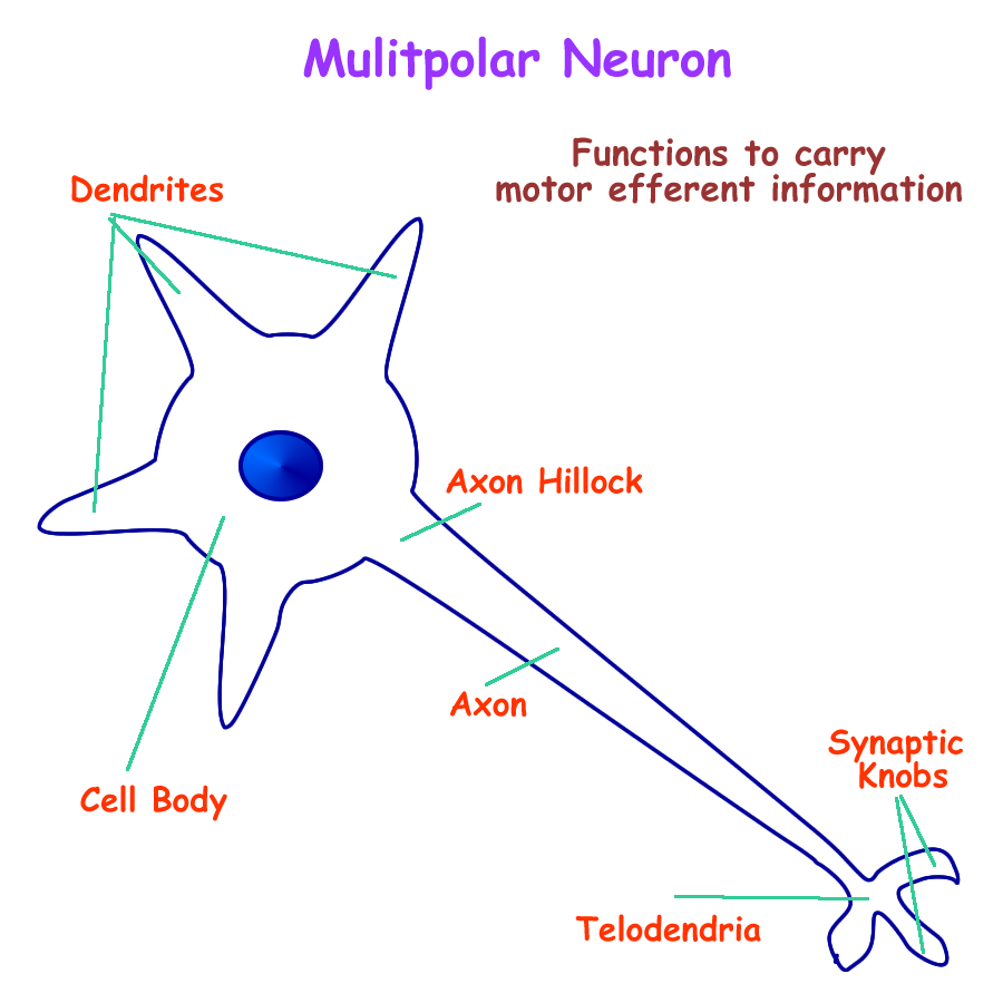
The neurons that form the PNS are:
Pseudounipolar neurons somatic sensory afferent
Bipolar neurons special sensory afferent
Multipolar neurons motor efferents
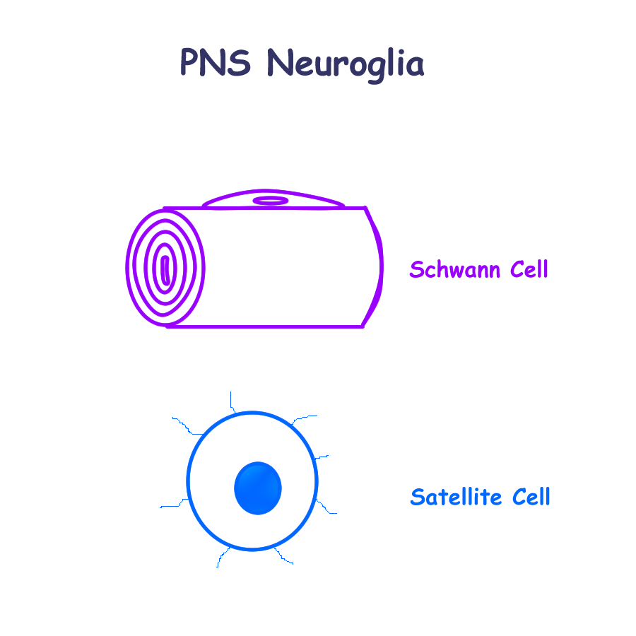
The neuroglial cells of the PNS are:
Satellite cells support and protect neuron cell bodies
Schwann Cells support, insulate, and +/- myelinate axons
Neuron Drawing : Pseudounipolar, Bipolar, Multipolar
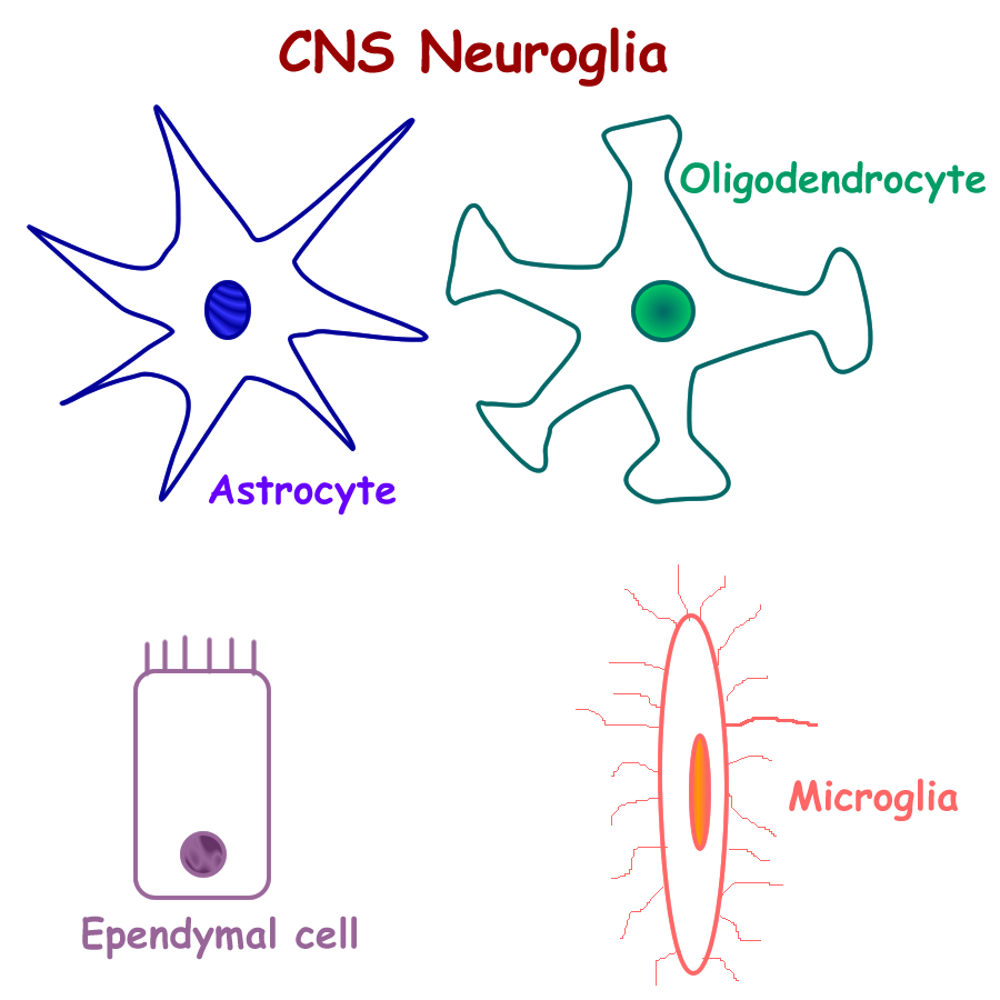
Neuroglial Drawing : CNS neuroglia, PNS neuroglia
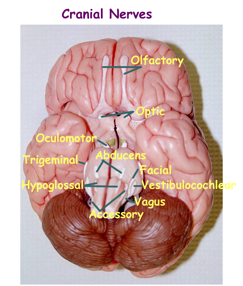
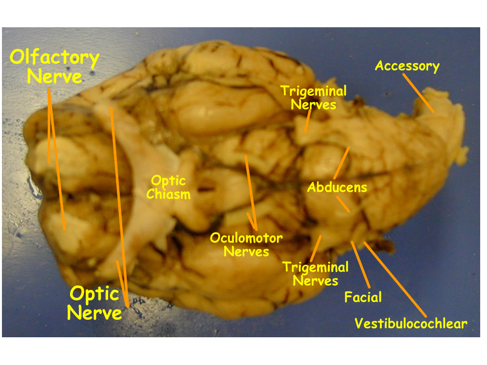
Cranial Nerves
There are twelve pairs of cranial nerves (CN) that form extensions from the brain. They are numbered in order of origin and are usually named by their function. Cranial nerves are bipolar for special sensory nerves, pseudounipolar for somatosensory, multipolar motor for efferent nerves, or a combination of both somatic sensory and motor known as a mixed nerve. The twelve pairs of cranial nerves are:
| CN I |
olfactory nerve
|
sensory
|
smell
|
| CN II |
optic nerve
|
sensory
|
sight
|
| CN III |
oculomotor
|
motor
|
eye muscle movements
|
| CN IV |
trochlear
|
motor
|
eye muscle rotational movements
|
| CN V |
trigeminal
|
mixed
|
sensory to the face, motor to the jaw
|
| CN VI |
abducens
|
motor
|
eye muscle movement for abduction
|
| CN VII |
facial
|
mixed
|
sensory to the front 2/3 of the tongue motor to muscles of the face |
| CN VIII |
vestibularcochlear
|
sensory
|
vestibular: equilibrium cochlear: hearing
|
| CN IX |
glossopharyngeal
|
mixed
|
glossal: sensory to the back 1/3 of tongue pharyngeal: motor to the throat |
| CN X |
vagus
|
mixed
|
sensory for pharyngeal taste buds motor for ANS parasympathetic |
| CN XI |
accessory
|
motor
|
muscles of the neck and shoulder
|
| CN XII |
hypoglossal
|
motor
|
muscles of the tongue
|
Tests are performed to check the integrity of the neuron pathways and help in brain assessment.
CN I smelling salts to test for smell
CN II visual tests: Snellen
CN III, IV, VI eye movements
CN V sensory to face
CN V, XII jaw and tongue movement
CN VII sweet, sour, salty taste sensations; facial expressions
CN VIII hearing, balance
CN IX bitter taste sensations; swallowing
CN X normal heart rate, normal respiratory rate, normal gut sounds
CN XI movement of skeletal muscles of the neck and shoulders
Spinal Nerves
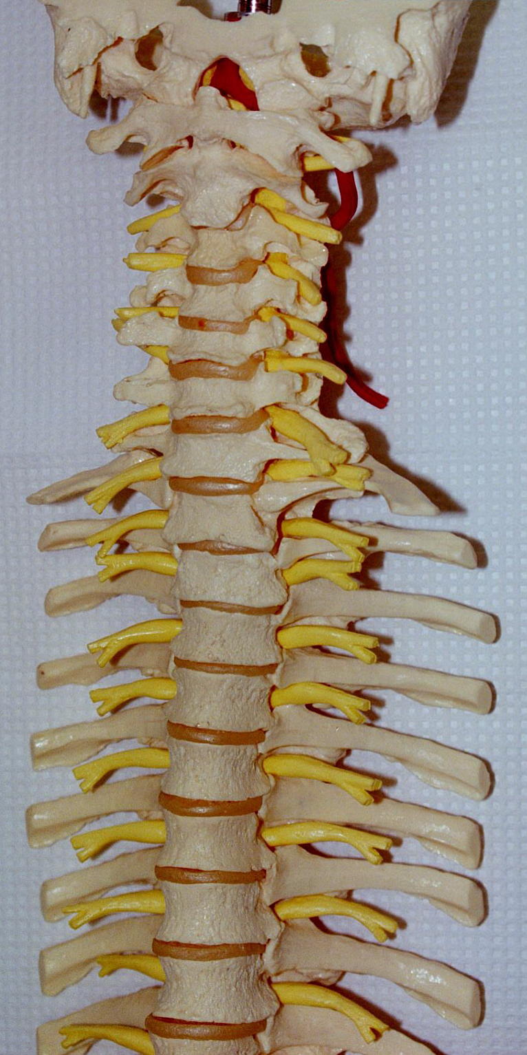
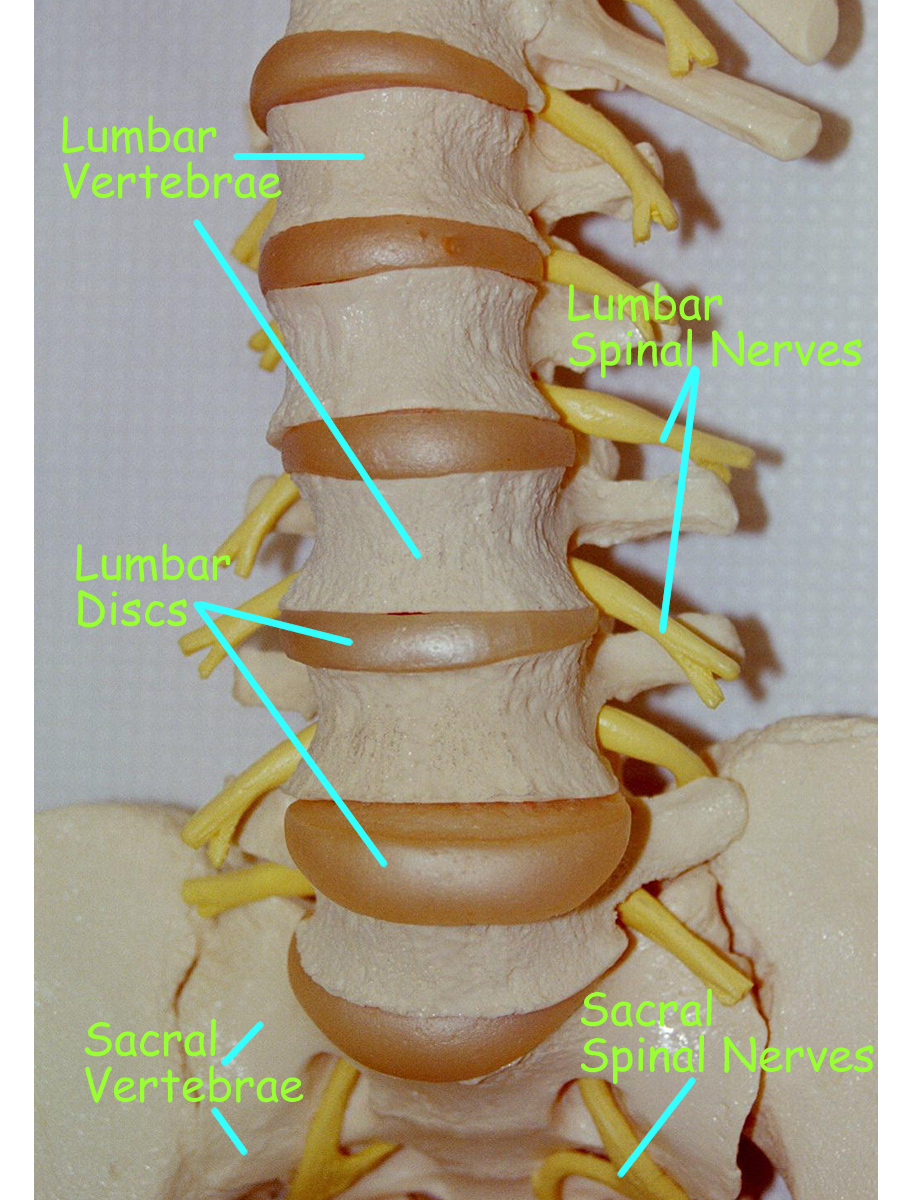
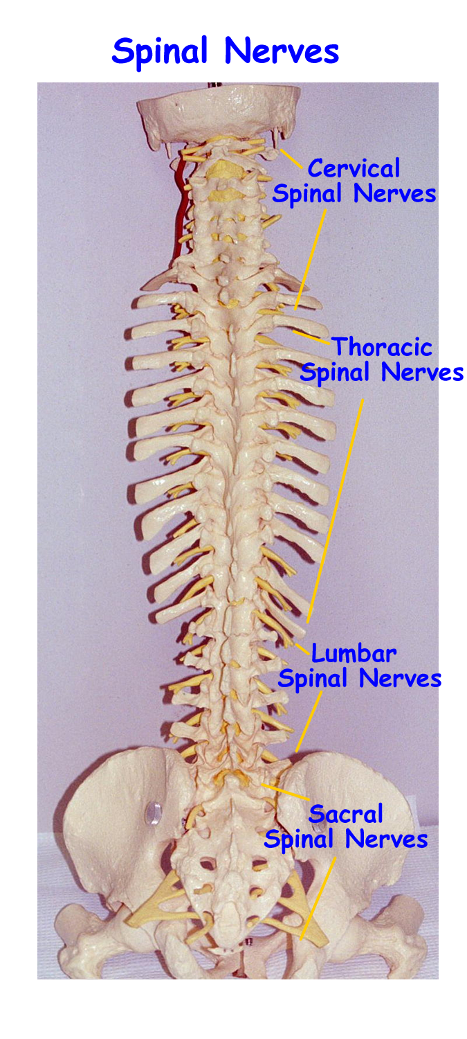
There are thirty-one pairs of spinal nerves that come from the spinal cord and are referenced by the spinal cord segments that pass through each vertebrae within each region.
In the cervical region, there are 8 pairs of spinal nerves labeled
C-1, C-2, C-3, C-4, C-5, C-6, C-7, C-8
In the thoracic region, there are 12 pairs of spinal nerves labeled
T-1, T-2, T-3, T-4, T-5, T-6, T-7, T-8, T-9, T-10, T-11, T-12
In the lumbar region, there are 5 pairs of spinal nerves labeled
L-1, L-2, L-3, L-4, L-5
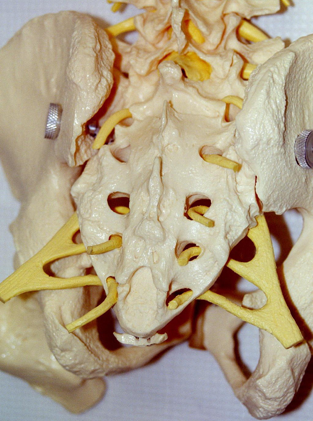
In the sacral region, there are 5 pairs of spinal nerves labeled
S-1, S-2, S-3, S-4, S-5
In the coccygeal region, there are on pair of spinal nerves labeled Cx-1
Combining the above, the spinal nerve formula for humans is
C 1-8, T1-12, L 1-5, S 1-5, Cx or C8, T12, L5, S5, Cx
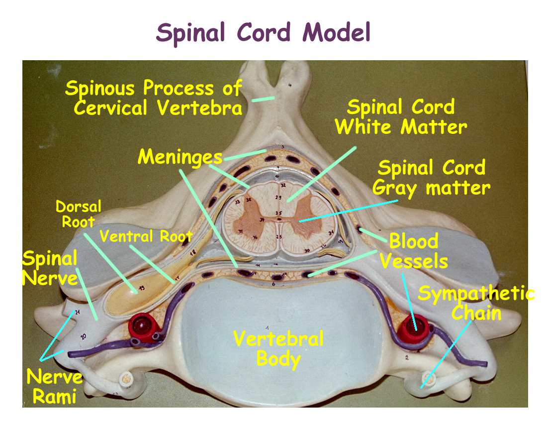
The spinal nerves are formed by a dorsal root that allows somatic sensory afferent information to enter the spinal cord and a ventral root that allows motor efferent information to leave the spinal cord. Because sensory information comes in through the spinal nerve to the cord and motor information leaves the cord through the spinal nerve, the spinal nerves are considered to be mixed nerves.
The spinal nerve divides into dorsal and ventral rami which carry information to their regional areas. The dorsal rami receives and sends information to the dorsum of the body and the ventral rami receives and sends information to the ventrum of the body. Since the ventrum is a larger area, the ventral rami divide again into branches that interconnect with other ventral rami branches from other spinal nerves.
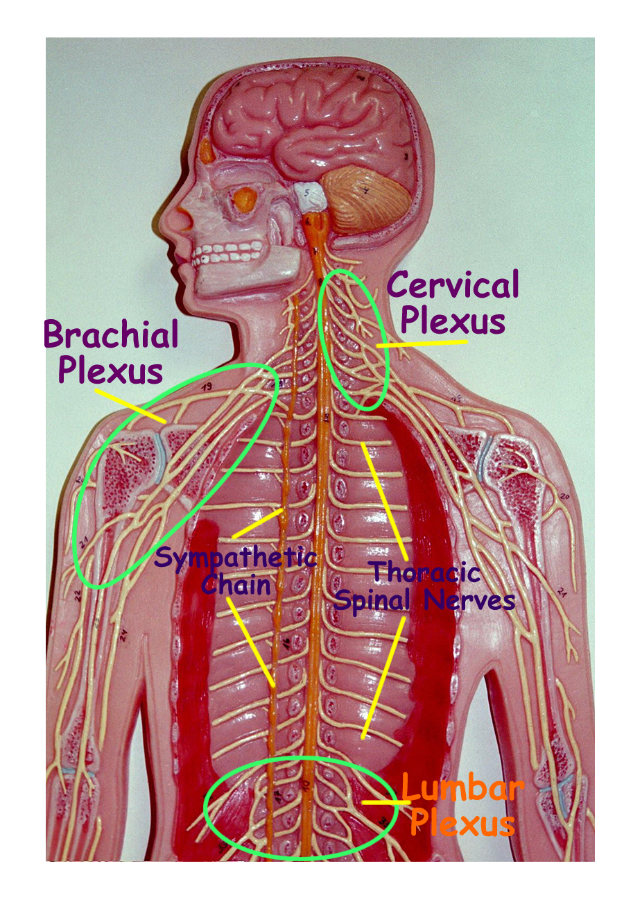
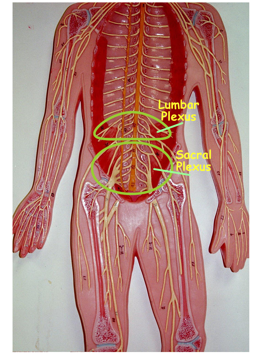
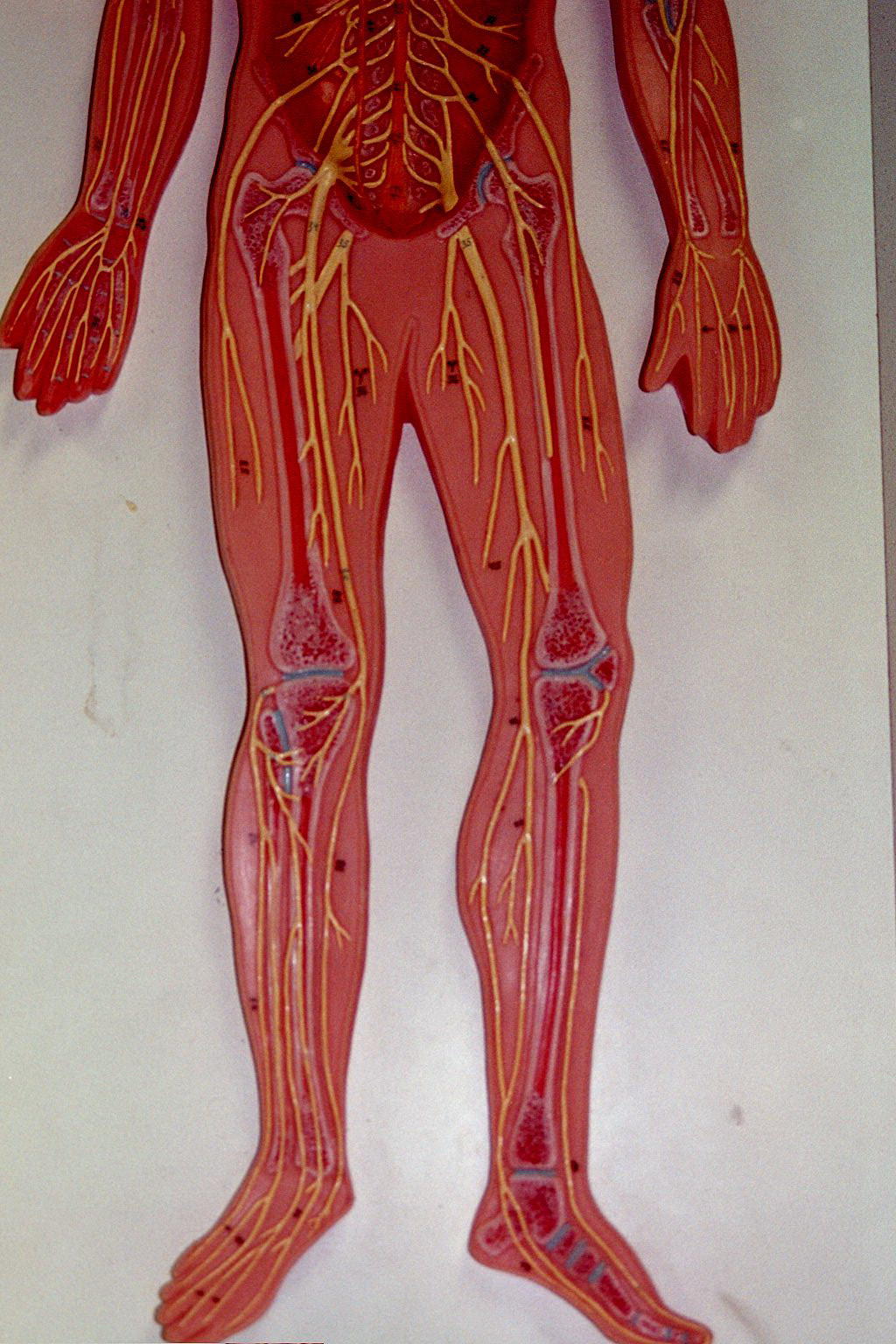
These interconnections of ventral rami branches form spinal nerve plexuses. Spinal Nerve plexuses occur in four vertebral areas and are named the cervical plexus, the brachial plexus, the lumbar plexus, and the sacral plexus. Since the spinal cord ends at L1 vertebra, the ventral roots and branches continue down the vertebral canal to form the cauda equina.
Spinal Nerve Plexus Ventral Branches Examples of Nerves from Plexus
Cervical Plexus C1-C4 Auricular nerve
Phrenic Nerve
Supraclavicular Nerves
Brachial Plexus C5-T1 Radial Nerve
Musculocutaneous Nerve
Median Nerve
Ulnar Nerve
Lumbar Plexus L1-L5 Femoral Nerve
Obturator Nerve
Lumbosacral Trunk
Sacral Plexus S1-S4 Sciatic Nerve
Gluteal Nerves
Pudendal Nerve
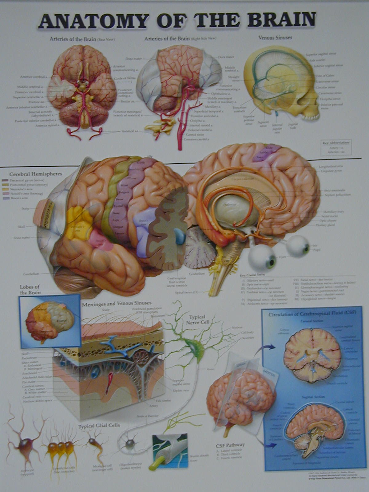
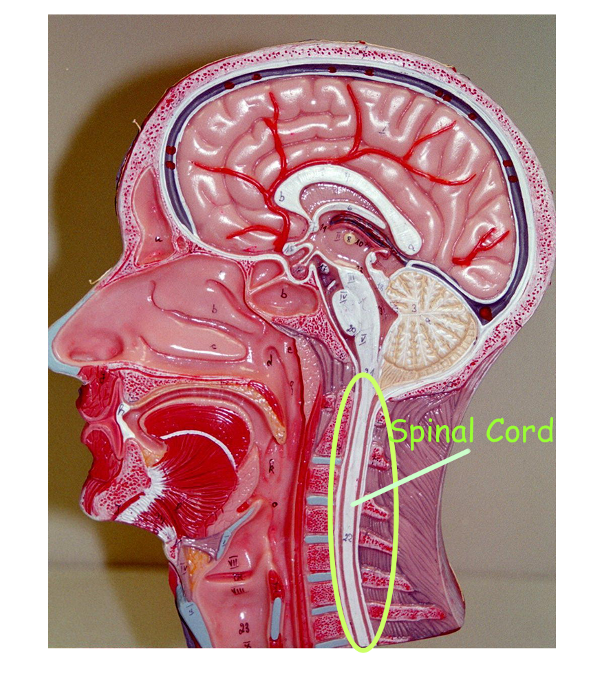
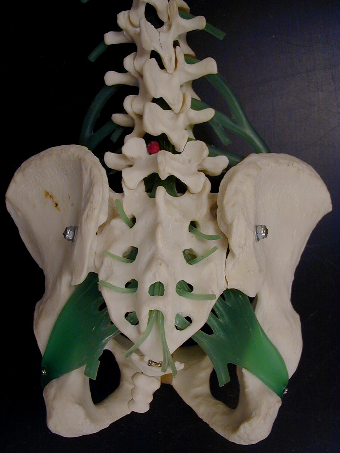
Spinal Cord on Sagittal Head Model
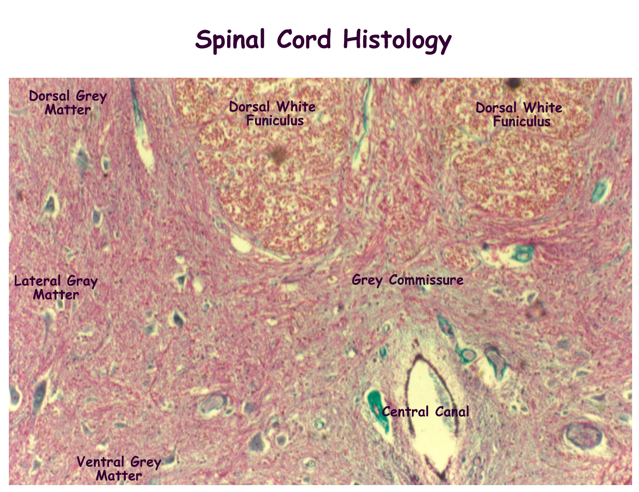
Spinal Cord Segment Drawing with roots and rami
Body Torso with Vertebral column
Plexus models: nerve man, vertebral column
Spinal Reflexes
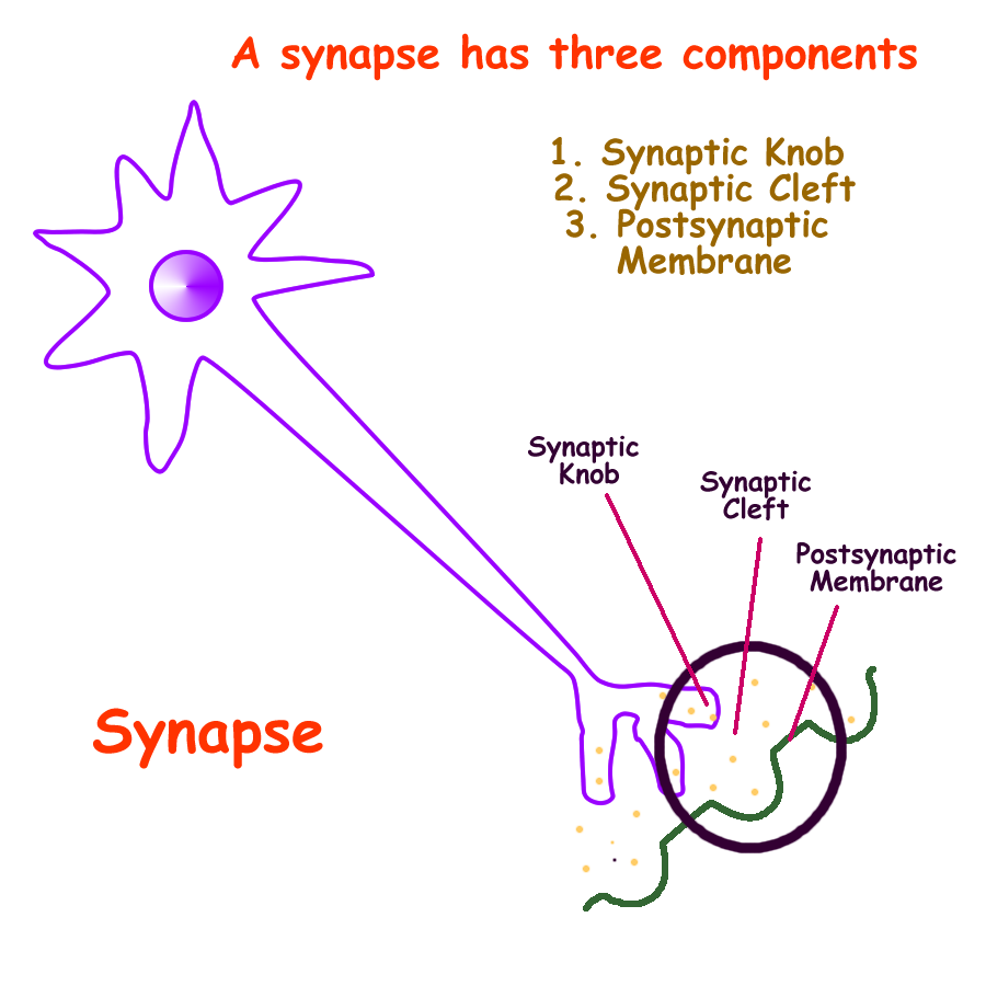
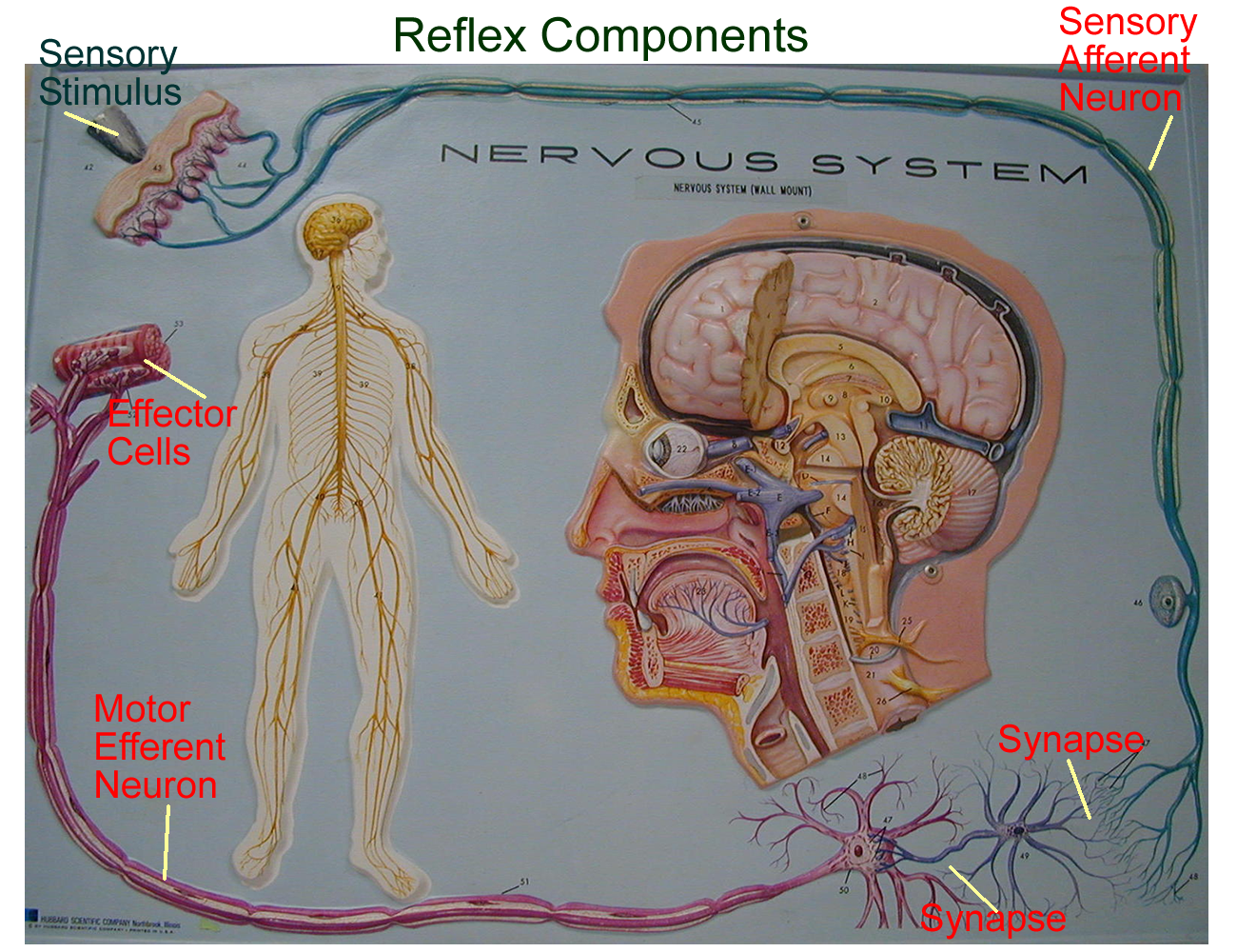
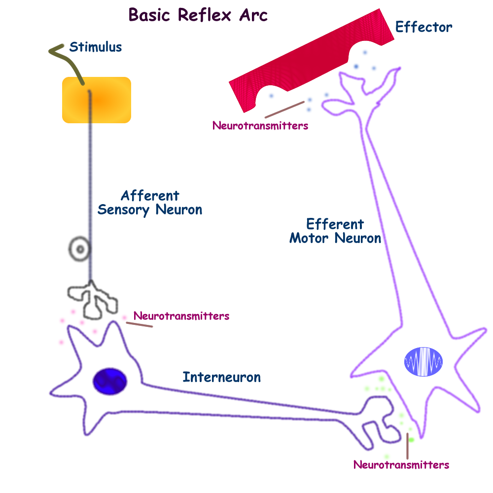
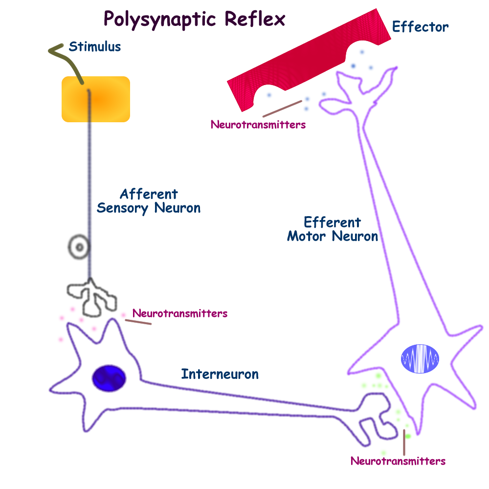
Once the anatomy of the spinal nerves and spinal cord is understood, pathways created by the rami of spinal nerves can be followed to detect nervous signals and proper responses. These pathways are called spinal reflexes. By definition, a reflex is an involuntary predictable motor response to a given stimulus. Most reflexes use the spinal cord to create simple cord responses. More complicated responses involve not only the spinal cord but several synapses to reach the brain and then return information to the spinal cord.
The basic components of any nervous system reflex are:
Sensory Stimulus
Sensory Neuron
Interneurons
Motor Neuron
Motor Response
Nervous System Wall Mount with Reflex
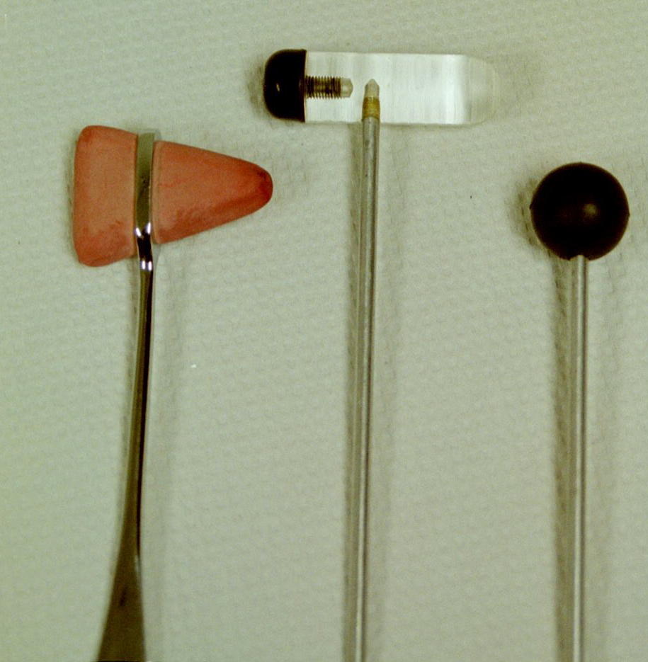
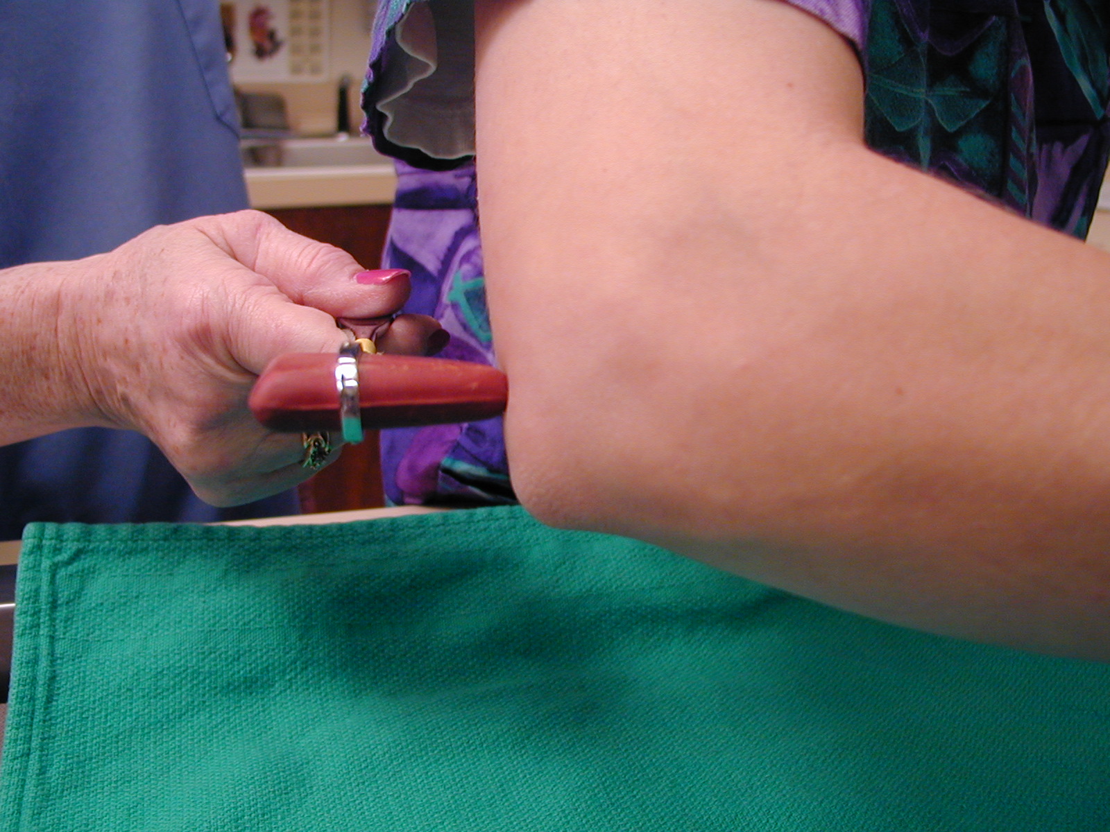
In simple cord reflexes there are no interneurons involved, so the reflex is also called a monosynaptic reflex. Examples of a simple, monosynaptic, cord reflex are the superficial tendon reflexes. A reflex hammer is used to test these muscle tendons that can be palpated through the skin. Examples of tendons used are biceps, triceps, patellar, and calcaneous tendons.
Once the pressure stimulus is given, the expected response relates to the muscle size and the tendons pull. Striking the Biceps Tendon should cause a slight flexion in the elbow. Striking the triceps tendon should cause a slight extension of the elbow. Striking the patellar tendon should cause a noticeable extension of the knee. Striking the calcaneous tendon should cause a moderate plantar flexion of the foot.
More complex reflexes involve interneurons and conscious awareness from the brain. Since multipolar interneurons are used, there are several more synapses in the pathway.
These more complex reflexes are called poly synaptic or multisynaptic reflexes. Examples of these more complex reflexes include the withdraw reflex, the crossed extensor reflex, and the Babinski reflex.
Drawing of polysynaptic reflex
Most reflex tests look primarily at the motor response. Motor reflex tests are graded 0 = normal, -1 and -2 as a decreased response and +1, +2 as an exaggerated response. Hypo- and Hyper- are also used to describe reflex results.
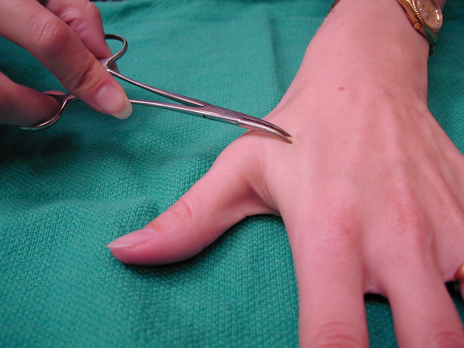
The sensory portion of the spinal reflex can be determined by analyzing the sensory neuron response. The dermatome test uses sensory stimuli for pain, pressure, and touch.
Each spinal nerve branch monitors sensory input from a particular body region. The map of spinal nerves and the body sections that they monitor is termed a dermatome. Sensory responses are also graded as to a normal response, an excessive sensory response known as hyperesthesia, or a decreased sensory response known as hypoesthesia.
Dermatome test: pinprick, hemostat
Autonomic Nervous System
The autonomic nervous system or ANS is the second major division of the PNS. It is responsible for sending sensory information from the internal body organs to the CNS and motor responses from the CNS to involuntary muscle and glands.
Sensory from the ANS is interepreted as pain, no matter what the original stimulus, even if it deals with temperature, pressure, touch, or position. This phenomenon called referred pain is due to the ANS sensory nerve tracts following somatic sensory for pain to the brain for interpretation. The brain interprets any visceral sensory input as skeletal muscle pain.
Several examples of referred pain include:
left arm pain (in males) heart attack
Back Pain kidney problems, usually an infection
Inner thigh pain urogenital problems, usually an infection
Neck pain lung problems, usually an infection
Right lower abdominal pain appendix problems, usually an infection
Right upper abdominal pain gall bladder problems, usually an infection
Upper, middle abdominal pain stomach problems, usually an ulcer
CNS motor efferents for the ANS go to cardiac muscle, smooth muscle, or epithelial glands. In the PNS, ANS motor efferents are divided into two pathways: sympathetic and parasympathetic. The ANS motor efferents differ from somatic motor efferents in organization.
Somatic motor efferents go to skeletal muscle using a one neuron pathway. These are usally large, myelinated multipolar neurons that make the neurotransmitter ACh.
ANS motor efferents go to involuntary muscles and glands using a two neuron pathway. The first ANS motor efferent neuron is called the preganglionic neuron and is usually myelinated. The second ANS motor efferent neuron is called the postganglionic neuron and is not myelinated. The axon lengths of the pre and post ganglionic neurons vary in each ANS motor division and this will cause changes in signaling and response time due to the axon length and the absence or presence of myelin.
Sympathetic motor pathways begin in the thoracic and lumbar regions of the spinal cord. Their responses are controlled in the brainstem and by higher centers such as the hypothalamus.
ANS sympathetic motor efferents have a generalized fight, flight, or freeze effect on the body’s internal organs. The primary neurotransmitter used by sympathetic neurons is norepinephrine.
Epinephrine is also made by the adrenal medulla and is used as a neurohormone for wide spread immediate sympathetic effects.
ANS parasympathetic motor efferents have very specific rest and digest effects on the body’s internal organs. The neurotransmitter used is ACh.
Parasympathetic motor pathways are carried in certain cranial nerves.
CN III carries parasympathetic to the eye for tear production and papillary muscle
CN V and VII carry parasympathetic to the salivary glands
CN X, the vagus nerve, carries parasympathetic to the thoracic and abdominal organs.
The sacral spinal nerves also carry parasympathetic motor information to the lower pelvic organs.
The following is a comparison of ANS motor efferent neurons:
|
Division |
Sympathetic |
Parasympathetic |
|
Generalized Body Effects |
Flight, Fight, Freeze |
Rest and Digest |
|
Neurotransmitter |
NE, EPI |
ACh |
|
Specific Organs: Heart |
Increase rate and force |
Decreased rate |
|
Lungs |
Increased rate and depth |
Decreased rate |
|
GI tract |
Decrease motility |
Increased motility |
|
Urinary |
Decreased urine production |
Normal urine production |
|
Pupils of Eye |
Dilate |
Constrict |
Since both ANS motor efferent pathways affect most body organs, these organs are said to have dual innervation, which means that both motor nerves can signal a response. Only one ANS motor division can dominate for long term homeostatic control. The ANS motor division that maintains homeostatic control produces the ANS tone for that organ.
For example, 24 hours a day you would most likely want your heart rate to be normal and the muscle contraction to pump enough blood to the tissues. If sympathetic division was in control, your heart would constantly race and pound and eventually wear out.
Since the parasympathetic is the better division to have for long term control, we say that the heart has parasympathetic tone.
Most organs have parasympathetic tone as sympathetic changes are only needed in an emergency which is hopefully short term. The only organ that has sympathetic control and therefore sympathetic tone are the smooth muscles of blood vessels.
In an emergency, blood flow needs to be immediately redirected and the sympathetic nervous system allows for such a quick response.
ANS reflex pathways are also in place. Recall that a reflex is a predictable motor response to a sensory stimulus. Examples of ANS reflexes are:
Pupillary light changes in pupil diameter with amount of light
Gut Sounds in response to sight, vision, and smell of food
Respiratory rate changes depending on blood gas and pH levels
Heart rate changes depending on pressure and flow
Nerve Man: Head, Thorax, Lumbar
ad, af to, toward ex, ef away from
multi- many -plegia paralysis
-asthenia weakness -esthesia feel, perceive
-ferent carry -fuge flee
ganglio- swelling grad- walk
neuro- nerve non- no, not
kinesi/o- movement ton/o- tone, tension
ID nerve pathways: Plexus, CN
Somatic Reflex tests: Patellar tendon, Calcaneon tendon, Crossed extensor
CN reflex tests: Gag reflex, Swallowing reflex
ANS Reflex tests: Pupillary light
Concept Map: Make a concept map of the PNS using its structures (gross and histological) anatomy, location, and physiological function. Include this map in your LAR lab report (if selected) as a document insert or as an additonal PDF document of the scan.
Gangliomas
Bell’s Palsy
Ataxia
Peripheral Neuritis
Spinal Cord Injuries: Paraplegia, Quadriplegia
Paresis
Sciatica
Anesthesiologist
anesthesia technologist
http://www.nlm.nih.gov/medlineplus/healthtopics.html
http://www.lumen.luc.edu/lumen/meded/histo/frames/histo_frames.html
http://www.kcmetro.cc.mo.us/maplewoods/Biology/Bio110/Labs.htm
http://www.medem.com/MedLB/article_detaillb.cfm?article_ID=ZZZB2KOBGJC&sub_cat=185
http://www.neurosurgery.org/health/patient/answers.asp?DisorderID=51
http://www.neurosurgery.org/health/patient/answers.asp?DisorderID=44
http://www.medem.com/MedLB/article_detaillb.cfm?article_ID=ZZZR9WH46JC&sub_cat=509
http://www.medem.com/MedLB/article_detaillb.cfm?article_ID=ZZZYUAM46JC&sub_cat=509 brain side view
http://www.medem.com/MedLB/article_detaillb.cfm?article_ID=ZZZCLJKBGJC&sub_cat=185
http://www.nlm.nih.gov/medlineplus/brainandnervoussystem.html
http://www9.biostr.washington.edu/da.html
http://www.vh.org/Providers/Textbooks/BrainAnatomy/TOC.html
http://www.vh.org/Providers/Textbooks/BrainAnatomy/BrainAnatomy.html
http://www.med.harvard.edu/AANLIB/home.html
http://www.uofs.edu/sheep/ieframerow.html
http://www.quia.com/fc/37303.html
http://www.leeds.ac.uk/chb/humbmods.html
http://www.stemnet.nf.ca/CITE/body.htm
http://www.neurologychannel.com
1. Name the divisions of the PNS.
2. Name 6 CN and their function
3. Explain how to test the 6 CN in question #2.
4. Define spinal nerve plexus and give two examples
5. Define reflex by using the anatomical structure
6. Name the two divisions of ANS motor and their general body effects
7. Define referred pain
8. Name the spinal nerve formula for humans.
9. Give the purpose of Schwann cells
10. Give examples of a simple cord reflex, complex reflex, and an ANS reflex