Biology 2404 A&P Basics Lab Exercise 8 Integumentary System Dr. Weis
| Objectives | Background | Medical Terms | Activities | Applications | Careers | WWW | Review Questions |
Students should be able to:
* Give the structure and function of the Integumentary system
* Give the components of the cutaneous membrane and the function
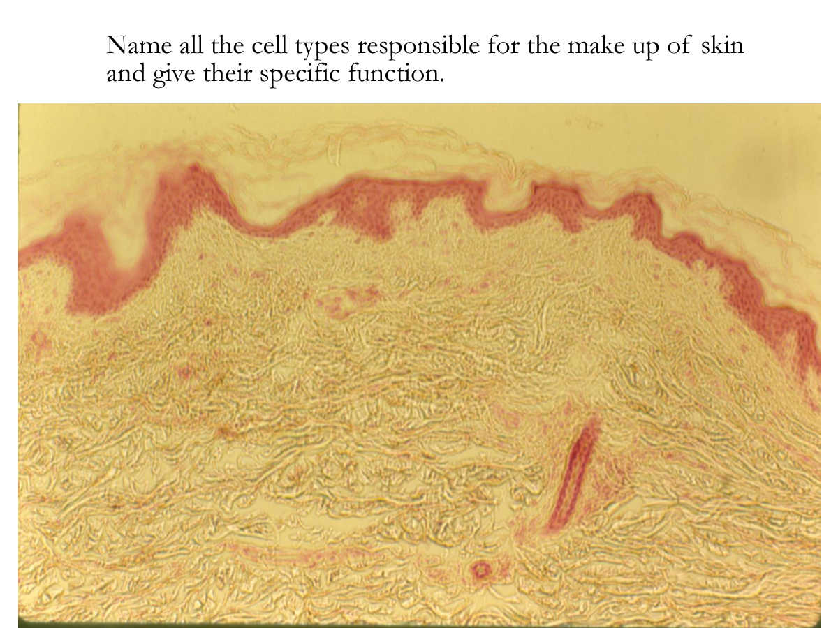
* Name the function of the various cells found in the skin
* Name the two layers of the dermis and its function
* Name the tissues that form the subcutaneous region
* Name the appendages of the skin and their function
Anatomy and Physiology Background
Read related textbook information
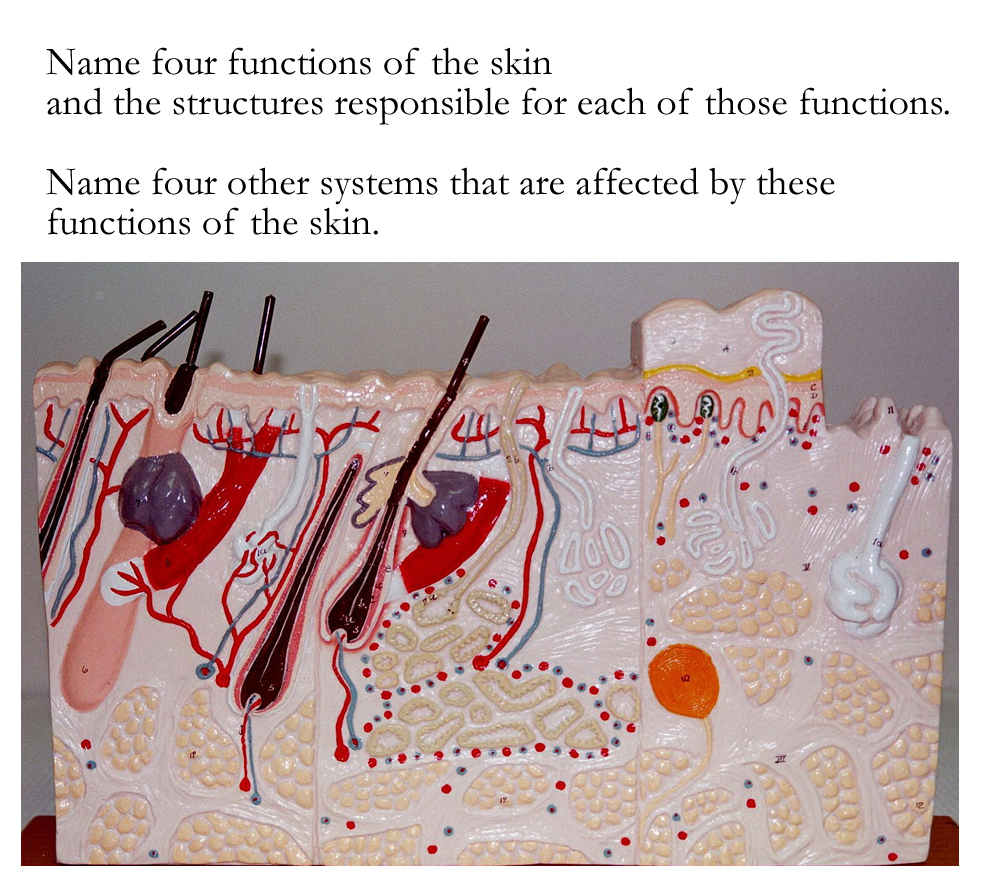
The Integumentary system is formed by the cutaneous membrane or skin, the subcutaneous region or hypodermis and the appendages or accessory structures such as hair and follicle sweat glands, sebaceous glands, and nails. As a system, the main function is to provide a protective covering for the body and provide secretions that aid in this function.
Additional functions are thermoregulation, somatic sensation, vitamin D production.
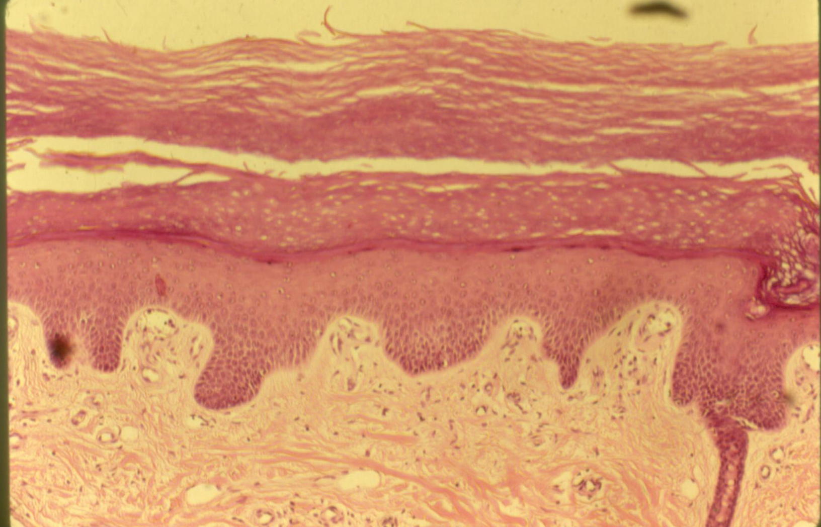
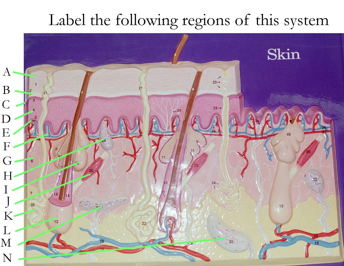
The skin or cutaneous membrane is formed by the epidermis and dermis. Together, they provide a protective barrier to prevent harmful substances from entering into the body.
The epidermis is a stratified squamous epithelium that is keratinized. There are several types of cells in the epidermis and the stratified layers are in five regions. The major cells of the epidermis are keratinocytes that secrete keratin, a water insoluble protein that fills the cells and replaces the normal organelles found in lower layers. Other cells include melanocytes that secrete a black-brown pigment called melanin, and Langerhans cells which are white blood cells that help provide defense.
The layers of the epidermis are named for their appearance. Starting at the bottom living layer and moving outward to the dead layers are:
|
Stratum Basale or Germinativum
|
single row of living cuboidal cells, regenerative | creates all other epidermal layers |
|
Stratum Spinosum
|
several layers of keratinocytes producing RNA | Melanocytes are also found here |
|
Stratum Granulosum
|
several layers of cells producing the protein keratin | Langerhans cells are found here |
|
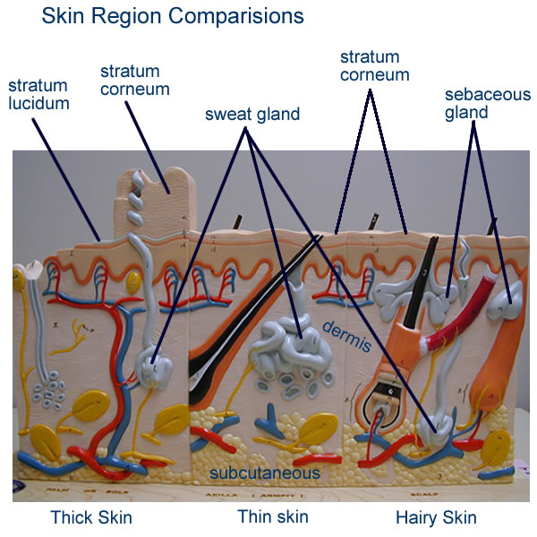
Stratum Lucidum
|
one-two layers of transparent dead cells | Present in thick skin of the palm and soles of feet |
|
Stratum Corneum
|
5-20 layers of dead cells, filled with keratin | Cells are flat or squamous |
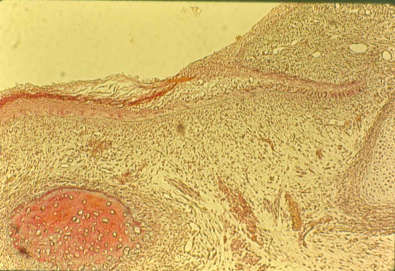
DERMIS
The dermis of the skin is connective tissue and is divided into two regions: the papillary region and the reticular region.
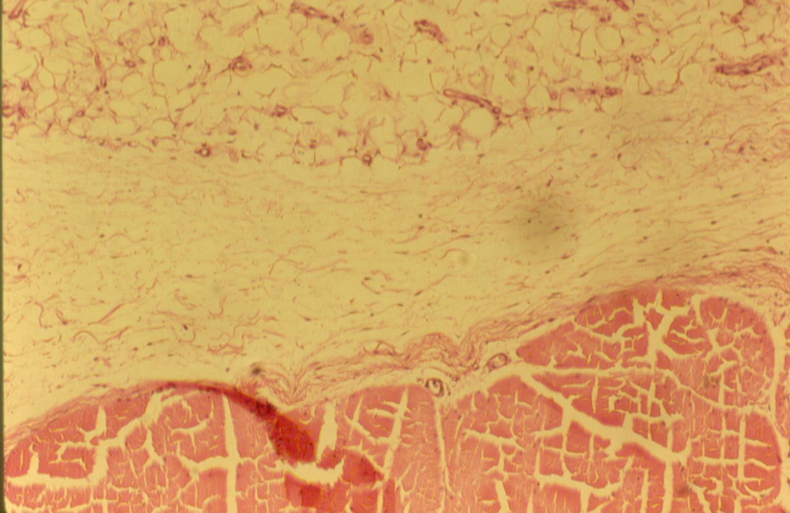
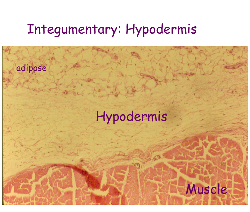
The subcutaneous region of the Integumentary system is also called the superficial fascia or hypodermis. It is primarily made of adipocytes (fat cells) that help connect the skin to the muscles below and also to provide cushioning, support, heat regulation, and energy. It also contains the larger blood vessels called arterioles and venules and nerve pathways for somatic (body) sensory information.
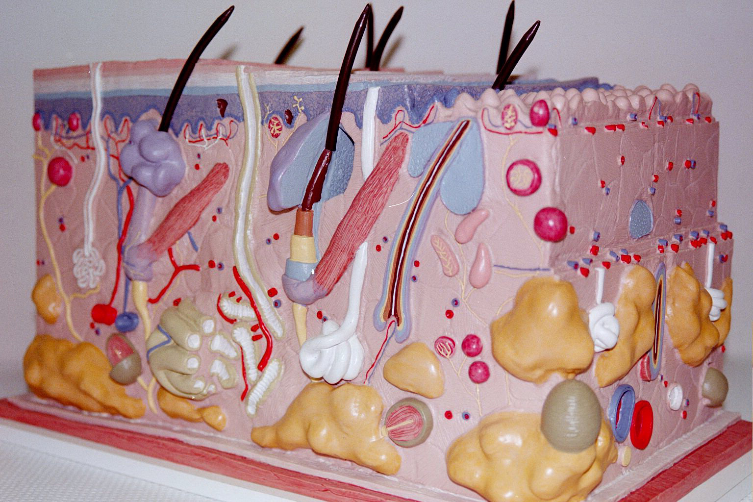
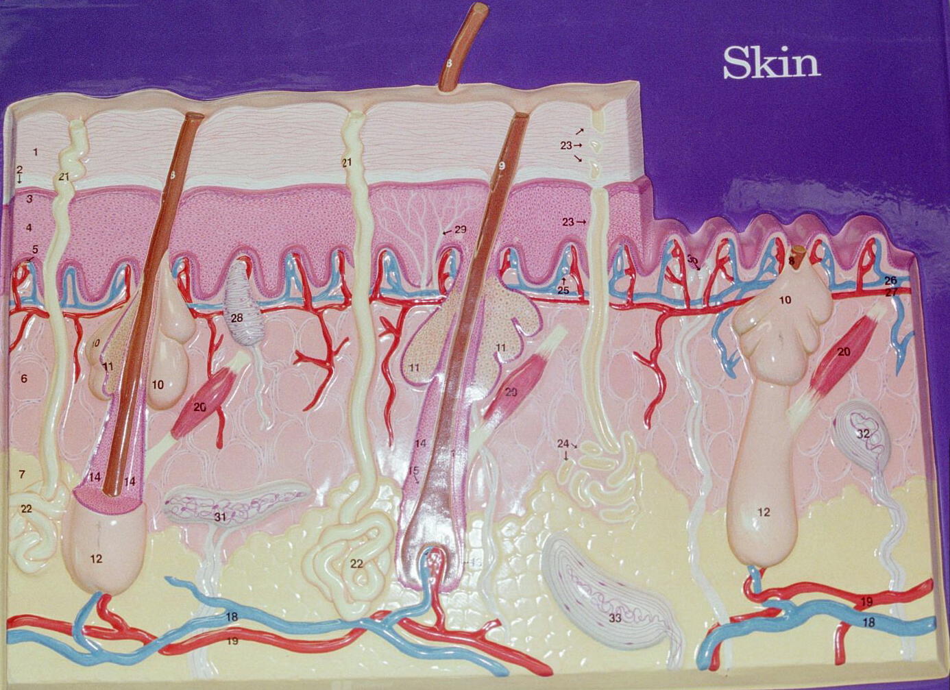
Drawing of Dermis and Hypodermis
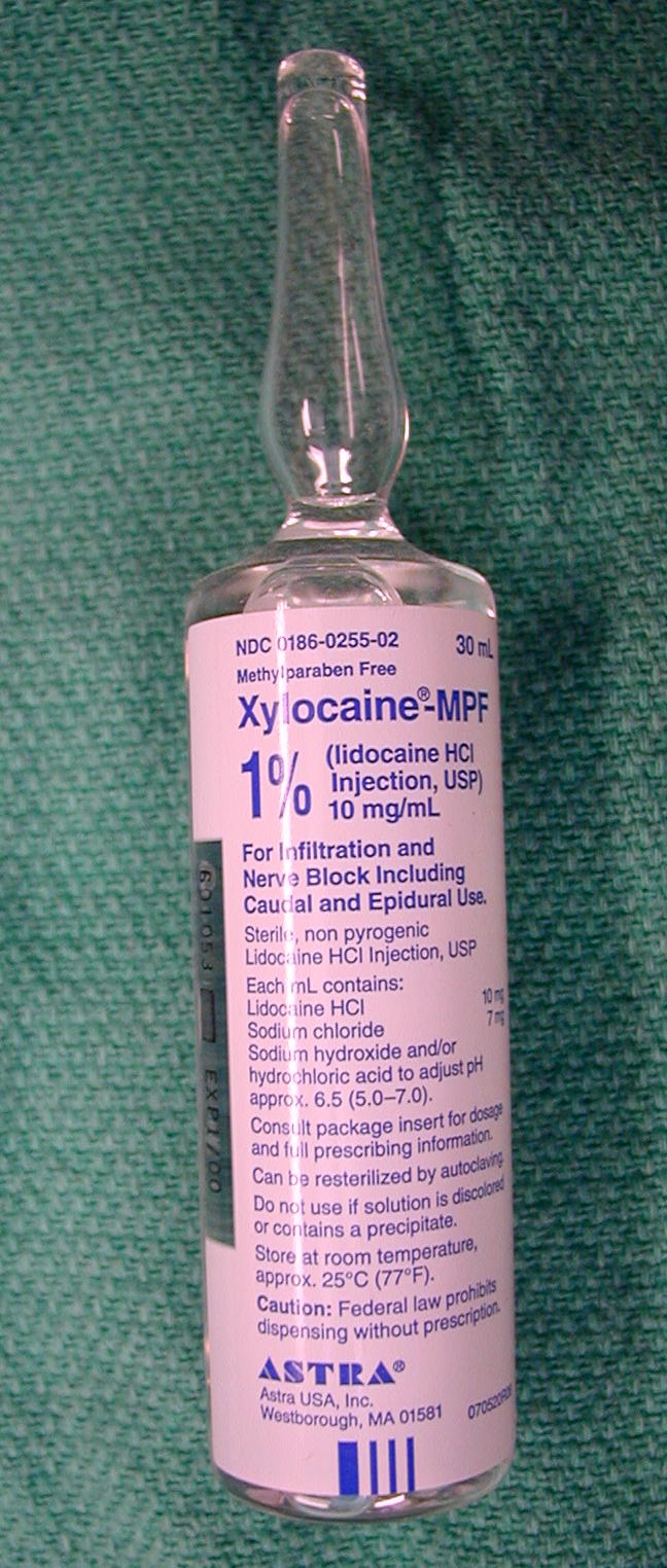
Drug used to deaden skin for surgery
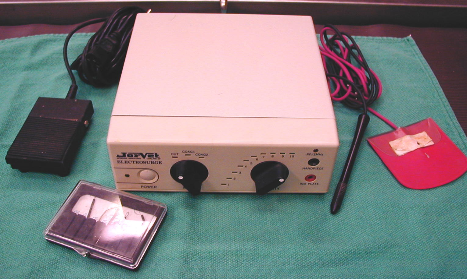
Electrocautery Unit used for surgery
Skin Staple gun used to close the skin after surgery
Accessory Organs of the Skin
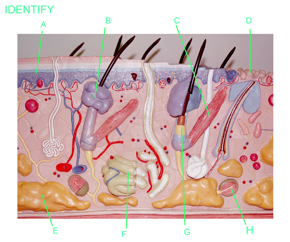
All accessory organs of the skin come from the epidermal regions of the stratum basale and stratum spinosum. They are also known as appendages and are found in the dermis. All accessory appendages have specific functions.
Hair follicles create a sheath and protect the hair which grows from the base of the follicle known as the papilla. The region of the hair that grows from the papilla is called the bulb or root. New epidermal hair cells of the root produce hard keratin (verses the soft keratin of the epidermis) to create the shaft or body of the hair. Melanocytes provide color to the hair based on genetic factors that control the amount of pigment produced. The follicle and chemical cross-linking of keratin determines whether the hair will be straight, curly, or tightly curled.
Hair growth occurs in three phases: growth, resting, and loss. Duration of the growth cycle depends on the hair location. Scalp hair grows for about three years, rests for about 1-2 years and is then lost when new hair begins to grow. Hair growth is affected by hormones and diet. There are different types of hair such as eyelashes, eyebrows, facial hair, axillary hair, perineal hair, truncal hair, and even fur and whiskers for some mammals. Each type of hair and its location provide a unique function for the body.
Attached to the follicle is a smooth muscle called the arrector pili muscle and provides movement of the follicle and therefore the hair to raise or lower the shaft. This movement provides temperature regulation for most mammals and can even provide information about the emotional state of the individual.
At the ends of the fingers and toes there are nail follicles formed from stratum basale and stratum spinosum that produce nails by mitosis at the nail root. The new cells produce hard keratin for the nail body and then die. The function is to provide protection to the distal bones of the phalanges and to protect the ends of the digits from mechanical trauma.
The nail bed that supports the visible nail body is living tissue and contains blood supply and nerves. Nails grow approximately 1 mm per week unless damaged or diseased. The white lunula or half moon that is seen is due to air that is mixed in the keratin matrix and its size is determined by genetics. The cuticle or eponychium is stratum corneum that extends out over the proximal end of the nail body and marks the area where the nail follicle arises.
Regeneration of a lost nail can take several months to almost a year.
Glands
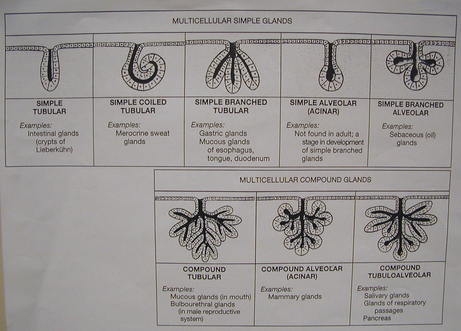
Exocrine glands are associated with the skin and produce secretions that are excreted through a duct system. The glandular appendages of the skin come from stratum basale and stratum spinosum. The cuboidal cells can produce a variety of secretions from watery clear solutions to opaque thick solutions.
Eccrine or merocrine glands that produce a watery, thin, clear secretion that aids in cooling the body. Sweat glands are found throughout the body, but they are found in higher concentrations on the palms and soles of the feet and forehead.
Apocrine sweat glands produce a viscous, cloudy secretion that provides a unique body odor. Apocrine sweat glands are found on the scalp, perineal, and axillary regions of the body. Bacteria metabolize the chemicals in the sweat and their waste products can create unpleasant body odors.
Mammary glands are modified sweat glands and are under hormonal control to produce milk for a viable offspring.
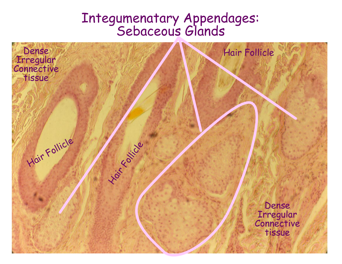
Sebaceous glands are found throughout the skin, except on the palms of the hand and soles of the feet. Their duct system can open into hair follicles or directly on the surface of the skin. The secretion is a thick, opaque product called sebum and it provides lubrication and protection to the stratum corneum. Sebaceous secretions are formed from entire cells dieing and secretions are controlled by hormones from the endocrine system.
Ceruminous glands are modified sebaceous glands found in the ear canal. Their secretion called ear wax or cerumen is thick and opaque and provides protection and lubrication to the outer ear canal.
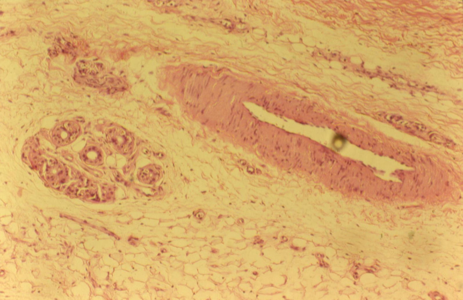
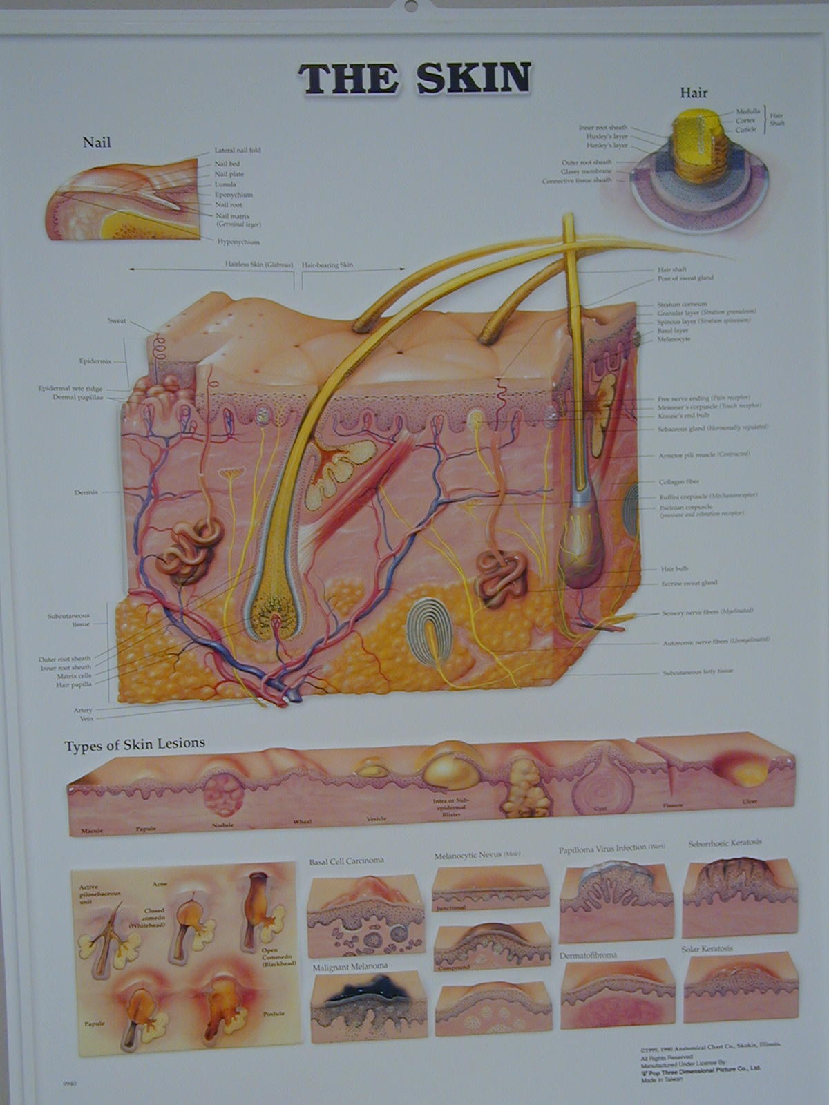
Hair and Hair follicle histology
Problems: Burns
Derma- skin kerat/o- hard, cornea
Intra- beneath inter- between
-itis inflammation liga- bind together
melan/o- black pachy- thick
per- through sub- under
dactyl- finger, toe syn- together
trans- across trich- hair
cyano/o- blue onych/o- nail
follicul/o- follicle xer/o- dry
pil/o- hair hidr/o- sweat
pulp- flesh kerato/- hard, skin
pell- skin xanth- yellow
Magnifying glass for pores, nails
Experiment: pain, temperature, touch, pressure
Concept Map: Make a concept map of the integumentary system using the structures (gross and histological) anatomy, location, and physiological functions. Include this concept map in your LAR lab report (if selected) as a document insert or as an additional PDF scanned document of this map.
Skin test for allergies
Skin patch
Injections
Sun screen
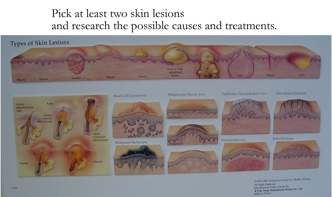
Skin diseases and conditions:
Impetigo
Eczema
Warts
Fever Blisters (cold sores)
Ringworm
Psoriasis
Shingles
Vitiligo
Papules, pustules, hives
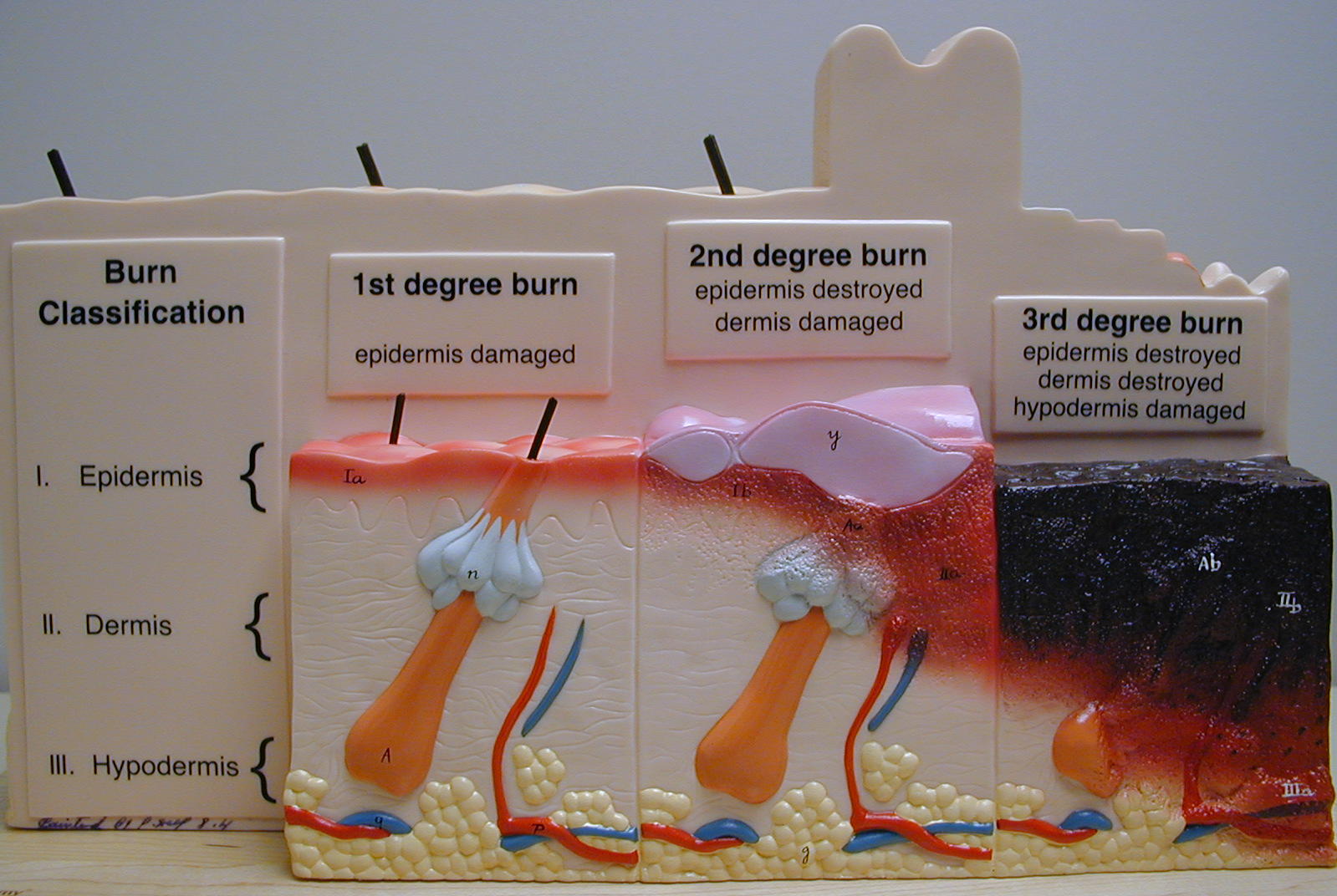
Burns: 1st degree, 2nd degree, 3rd degree
Cancers: basal cell carcinoma, squamous cell carcinoma, melanoma
Urticaria (Hives)
Acne Vulgaris
Alopecia
Furuncles and Carbuncles
Pediculosis
Dermatophytoses
Corns, Calluses
Discoid Lupus Erythematosus (DLE)
Scleroderma
Dermatitis : Seborrheic, Contact, Atopic (Eczema)
Dermatologist
Dermatopathologist
http://starnet.esc20.net/anatomy/integumentquiz.htm
http://www.dermnet.org.nz/index.html
http://www.science.ubc.ca/~biomania/tutorial/skin/outline.htm
http://www.science.ubc.ca/~biomania/tutorial/exogland/outline.htm
http://www.kcmetro.cc.mo.us/maplewoods/Biology/Bio110/Labs.htm
http://www.medem.com/MedLB/article_detaillb.cfm?article_ID=ZZZQYMPCGJC&sub_cat=98 skin
http://www.nlm.nih.gov/medlineplus/skinhairandnails.html
1. List four functions of the Integumentary system
2. Name the five possible layers of the epidermis
3. Name the glandular accessory structures of the skin and their function.
4. Name the cells of the skin and their function.
5. Give the function of hair/hair follicles and nails
6. If all people have the same number of melanocytes in their skin, explain how different races have different colors.
7. What sensations are picked up by the skin?
8. How is thermoregulation achieved?
9. How does the Integumentary system affect other body systems?
10. What are some aging changes that occur in our skin?