Biology 2404 A&P Basics Lab Exercise 10 Muscular System Dr. Weis
| Objectives | Background | Medical Terms | Activities | Applications | Careers | WWW | Review Questions |
Students should be able to
* compare and contrast the three types of muscle tissue
* list the functions of skeletal muscles
* give the structure of the sarcomere
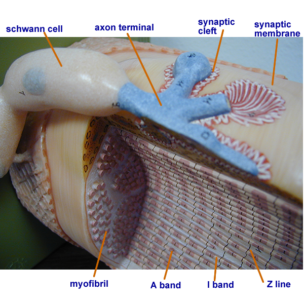
* give the structure of the neuromuscular junction
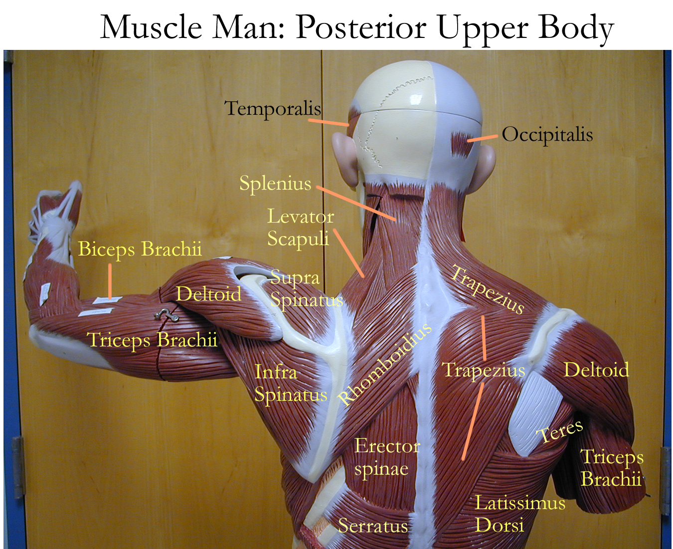
* identify the superficial muscles of the body
* define related muscular terms
Anatomy and Physiology Background
Read related material in the textbook
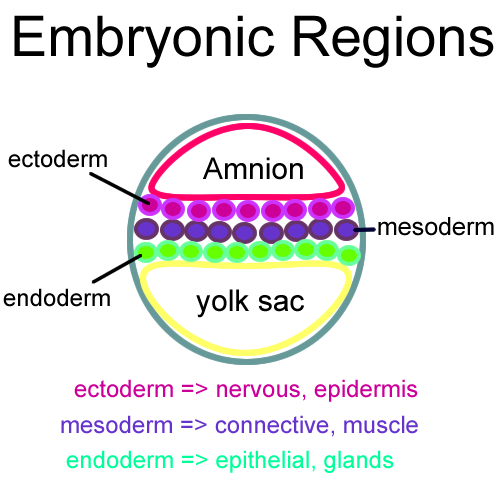
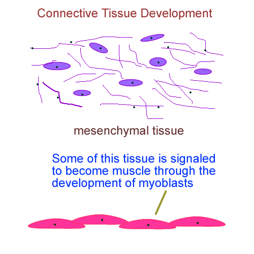
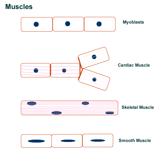
All muscles develop from myoblasts of the embryonic mesoderm and are specialized for contraction. The three types of muscle that develop are skeletal, smooth, and cardiac muscle. Most of the body’s muscle is located on the skeleton and therefore called skeletal muscle. Due to its specialization, muscle cell parts are renamed.
The outer cell membrane is called the sarcolemma, the cell’s cytoplasm is called sarcoplasm. Some of the cells organelles are renamed. Smooth endoplasmic reticulum becomes a specialized structure for calcium storage called the sarcoplasmic reticulum. Inside the muscle cell are contractile proteins called actin and myosin as well as another protein for storing oxygen called myoglobin. Mitochondria are also need to produce energy in the form of ATP.
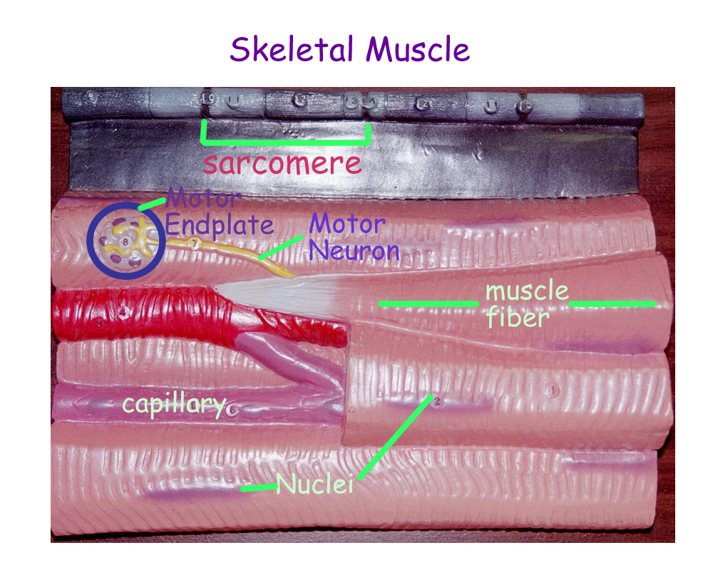
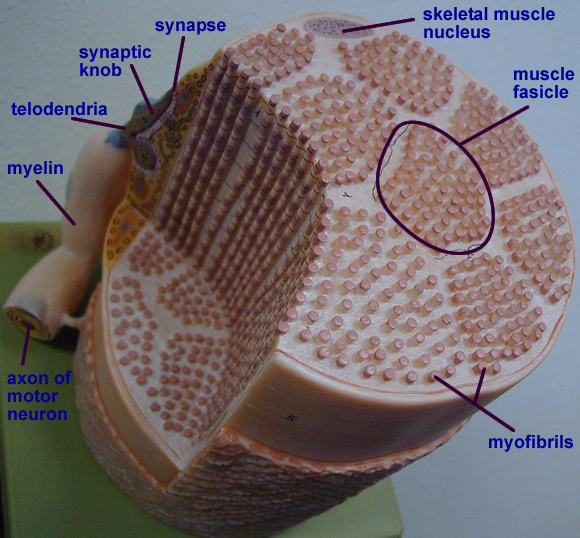
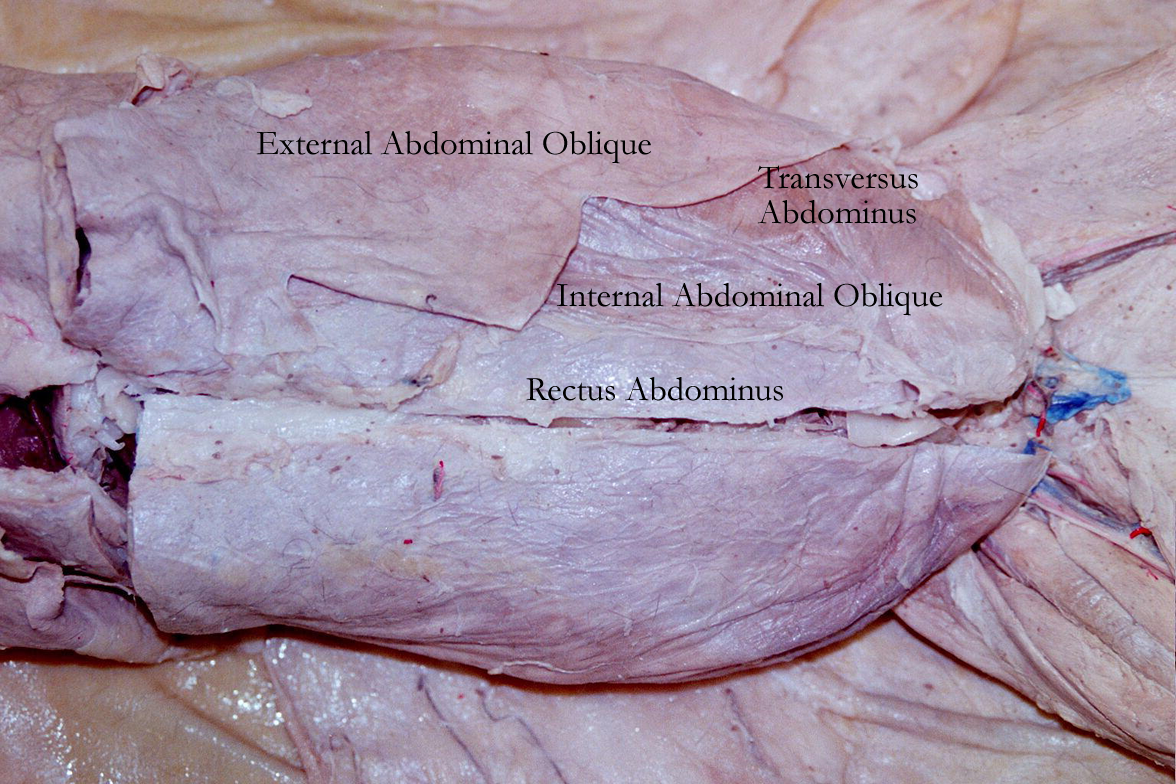
Skeletal muscle has several connective tissue divisions. Around the sarcolemma is a connective tissue called the endomysium. This connective tissue layer allows muscle cells to pull on it, instead of themselves, when they contract. Muscle bundles are then created and are bound by a second connective tissue called perimysium. This connective layer allows for support and protection for blood vessels and nerves. Recall that skeletal muscle is under voluntary control from the nervous system and cannot contract without a nervous system signal. The outer layer of connective tissue that defines a muscle group is called the epimysium. The epimysium also continues as the muscle’s tendons of origin and insertion. As the muscle contracts, tension increases in the muscle and the tendon to pull on bones across joints or soft tissue. Muscle tendons that cross at Synovial joints can move bone in certain planes and ranges.
Skeletal Muscle End (Histology)
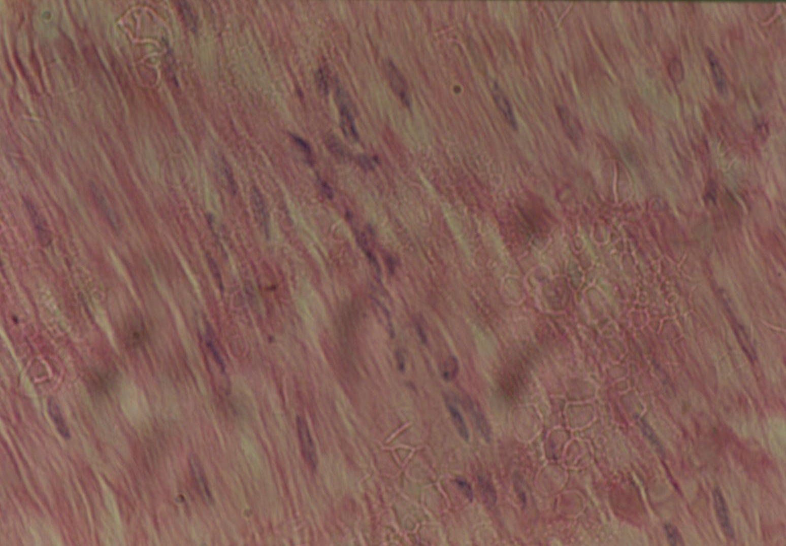
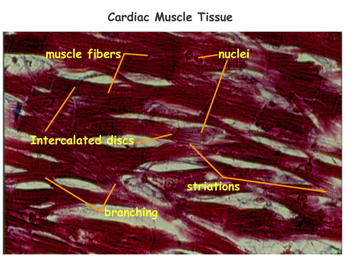
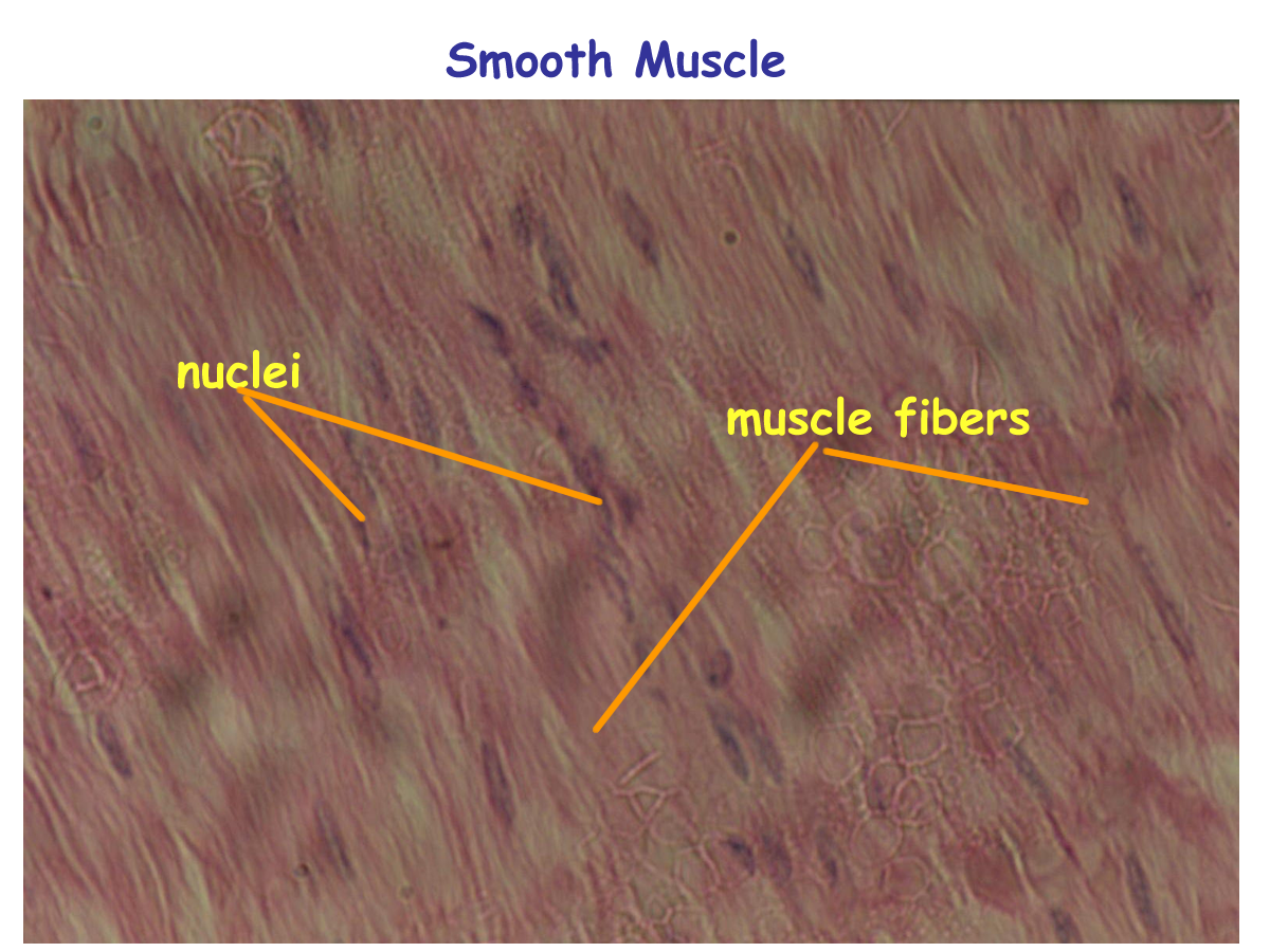
Cardiac and Smooth muscle have some differences when compared to skeletal muscle. The sarcoplasmic reticulum in both of these muscles is less developed and calcium stores are limited. Both have actin and myosin for their contractile proteins.
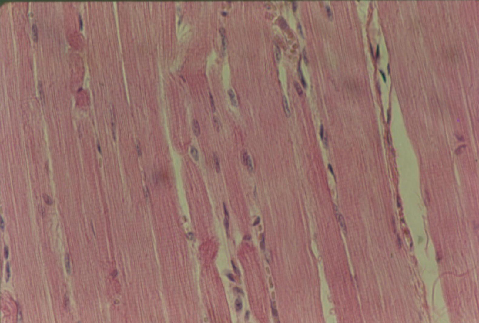
Cardiac muscle’s actin and myosin are arranged to create bands or striations like skeletal muscle. Smooth muscle’s actin and myosin are not regularly arranged in a banding pattern. Recall that both cardiac and smooth muscle are considered involuntary and are supplied by the autonomic division of the nervous system. Both have single nuclei within their cells and their fiber direction changes depending on the area within the organ.
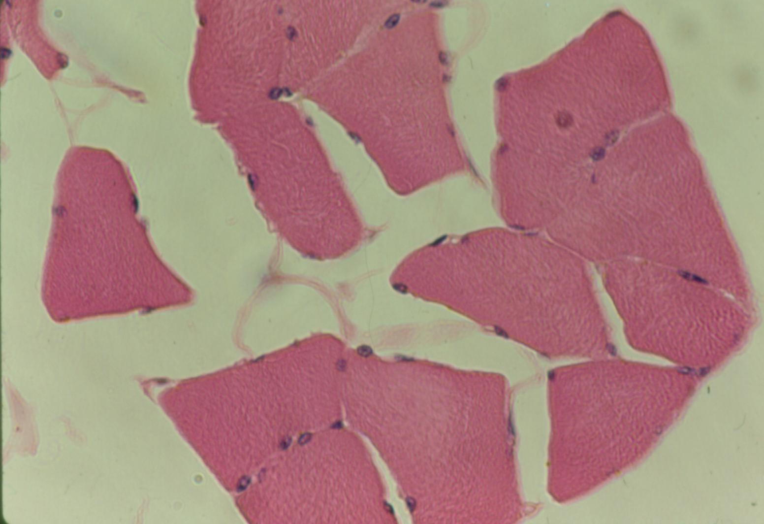
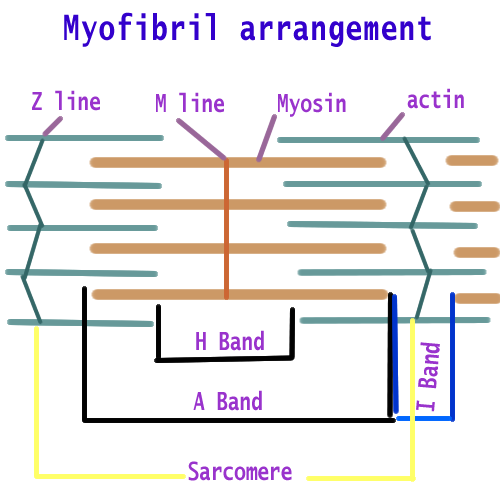
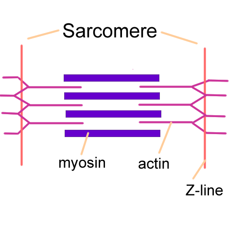
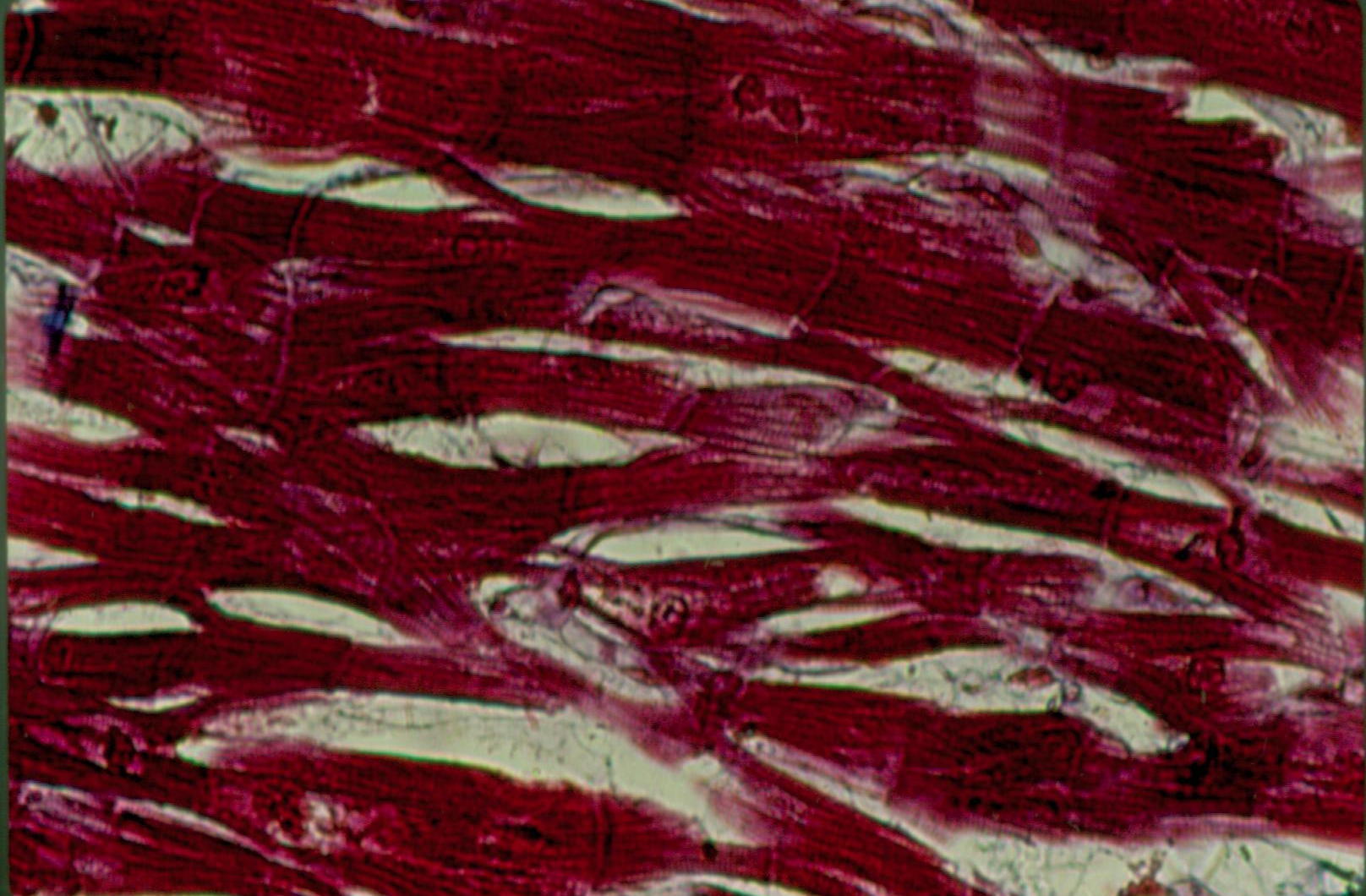
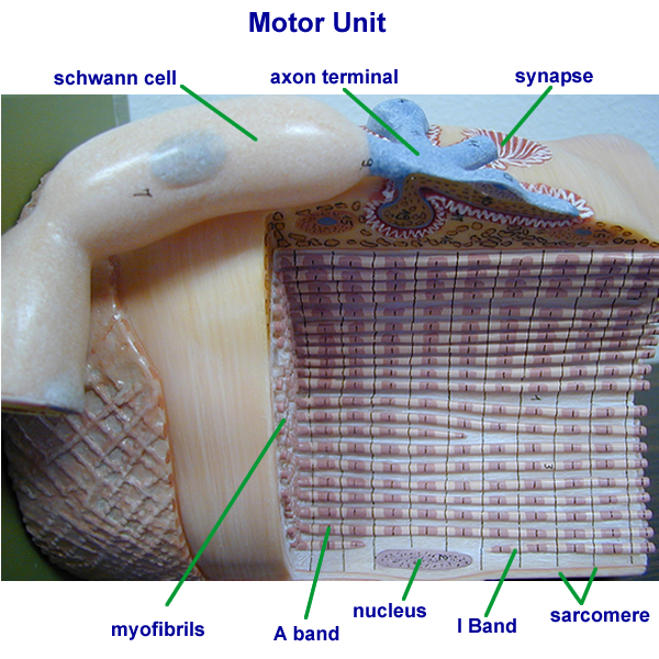
The histology of skeletal muscle also reveals a multinucleated cell fiber with striations. Striations are alternating light and dark bands due to the arrangement of contractile proteins. The bands have names based on their appearance and ability to bend light.
The dark bands are called A bands, the light bands are called I bands. Actin and myosin are kept in a regular arrangement by other proteins. The M line keeps myosin properly arranged and the Z line keeps Actin properly arranged. Based on the area between two consecutive Z lines, muscle’s functional contractile unit is identified and called the sarcomere. Individual sarcomeres of skeletal muscle all shorten and add together to produce overall muscle length shortening.
Skeletal muscle sarcomere shortening that produces an increase in tension but no movement is called an isometric contraction. Skeletal muscle sarcomere shortening that produces an increase in tension and movement is called an isotonic contraction.
Muscle Histology
Muscle Contraction
Contraction is activated by neurotransmitters from the nervous system.
At the neuromuscular junction for skeletal muscle, the neurotransmitter is acetylcholine, abbreviated as ACH or ACh.
ACh binds to special receptors on the sarcolemma and triggers Na+ channels to open. Sodium ion entry into the muscle changes the resting electric potential which now must reach threshold before it can signal a contraction. Contraction events begin when sodium movement reaches threshold and triggers calcium release within the skeletal muscle cell. Calcium then binds to its receptor on actin and allows an interaction between actin and myosin. Some physiologists call these contraction events the walking filament or sliding filament theory. As long as ACH is released, the calcium is available and the contraction is maintained. When ACh is no longer released and is removed from the skeletal muscles sarcolemma receptors by the enzyme acetylcholinesterase, the sodium ion channels close, and without sodium, calcium ion is pumped back into its stores in the sarcoplasmic reticulum. During this process of ions returning to their normal locations, this movement is marked by the physiologic process called Repolarization and it signals the relaxation of the skeletal muscle.
Energy Sources for Muscle
Energy sources are also needed in the contraction event. Energy in the form of ATP from the mitochondria is needed to help create part of the myosin head and is used to release actin from myosin during the contraction events. The skeletal muscles use aerobic and anaerobic means to generate ATP. Glucose is used as an energy source to create ATP during active use of muscles, while fatty acids are used as the energy source to create ATP when muscles are at rest. Oxygen is needed for aerobic pathways to occur.
Energy stores also occur in muscles by the chemical creatinine phosphate which enables ADP to be regenerated to ATP for the myosin head. Oxygen for aerobic metabolic pathways is made available by hemoglobin of red blood cells or myoglobin within the muscle cell. When the muscle runs out of oxygen, anaerobic pathways are used and create lactic acid which causes muscle fatigue. The oxygen debt must be “repaid” so that lactic acid can be converted back to pyruvic acid in the liver. Pyruvic acid in the presence of oxygen can entice the aerobic pathways to generate adequate amounts of ATP.
Muscles are never truly at rest and are at a state of slight contraction known as muscle tone. Muscle tone is controlled by the nervous system and maintains the health and ready state of muscles. Without nervous system stimuli, skeletal muscle cells will degenerate or atrophy and eventually all that is left is connective tissue which may result in contractures.
All of the muscle cells or fibers that are innervated by one motor neuron are called a motor unit. Motor unit recruitment can create several different responses that can be mapped on a graph. A single muscle contraction and relaxation is called a twitch. When muscles are stimulated during their entire physiologic cycle, tetanus or sustained contraction can be created. The more motor units that are recruited, the stronger the muscle contraction as more muscle cells are involved and this can be depicted by the graph called treppe.
Muscles are grouped into functional arrangements to provide movement. The two types of functional groups are synergistic and antagonistic. Synergistic muscles are those that work together to perform a certain function. There is one prime mover within the synergistic group. Others help stabilize or steady a joint to make a more precise movement possible. Antagonistic muscles oppose the movement created by the synergistic muscles. Muscles can pull, but not push. If one group flexes a joint, another group extends the joint. Muscle sense or proprioception is the brain’s ability to determine where our muscles are and what they are doing. Stretch receptors and specialized muscle spindle fibers detect changes in the muscle length as it is stretched.
Impulses are generated by these receptors and sent to the brain for interpretation and action.
Muscle Names
Muscles are named for several reasons: location, action, shape, size, divisions, origin, insertion, fiber direction, and Latin root word. Sometimes several references are used. Examples are:
Location Subscapularis
Direction and location Rectus Femoris
Location and size Peroneous Brevis
Shape Deltoid
Shape Trapezius
Division and location Biceps Brachii
Origin and insertion Sternocleidomastoid
Action and location Flexor carpi ulnaris
Root word Gracilis (slender)
Root word Sartorius (strap like)
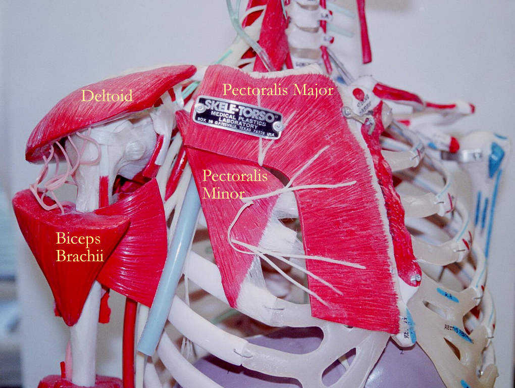
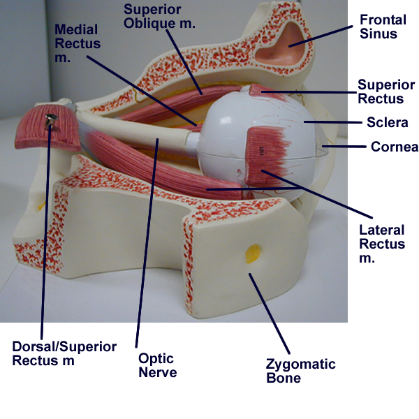
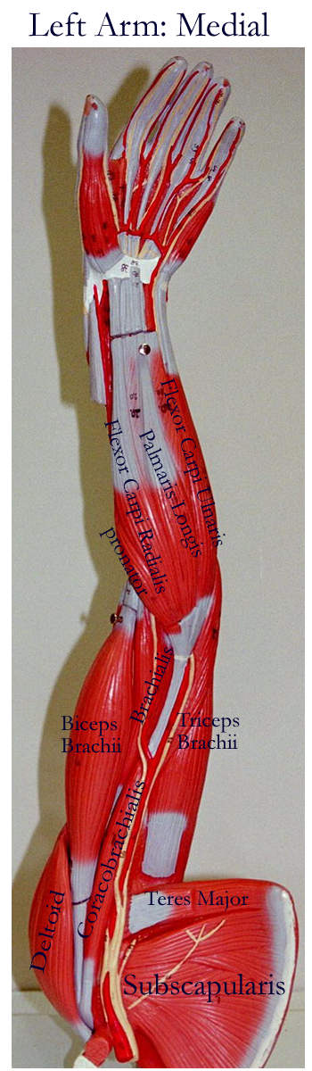
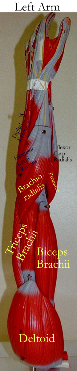
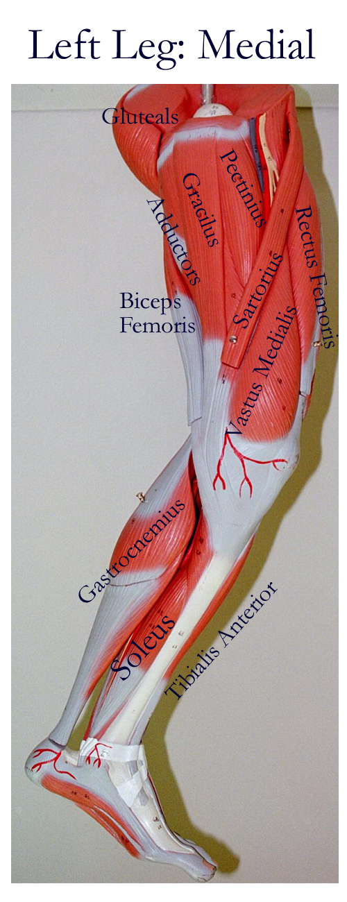
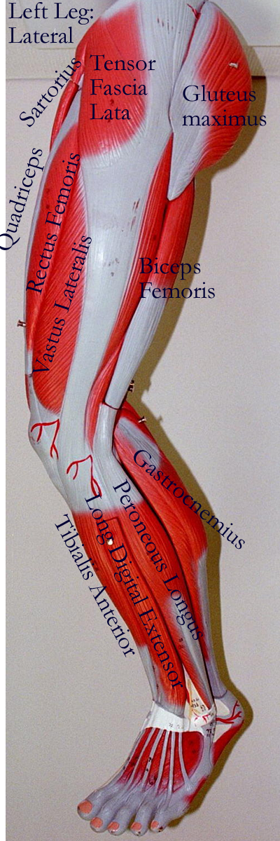
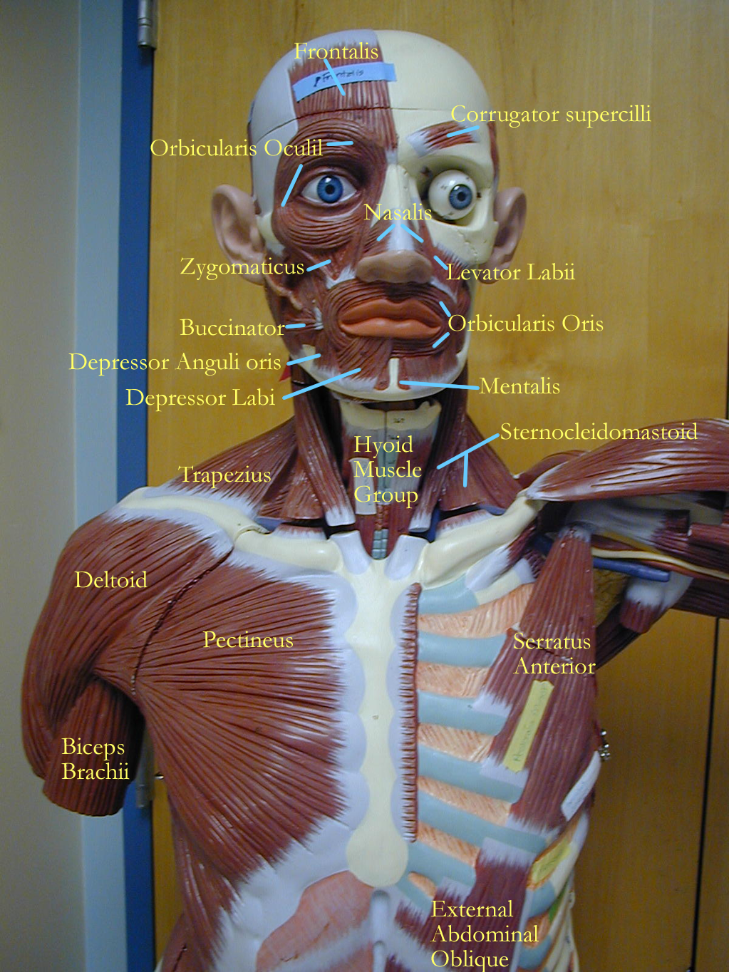
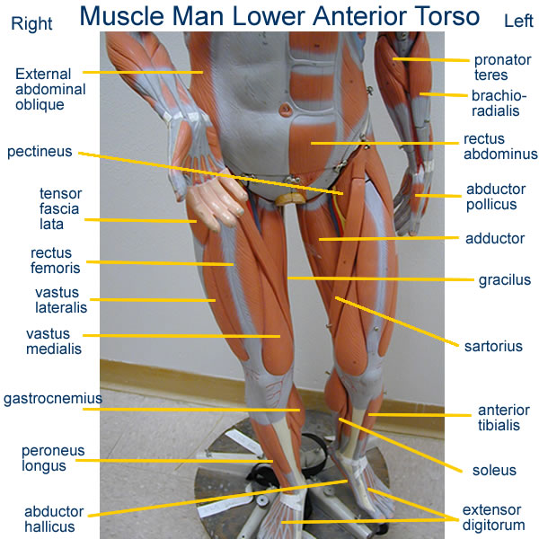
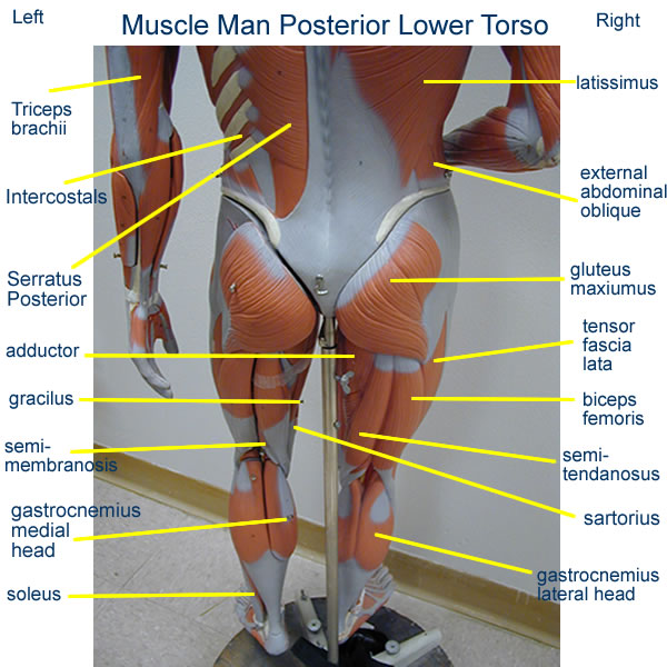
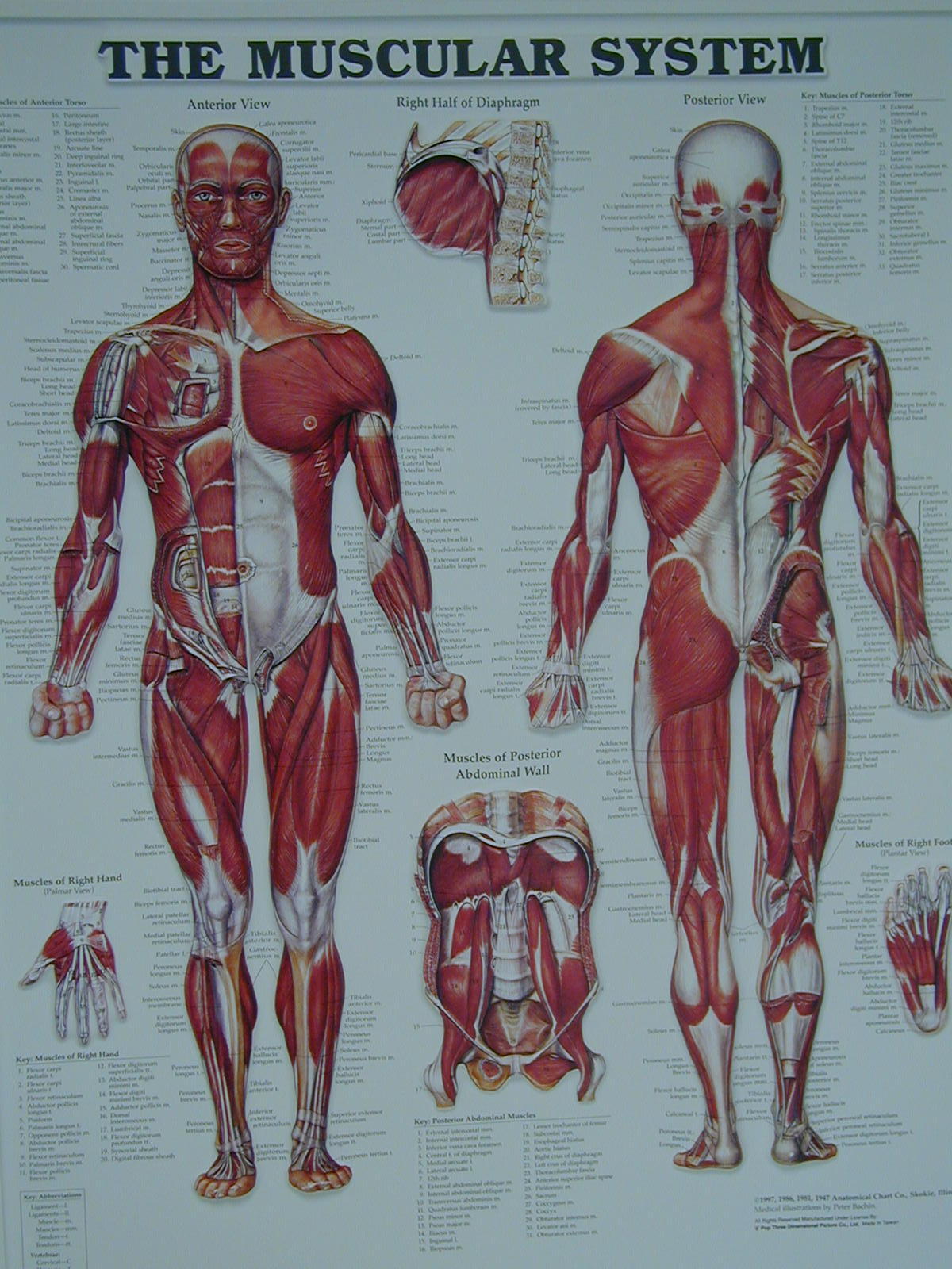
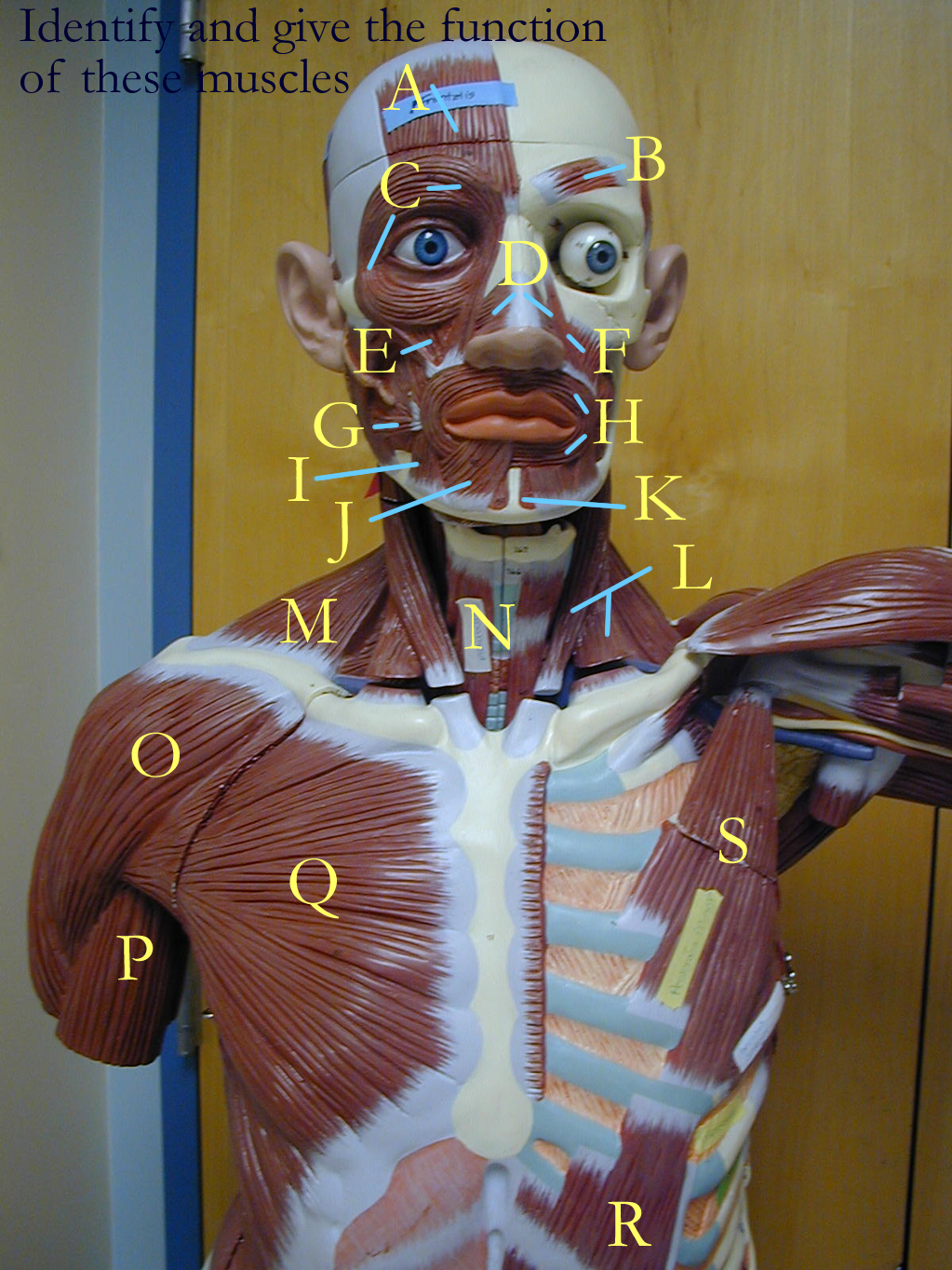
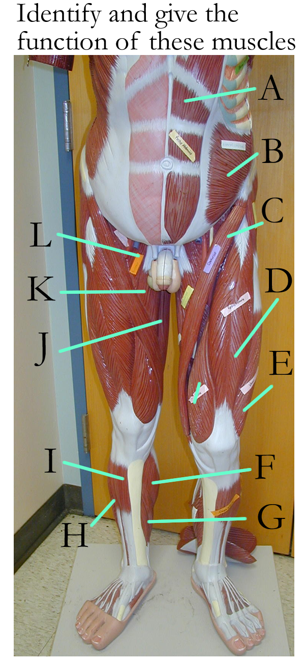
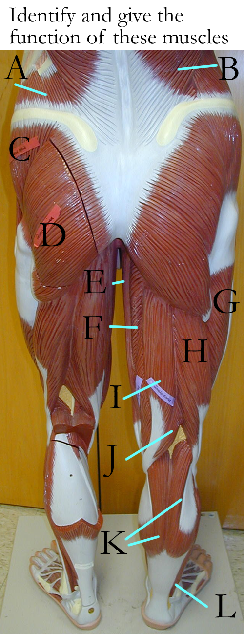
Superficial Muscles of the Body
Head and Neck : Anterior, Lateral, Pectorals
Eye: Laterial view
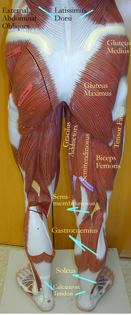
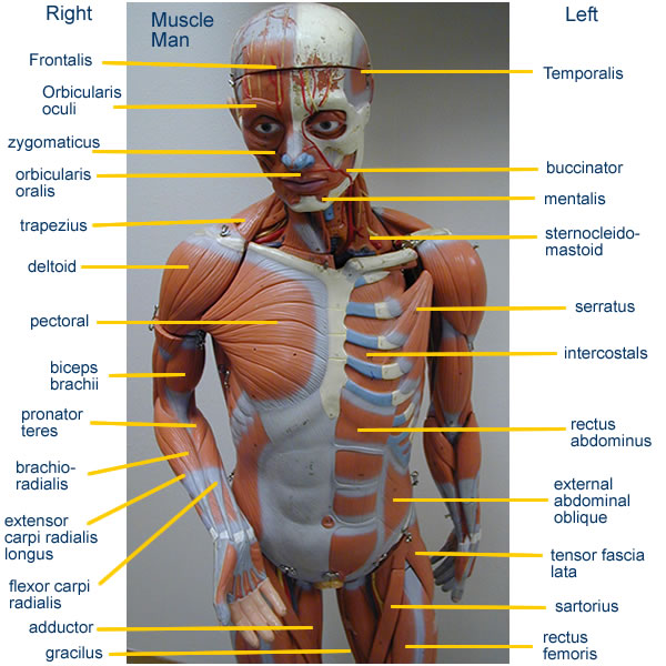
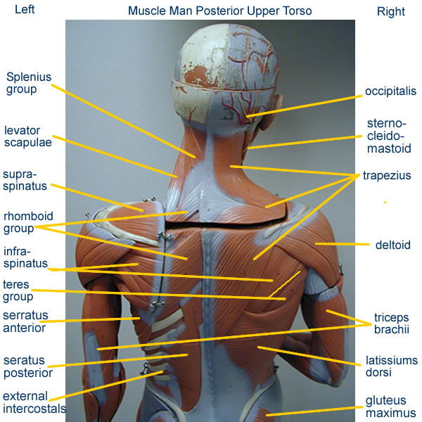
Large Muscle Man :
Small Muscle Man:
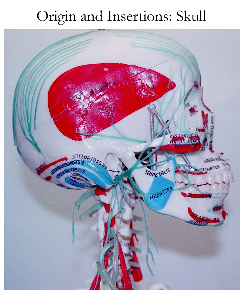
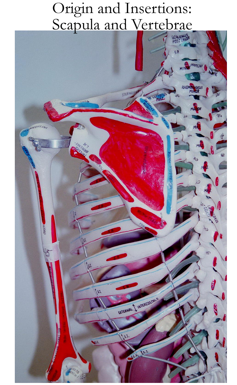
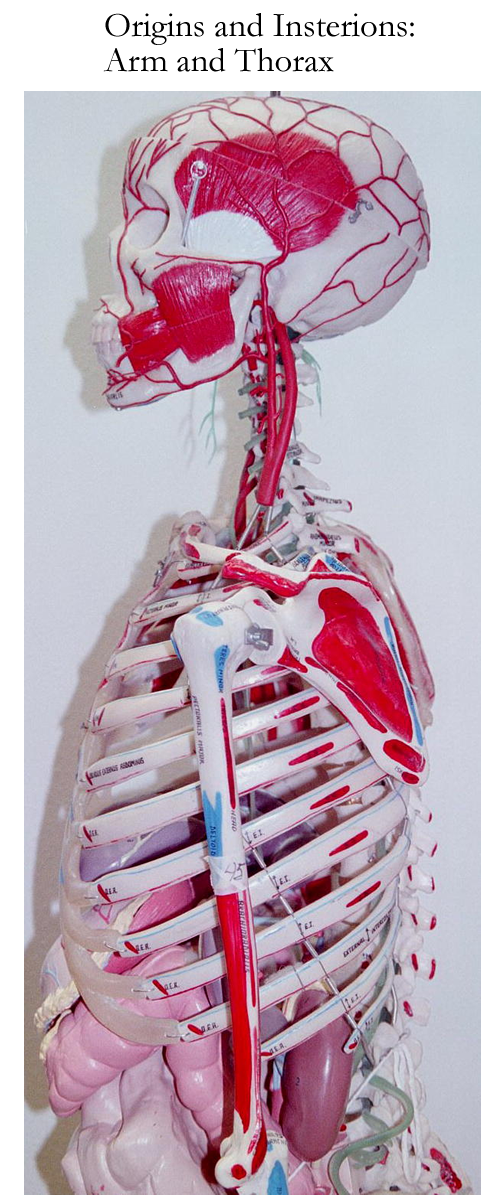
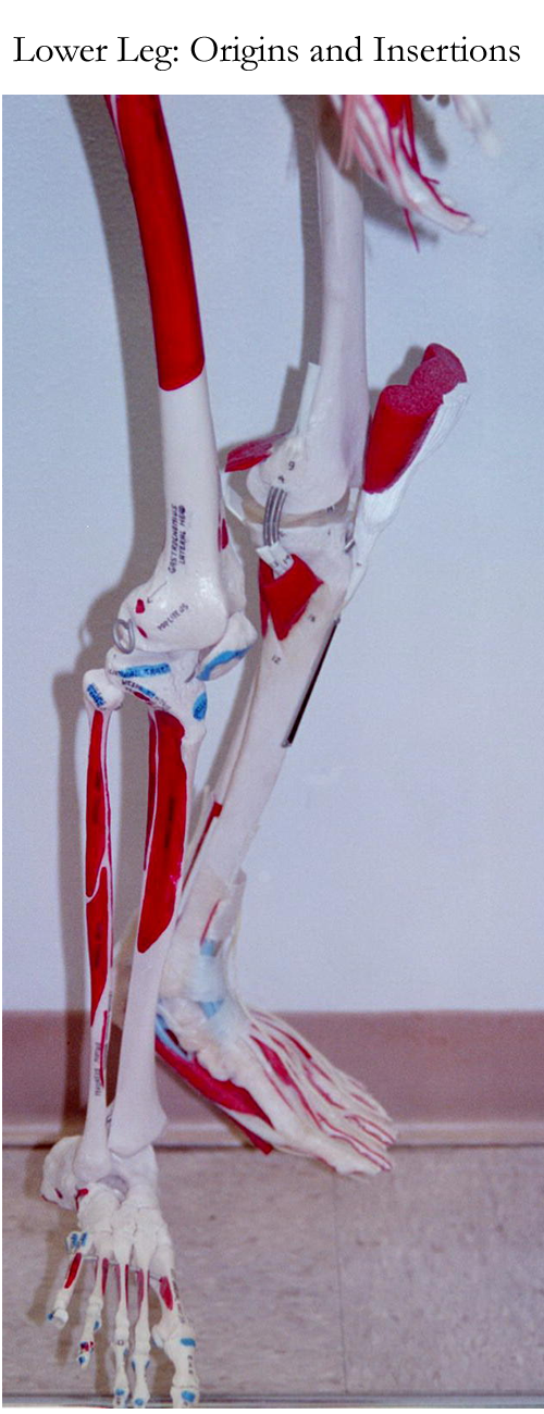
Skeleton with origin/ insertion: Skull, Vertebrae, Upper Thorax, Leg
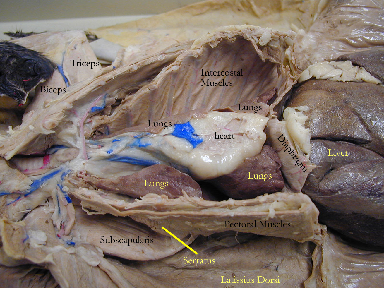
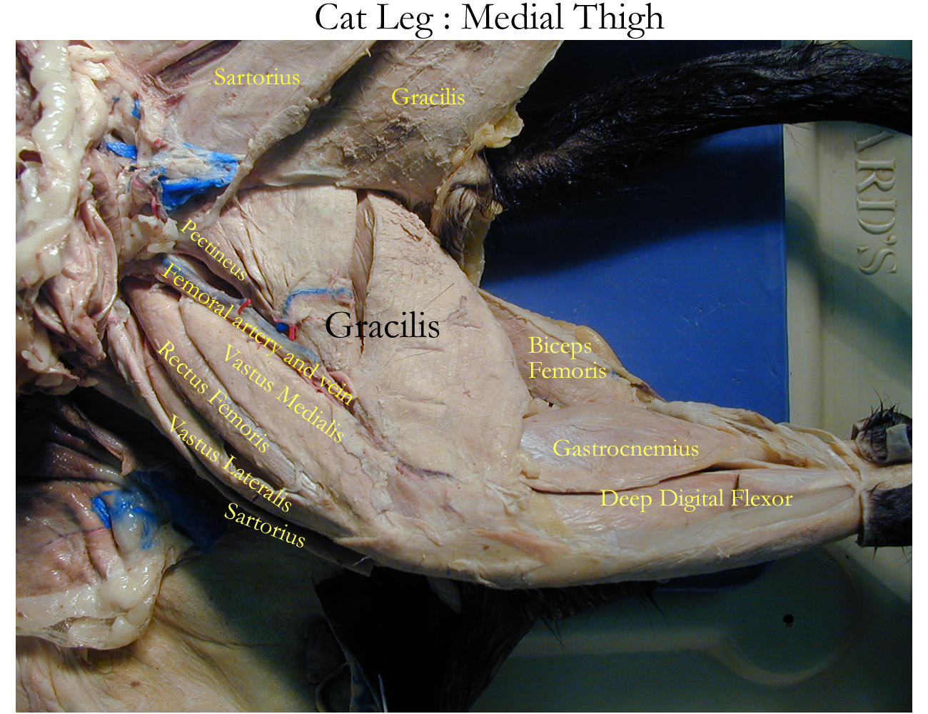
Specimens : Cat : Thorax, Abdomen, Medial Leg
Histology
Anti-, contra- against grav- heavy
Bi-, di- two, twice labi- lip
Gloss/o- tongue tens- strength
Infra- below -asthenia weakness
Iso- equal ten/o, tendin/o tendon
Kin/o-, -kinnin move muscul/o- muscle
-lemma husk, covering my/o-, myos/o- muscle
-meter measure ligament/o- ligament
peri- around fasci/o- fascia
sarc/o- flesh dys- painful
-trophy nourish a-, an- without
desmo- band faci- face, fascia
Muscle Use : click here for experiments
Isotonic exercise : Place your textbook on a table and pick it up using one hand. Why were you able to lift the book? Now place another book on top of your textbook and repeat the experiment. Were you able to lift the two books? Why was it harder the second time? Think about muscle fiber recruitment and motor units.
Isometric exercise: Using a chair (i.e. kitchen, folding, dining room) take one finger and lift it. Were you able to accomplish the task ? Why or why not ? Repeat the experiment noticing the tension in your muscles building. Now use one hand to lift the chair. Was it easier ? Think about muscle fiber size, strength, and number of muscles used.
Muscle
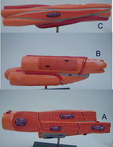
Types: Identify the following muscle types
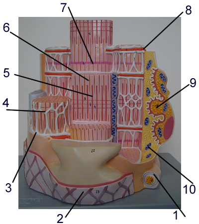
Sarcomere: Identify the following structures of the motor unit.
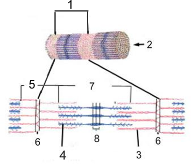
Contractile Unit: Identify the labeled structures.
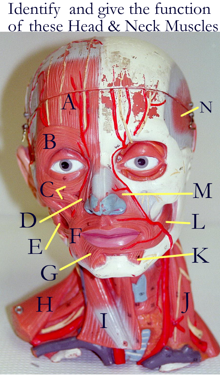
ID muscles:
Upper Head Torso and Arms: Anterior View
Upper Head, Torso and Arms: Posterior View
Muscle Parts: Obtain a cooked (boiled, baked) chicken leg. Identify
the following structures. (If completing this exercise for an LAR report, take
a photo of the leg with parts identified or draw a picture with labels.)
Aerobic vs Anaerobic exercises:
Explain the differences between these two exercises and be able to give examples
of both.
Which type of muscles are used (color, twitch, metabolic class) for each of
these types of exercises?
Muscle
review and quiz : click on muscle tissue
and muscle system to begin
Concept Map: Make a concept map for the muscular system (gross and histo) anatomy as well as functional groups regarding movement and nerve supply. Include this concept map in your LAR lab report (if selected) as a document insert or as an additional PDF scanned document of this map.
Space Flight changes
Intramuscular Injections
Cramps
Muscular Dystrophy
Myasthenia Gravis
Atrophy
Hypertrophy
Myositis, polymyositis
Myalgia
Botulism
Tendonitis
Occupational Therapist
Physical Therapist
Sports Medicine
http://www.nlm.nih.gov/medlineplus/healthtopics.html
http://www.lumen.luc.edu/lumen/meded/histo/frames/histo_frames.html
http://www.gen.umn.edu/faculty_staff/jensen/1135/webanatomy/
http://www-medlib.med.utah.edu/WebPath/HISTHTML/ANATOMY/ANATOMY.html
http://www.ohsu.edu/cliniweb/A2/A2.html
http://rpiwww.mdacc.tmc.edu/ap/frontAnatomy.html
http://www.ptcentral.com/muscles/
http://www.gwc.maricopa.edu/class/bio201/muscle/mustut.htm
http://www.ultranet.com/~jkimball/BiologyPages/M/Muscles.html
http://www.kcmetro.cc.mo.us/maplewoods/Biology/Bio110/lab3.htm
http://calloso.med.mun.ca/%7Etscott/second.htm
http://www.track0.com/canteach/links/linkbodysystems.html
http://www.carr.lib.md.us/schs/science/anatomy/systems.html
http://www.medem.com/MedLB/article_detaillb.cfm?article_ID=ZZZ5WGJU8JC&sub_cat=258 muscles of the back
http://www.medem.com/MedLB/article_detaillb.cfm?article_ID=ZZZPUF8BGJC&sub_cat=258 muscles of the front
http://www.medem.com/MedLB/article_detaillb.cfm?article_ID=ZZZ1PUCBGJC&sub_cat=258 muscles of the side
http://www.nlm.nih.gov/medlineplus/bonesjointsandmuscles.html
1. Give the types, location, and specific function of each type of muscle
2. Name the three connective tissue layers that help divide and support skeletal muscle
3. Name the two major bands and the two lines seen on microscopy of skeletal muscle.
4. Define sarcomere
5. Name the nervous system control and the neurotransmitter for the skeletal muscle motor end plate.
6. Define physiology terms involved with excitation- contraction coupling events:
a) Resting membrane potential
b) Depolarization
c) Threshold
d) Repolarization
7. Define contraction and the types of graphs that can be used to show contraction events.
8. Give the possible energy sources for skeletal muscle
9. Explain how muscles are named and give examples.
10. Be able to identify superficial muscles of the body
11. Define synergist, prime mover, antagonist in relation to skeletal muscles
12. Name the contractile proteins of muscle
13. Explain how calcium is used in muscle contraction
14. Define muscle tone.
15. Explain the difference between isometric and isotonic contraction.