Biology 2404 A&P Basics Lab Exercise 14 Blood Vessels Dr. Weis
| Objectives | Background | Medical Terms | Activities | Applications | Careers | WWW | Review Questions |
Students should be able to:
* Describe the differences in the major vessel types
* Define anastomosis and give examples
* Define blood pressure; know how it is measured; normal human values
* Define pulse and be able to identify several pulse points
* Know the blood flow pathways for the
Pulmonary Circuit
Systemic Circuit
Aortic branches
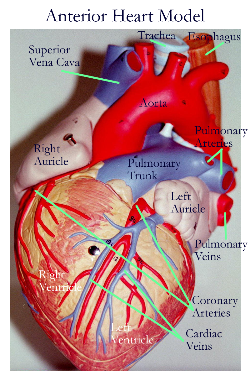
Coronary Circuit
Circle of Willis
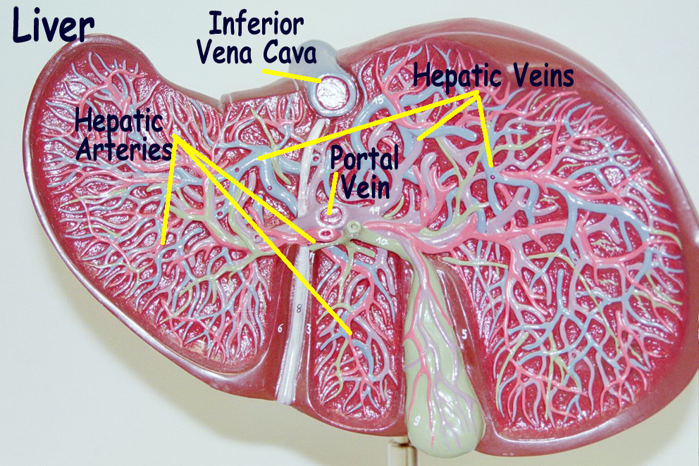
Hepatic Portal System
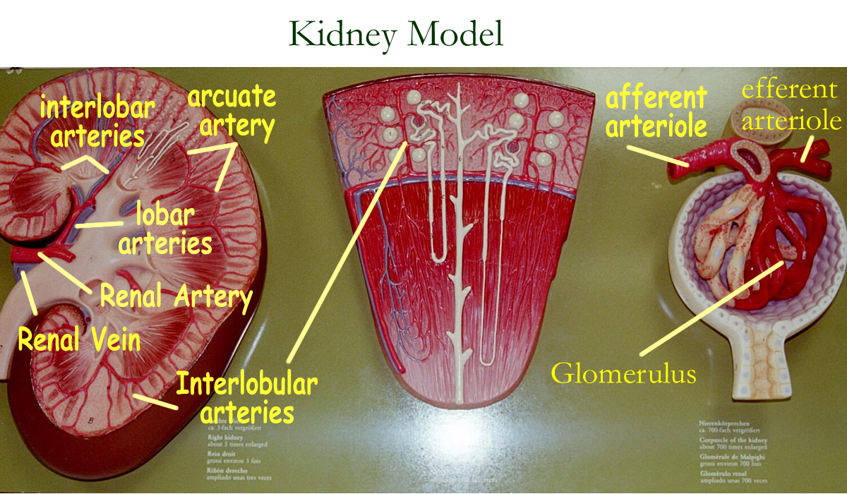
Renal Circuit
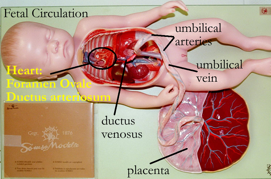
Fetal Circulation
and General Circulation
* define related terms such as constriction, dilation, filtration, reabsorption, edema
Read related information in textbook
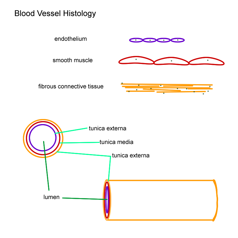
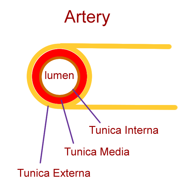
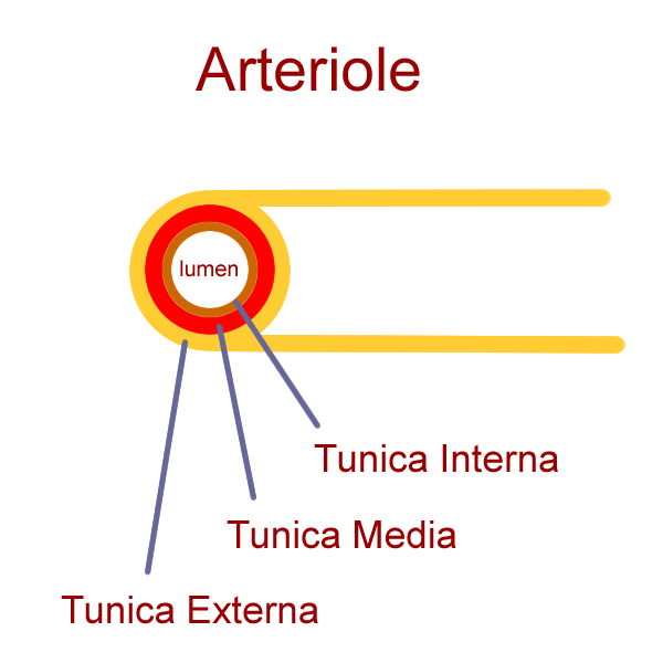
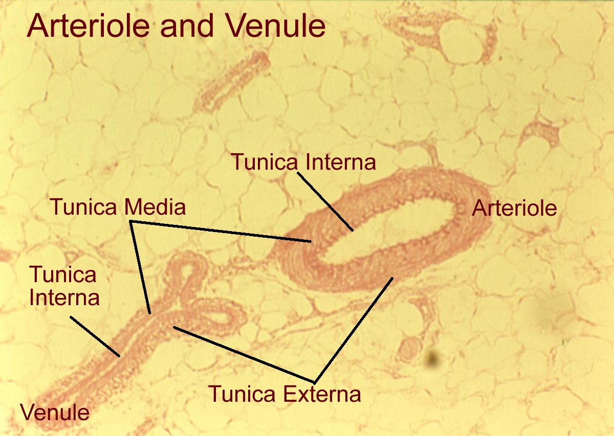
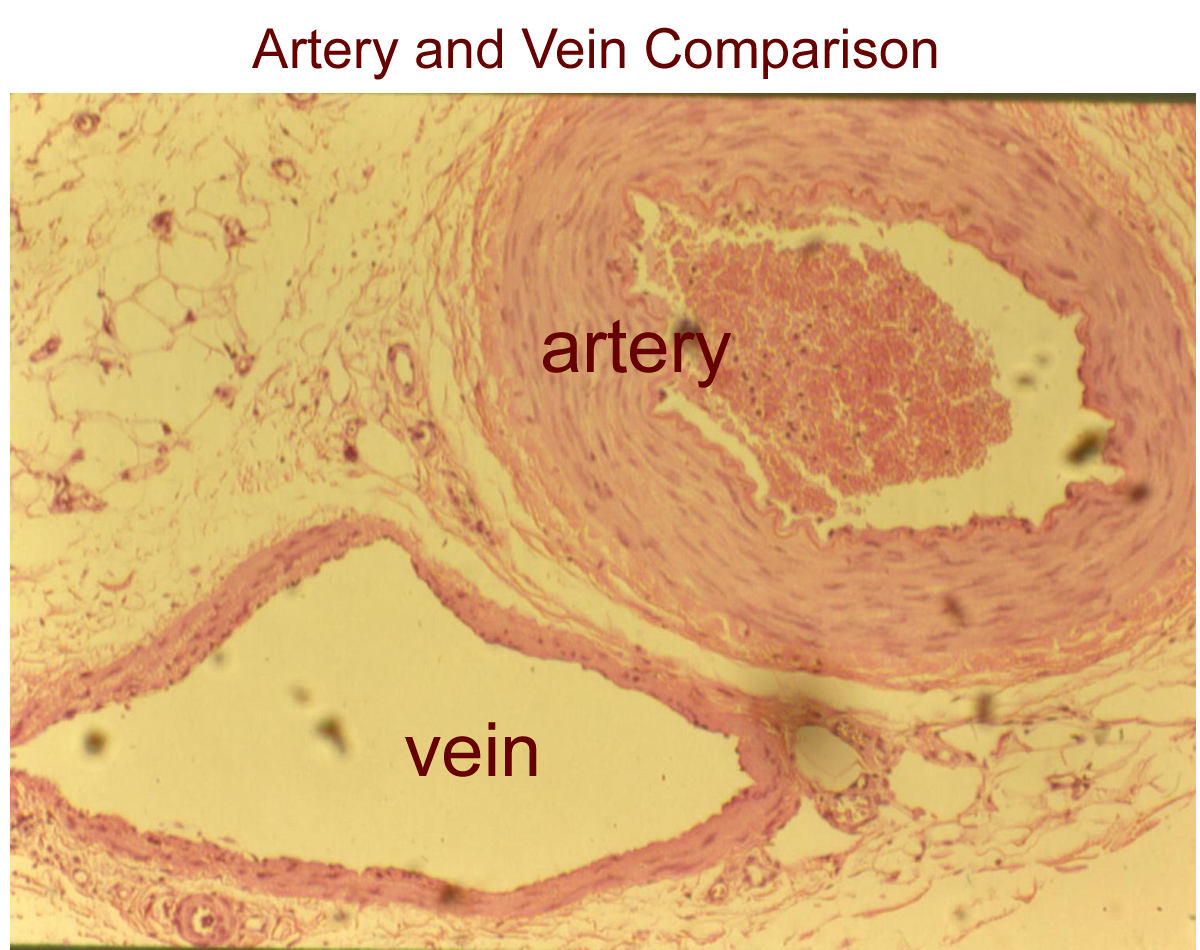
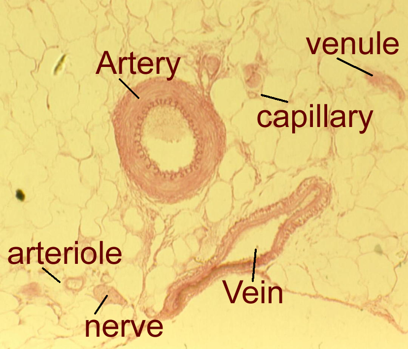
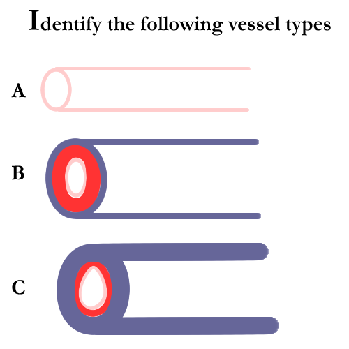
Blood vessels form a closed tubular network that begins and ends at the heart. The role they play involves blood flow and its distribution to and from body organs and tissues. Vessels can have up to three histological layers to give unique structure and function for the five major types of vessels seen: arteries, arterioles, capillaries, venules, and veins.
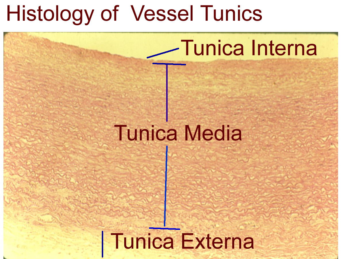
The histological layers are:
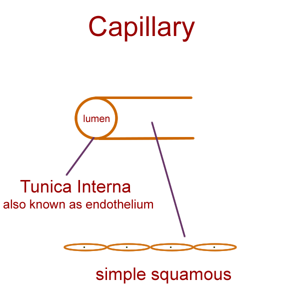
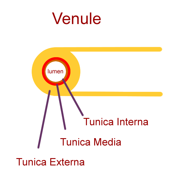
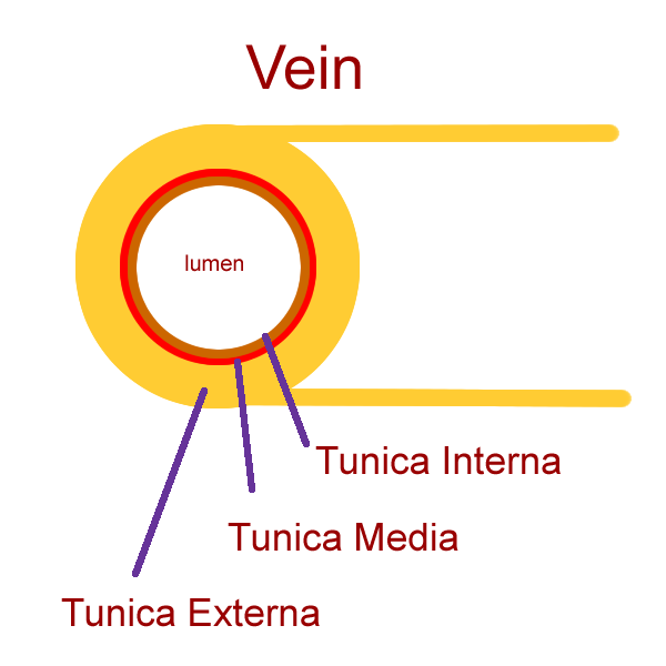
a) Tunica Interna (Tunica Intima) simple squamous epithelial lining, also known as the endothelium
b) Tunica Media smooth muscle in a circular plane
c) Tunica Externa (Tunica Adventitia) connective tissue outer covering
Arteries and arterioles carry blood away from the heart. Arteries have all three tunics and the tunica media (TM) is the thickest of the three layers. For the arteries that leave the heart, elastic connective tissue is also present to allow for recoil due to pressure changes as the heart contracts to pump blood to the body organs, some of which are superior to the heart. Most arteries are called muscular or distributing vessels since smooth muscle in the tunica media predominates in these vessels. As arteries decrease in size, so does the amount of smooth muscle in the tunica media.
Arterioles are small arteries and will still have the three histological layers, with the Tunica Media in decreasing amounts.
The metarteriole is the final distributing vessel on the arterial side and has only one layer of smooth muscle called the precapillary sphincter that surrounds the tunica interna. This vessel, by means of the capillary sphincter, controls blood supply into the capillary bed.
The ANS sympathetic division controls the smooth muscle in the arterial vessels and can signal for contraction changes that result in dilation or constriction to help change pressure and flow within the vessel and to regulate proper amounts of blood to organs depending on nutrient (primarily O2) demands.
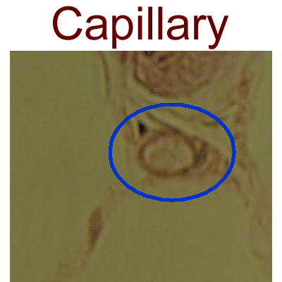
Capillaries consist of tunica interna only. The simple squamous lining can have several differences to create three different types of capillaries. All capillaries allow for some type of exchange, such as gas exchange, and nutrient / waste exchange by means of diffusion.
The three types of capillaries are continuous, fenestrated, and sinusoidal.
Continuous capillaries are found in the brain and limit the exchange of water soluble molecules. Regulation of these capillaries is controlled by neuroglial cells known as astrocytes. Sinusoidal capillaries are found in the liver, spleen, red bone marrow, and pituitary which allow widespread exchange of substances, including red blood cells, through the basement membrane that links the squamous cells together. Fenestrated capillaries are found in most other tissues and have selective exchange of substances due to the small openings in the squamous plasma membranes. Besides exchange of substances, capillaries also join arterioles to venules.
Veins and venules return blood back to the heart and are formed by all three vessel tunics, with the tunica externa (TE) predominating. Venules are smaller veins and have less TM and TE than veins. The endothelial lining of veins is also folded into flaps that form valves. Just as in the heart, the valves of veins prevent backflow and are regulated by pressure gradients as blood is moved back to the heart by skeletal muscle contraction.
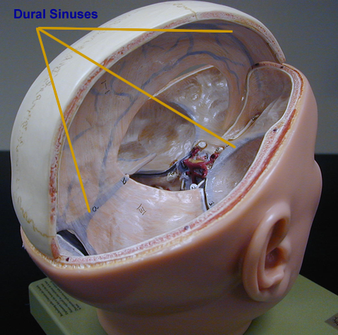
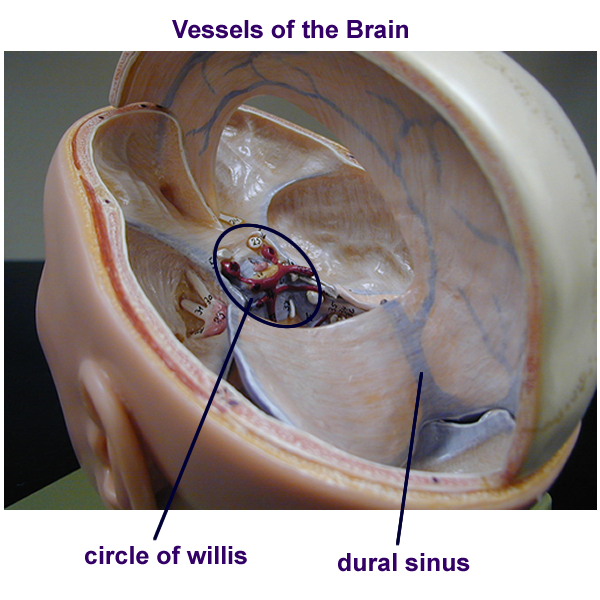
Specialized veins are found in areas of very low pressure. These veins, called sinuses, resemble large capillaries and function in returning blood from areas of very low pressure. Sinuses are found in the brain as the dural sinuses and in the heart as the coronary sinus.
An anastomosis is the joining of two structures not normally joined together. In a vascular anastomosis, vessels are joined to provide alternative blood flow pathways.
Examples are arterial anastomosis where two arteries are joined, venous anastomosis where two veins join together, and arterio-venous anastomosis that bypass capillary beds.
Blood Vessel General Histology Drawing
Blood Vessels Drawing : Artery, Arteriole, Capillary, Venule, Vein
Blood vessel Histology
Blood Vessel Physiology
Blood vessel physiology deals with blood flow to and from the capillary and the exchange that happens at the capillary level. To maintain the proper pressure to aid diffusion and filtration, resistance factors to flow need to be overcome. The proper blood consistency needs to be maintained to ensure flow as the vessel diameter can change by smooth muscle contraction or relaxation, mediated by the sympathetic division of the ANS.
Proper pressures ensure capillary wall integrity and create the movement of substances across the lining. Movement of substances from the capillaries to the tissues is known as filtration and movement from the tissues back into the blood vascular spaces is called reabsorption.
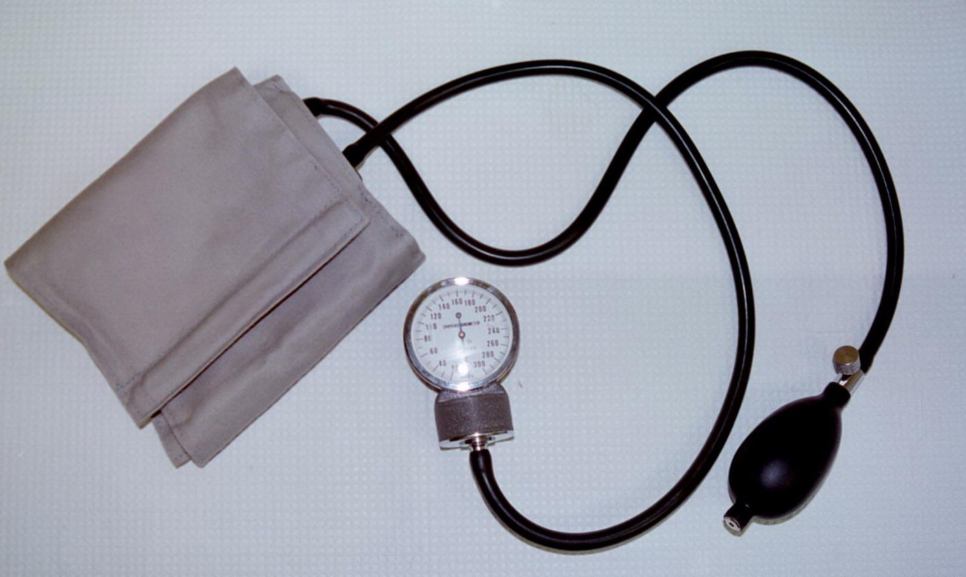
The pressure that drives this exchange is the result primarily from the ventricular cardiac contraction. Blood pressure can be measured in the arterial system by indirect means and in the venous system by direct means. Pressures are measured in millimeters (mm) of Mercury (mmHG) for the metric system. As the heart contracts, a systolic pressure can be determined within the vessels of the arterial system. As the heart relaxes, a diastolic pressure can also be determined. This forms the common notation of blood pressure as Systolic divided by Diastolic or S/D. Commonly seen numbers for healthy arterial blood pressures are 120/80 (upper limit of acceptable normal ranges).
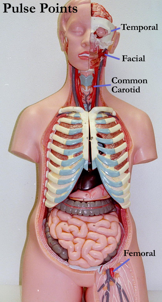
A pulse is a pressure wave is created by the differences in the systolic and diastolic pressure (S-D). Pulse points can be found where arteries are more superficial. Examples of areas where pulse waves can be felt include the temporal, facial, carotid, brachial, radial, femoral, popliteal, posterior tibial, and dorsal pedal arteries.
Regulation of blood pressure is dependent on ANS sympathetic short term influence on the smooth muscles of the tunica media. Longer duration of control of blood pressure is reflected in hormonal interaction and renal function. The protein albumin, which is made in the liver, plays an important role in allowing reabsorption and reclaiming of the tissue fluid back into the venous end of the cardiovascular system.
Not all the fluid pushed out of the capillaries to bath the cells is brought back into the blood vessels. Extra tissue fluid (leftover interstitial fluid) will then enter the lymphatic system and will eventually be returned to the cardiovascular system in lymphatic vessels.
Pulse pressure points: Body Torso
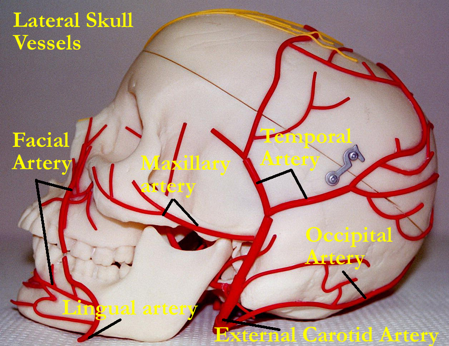
Skull with vessels: Anterior, Lateral
Blood Vessel Pathways
(See textbook for pictures and detail of blood vessel routes)
Blood vessels form two major circular pathways called circuits. These are the pulmonary and systemic circuits. Special pathways occur in several areas and most of these involve anastomosis. Examples of these special pathways are:
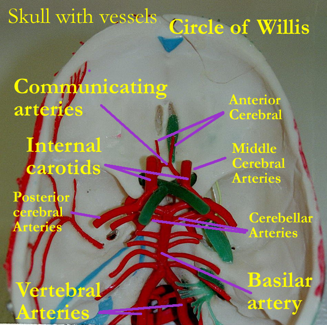
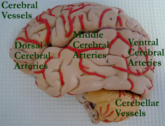
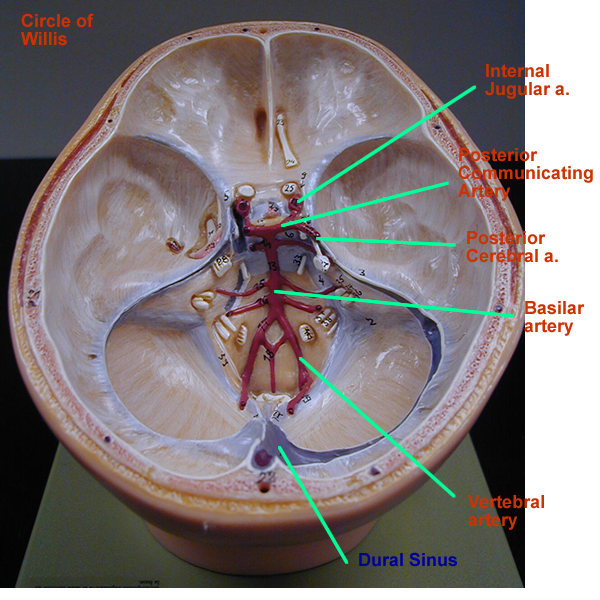
Circle of Willis in the brain arterial anastomosis
Hepatic Portal System in the liver venous anastomosis
Renal circuit in the kidney capillary/arterial anastomosis
Coronary circuit in the heart arterial anastomosis
Fetal circuit in the fetus bypasses for lungs & liver
Drawing of Systemic Pathways:
Skull with vessels: Anterior, Posterior
Brain with vessels: Dorsal View, Lateral View
Head model: Circle of Willis, Dural Sinuses, Circle and Sinuses
Liver model with vessels , Clear Liver Model with vessels
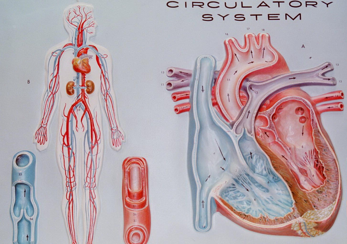
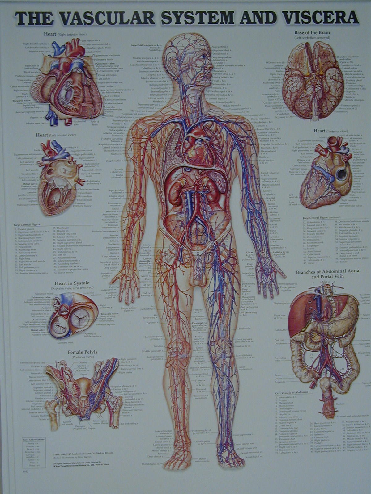
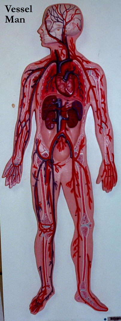
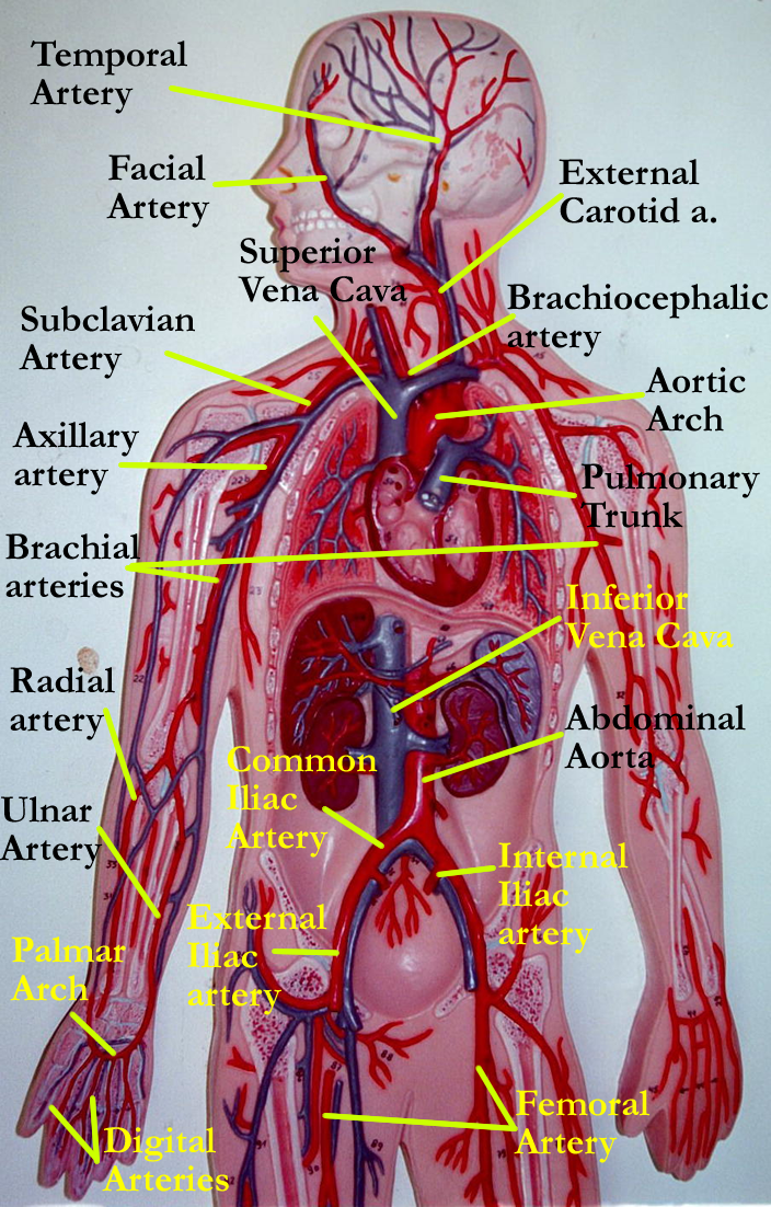
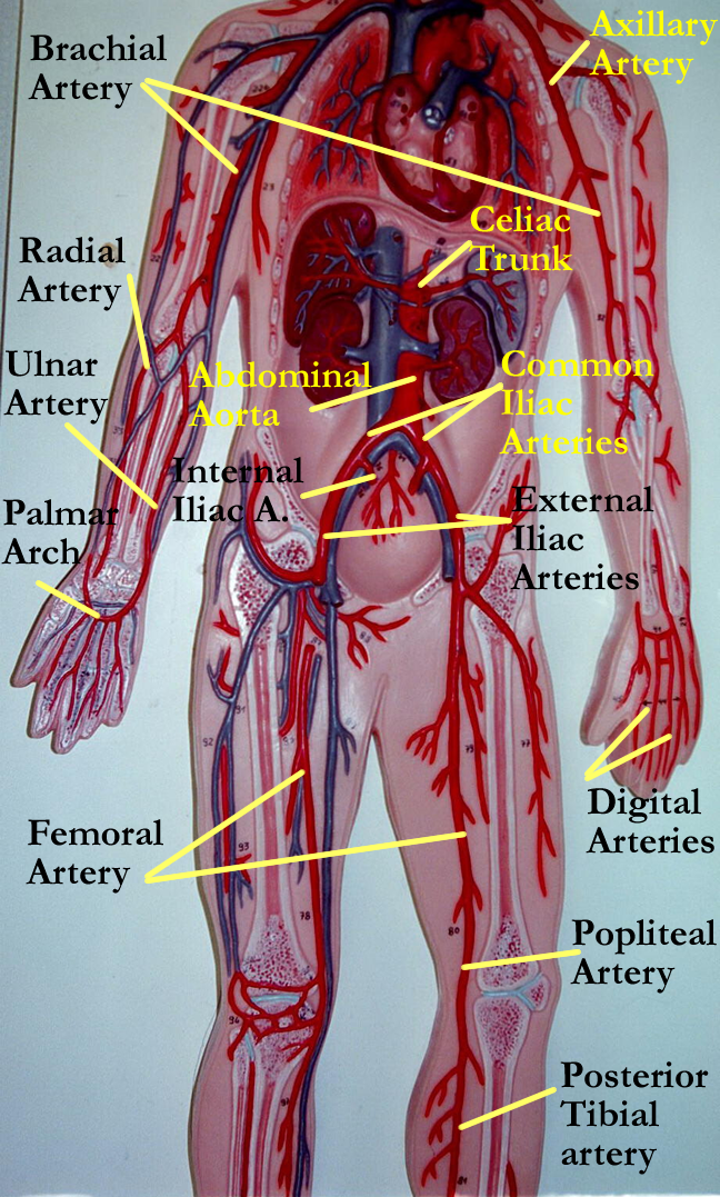
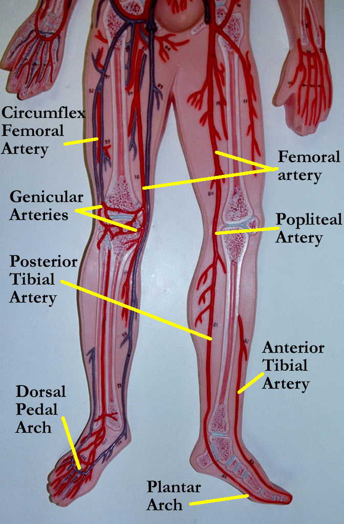
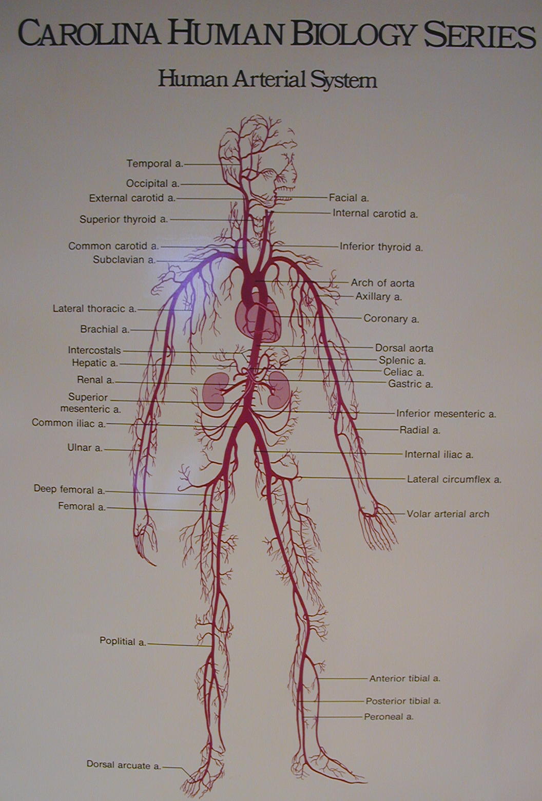
Vessel man : Whole view, Upper Portion, Middle Portion, Lower Portion
Artery Poster: Whole, Upper, Middle, Middle-Lower, Lower
Vein Poster: Whole, Upper, Middle, Lower
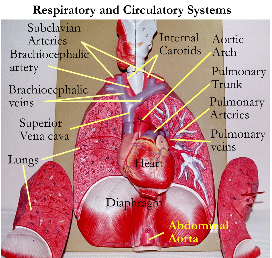
Pulmonary Circuit
Right ventricle -> pulmonary semilunar valve -> pulmonary trunk -> right and left pulmonary arteries-> pulmonary arterioles-> pulmonary capillaries -> pulmonary venules -> pulmonary veins -> left atrium
Systemic Circuit
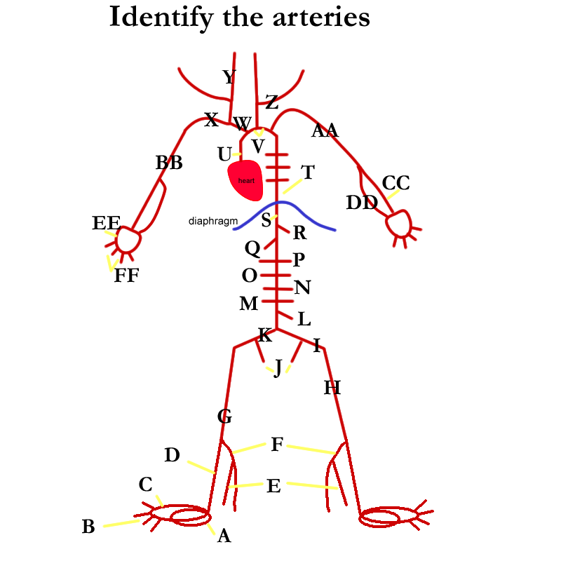
Left ventricle -> aortic semilunar valve -> aorta -> body organs & tissues -> right atrium
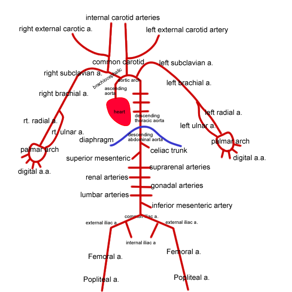
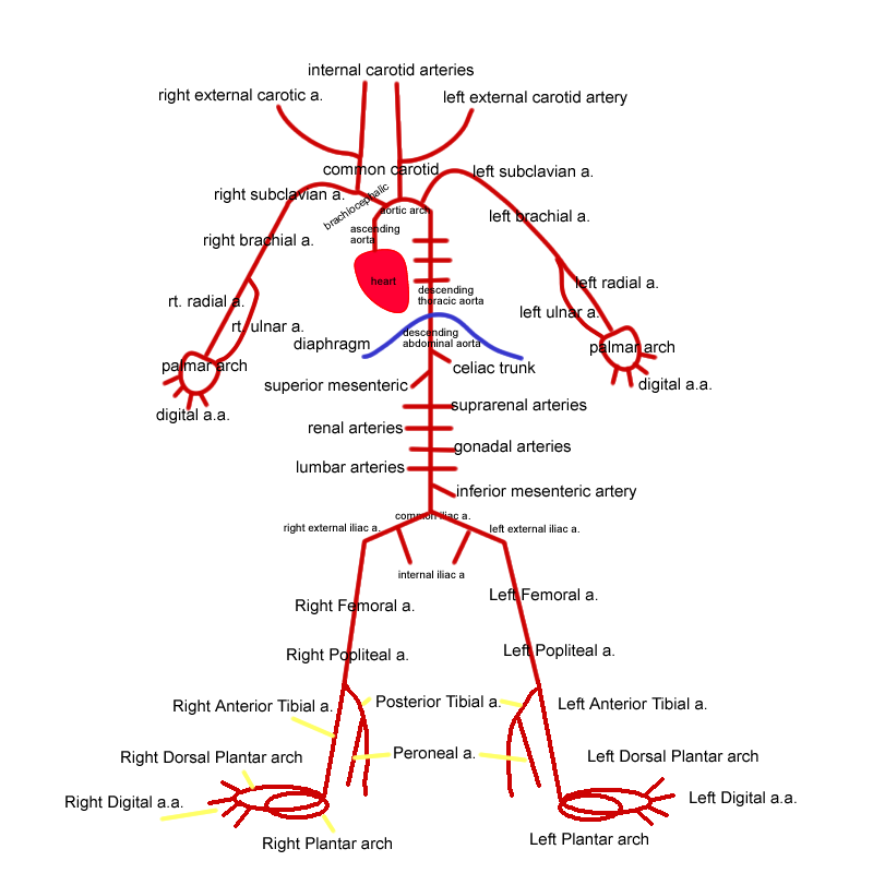
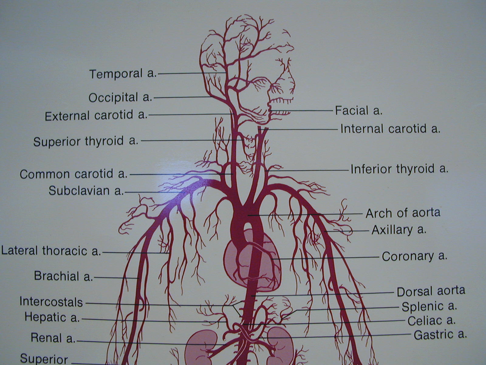
Aorta regions and branches.
Brachiocephalic arteries divide into the right common carotic artery and the right subclavian artery. After that, right and left sides mirror image one another.
Subclavian arteries -> brachial arteries -> radial and ulnar arteries -> palmar arches
Common carotids -> external and internal carotid arteries
External carotids supply the external face
Internal arteries supply the brain and are involved with the Circle of Willis
Descending thoracic aortic branches: provide arterial blood to thoracic organs.
Major branches are mediastinal, esophageals, bronchials, pericardials, intercostals, phrenics.
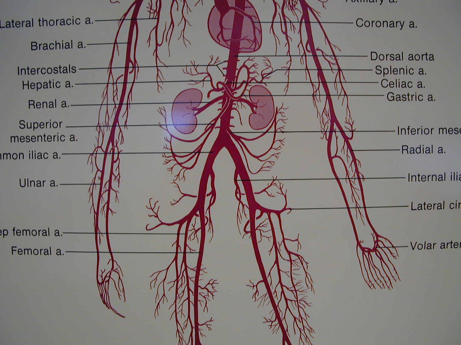
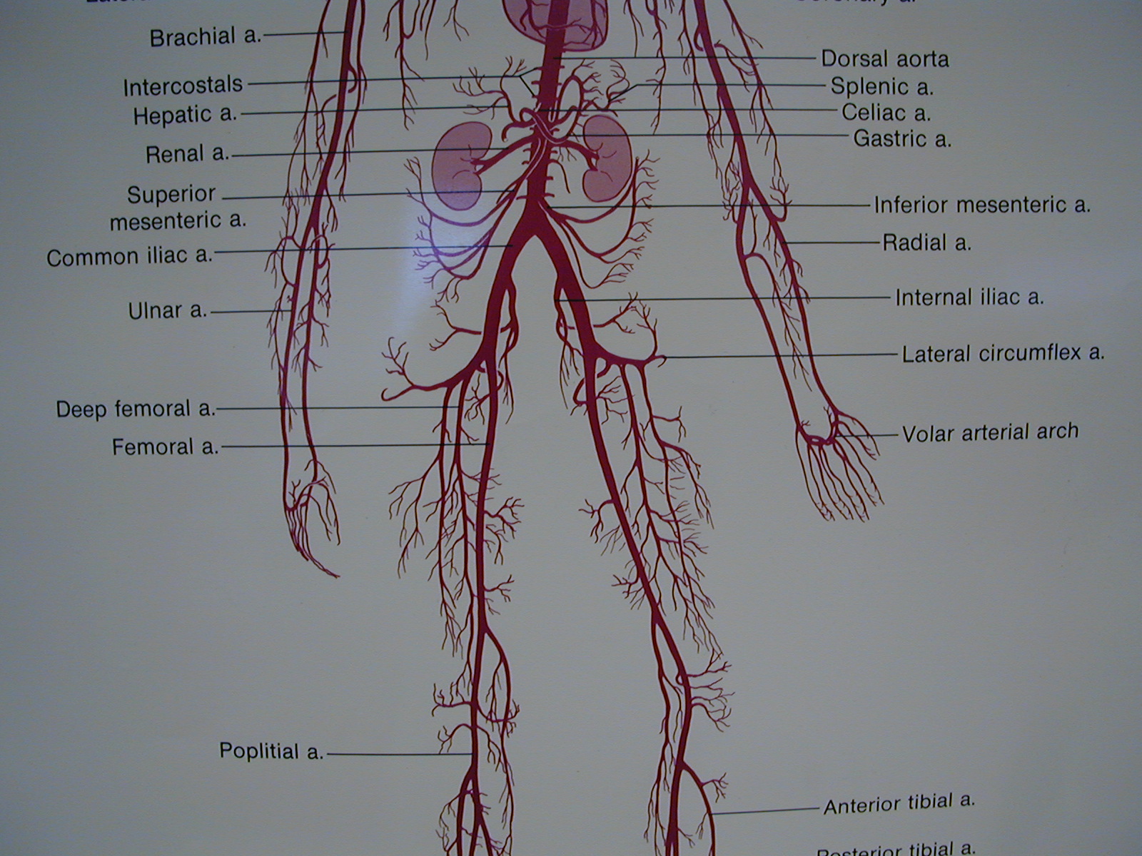
Descending abdominal aortica branches provide arterial blood to abdominal organs.
Major branches are celiac trunk, superior mesenteric, adrenals, renals, lumbars, gonadals, and the inferior mesenterics.
Descending abominal aorta then divides in the right and left common iliac arteries.
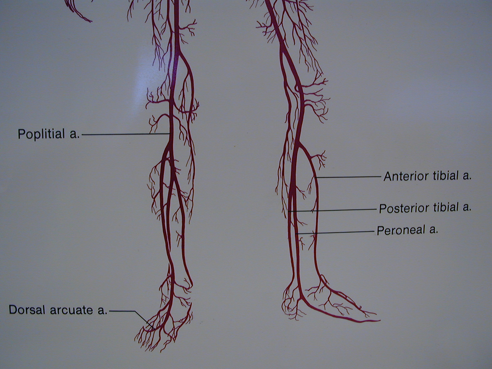
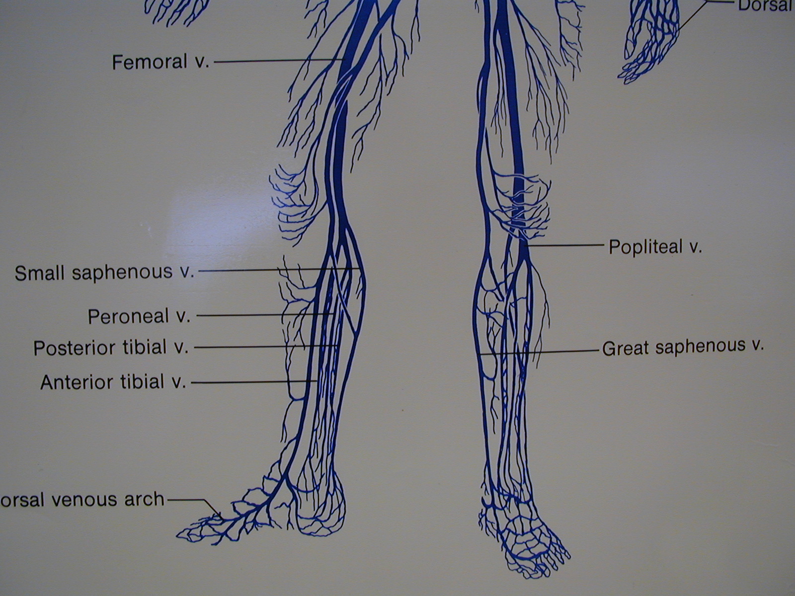
Common iliacs divide into internal and external iliac arteries. Internal iliacs provide blood supply to the pelvic area. External iliacs supply blood to the lower limbs as the femoral artery -> popliteal artery -> anterior and posterior tibial artery -> dorsal pedal artery with the plantar arteries.
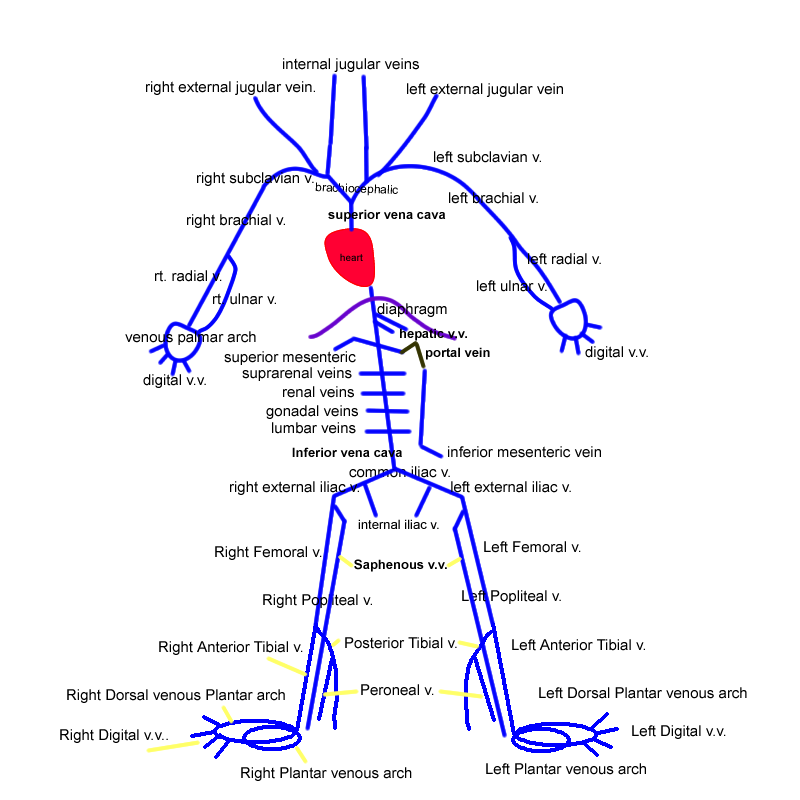
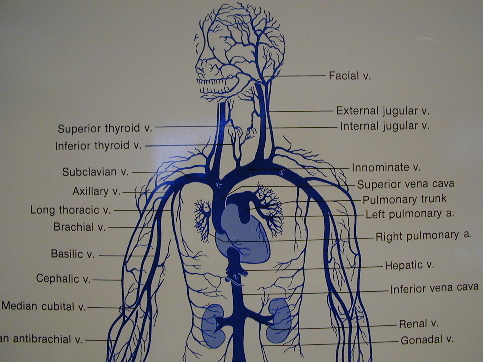
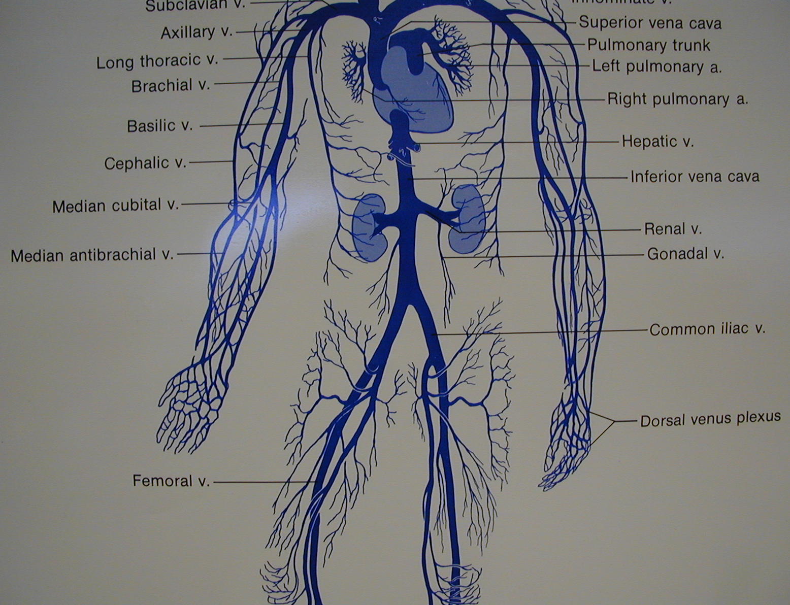
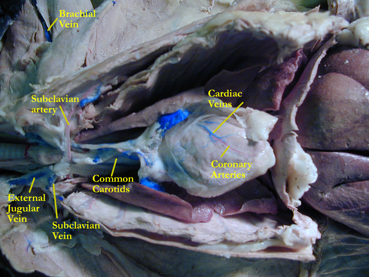
Deep veins follow arteries and usually have the same name. Unpaired superficial veins allow for alternate return of blood back to the heart and contribute to the venous pool of blood available if needed in emergency situations. When paired vein names differ from their arteries, it usually means that there has been an anastomosis in the pathway.
Cat specimen with injected vessels
Arteri/o- artery bar/o- pressure
Ven/o- vein phleb/o- vein
Capill/o- capillary -al pertaining to
Angi/o- vessel vas/o- vessel
Sten- narrow vascul/o vessel
I. Pulse points : Locate several pulse points using your index or middle finger and take your pulse in these locations. Identify the artery involved. If possible, correlate your pulse to your heart rate using a stethoscope to listen to your heart while you feel your pulse. Note the strength of each pulse and any missed beats. Take the pulse of someone else (with their permission). Do you notice any difference ? What would a weak, thready pulse mean ? How does your pulse relate to the systolic and diastolic portion of the cardiac cycle ?
II. ID vessels
A Drawings : Vessel type, Pathway
C Model
III. Veins: Since you know arterial blood supply, name some deep veins that run with the arteries you have learned. Name three superficial veins (veins not running with arteries). Why do we have more veins than arteries ? Why do veins below the heart need valves ? Why do some vein names not match their corresponding arteries (i.e carotid artery with jugular veins or coronary arteries and cardiac veins)?
IV. Blood pressure : If you have access to a sphyngomanometer, take the blood pressure of someone (with their permission) while seated in a chair. What results did you obtain ? Now ask the person to lay down and repeat the blood pressure procedure. What results did you obtain ? Were they different from the original reading? What is the term for high blood pressure and the term for low blood pressure ? Why is blood pressure recorded based on systolic and diastolic phases of the cardiac cycle? What organs of the body rely heavily on adequate blood pressure? Do you know (remember) any hormones that had their effects on blood pressure ? If so, which one(s) and how did they affect blood pressure ?
Concept Map: Make a concept map of the blood vessels (gross and histo) anatomy and physiology for major arteries, veins, and capillaries (3 types). Include this map in your LAR lab report (if selected) as a document insert or as an additional PDF document scan.
Atherosclerosis arteriosclerosis
Hypertension hypotension
Shock aneurysm
Phlebitis coronary thrombosis
varicose veins hemorrhoids
Perfusionist
Phlebotomist
http://www.gen.umn.edu/faculty_staff/jensen/1135/webanatomy/wa_cvs/
http://www.andrews.edu/~murdick/write/fluid/system.htm
http://www.usouthal.edu/biology/shardo/bly152/cardiovascular.html
http://www.clt.astate.edu/radsci/pfgs311cvsimages.htm
http://www.student.loretto.org/anatomyphys/chp43.htm
http://www.crha-health.ab.ca/hlthconn/items/cvsystem.htm
http://cellbio.utmb.edu/microanatomy/cardiovascular/cardiovascular_system.htm
http://www.medem.com/MedLB/article_detaillb.cfm?article_ID=ZZZ2O6756JC&sub_cat=510 circulatory system – arterial
http://www.medem.com/MedLB/article_detaillb.cfm?article_ID=ZZZG57C56JC&sub_cat=510 circulatory system – venous
http://www.nlm.nih.gov/medlineplus/heartandcirculation.html
1. Name the 5 major types of vessels and their function.
2. What is the precapillary sphincter?
3. Name the three types of capillaries.
4. Define anastomosis and give vascular examples.
5. Define blood pressure, explain the control of pressure, and give the normal values.
6. Follow the blood flow for the pulmonary circuit.
7. What and where is the circle of Willis?
8. Explain the function of the hepatic portal system.
9. Name the major arterial blood supply to the arm starting at the subclavian artery.
10. Name the aortic regions and major branches.
11. Name the major arterial blood supply the leg starting at the external iliac artery.
12. How is fetal circulation different than adult circulation?