Biology 2404 A&P Basics Lab Exercise
19 Digestive Accessory
| Objectives | Background | Medical Terms | Activities | Applications | Careers | WWW | Review Questions |
Students be able to:
* Identify, give the location of GI accessory organs
* Give the functions of the GI accessory organs
* Name the major enzymes produced by accessory glands and their function
* Give the source, storage, composition, and function of bile
* Define related terms
Read related material in textbook
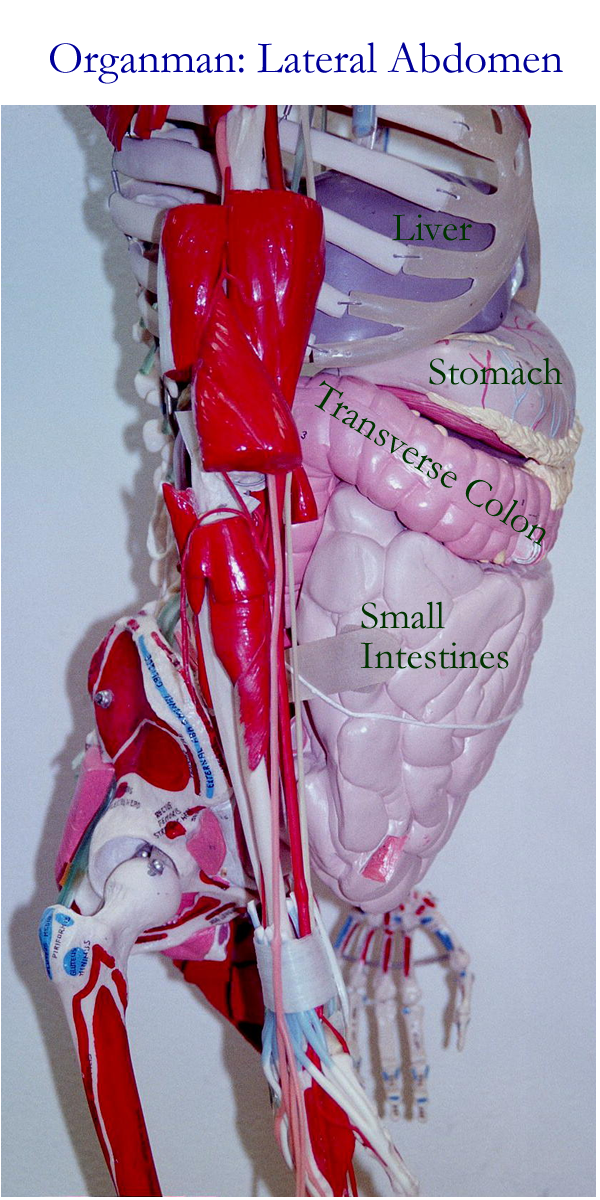
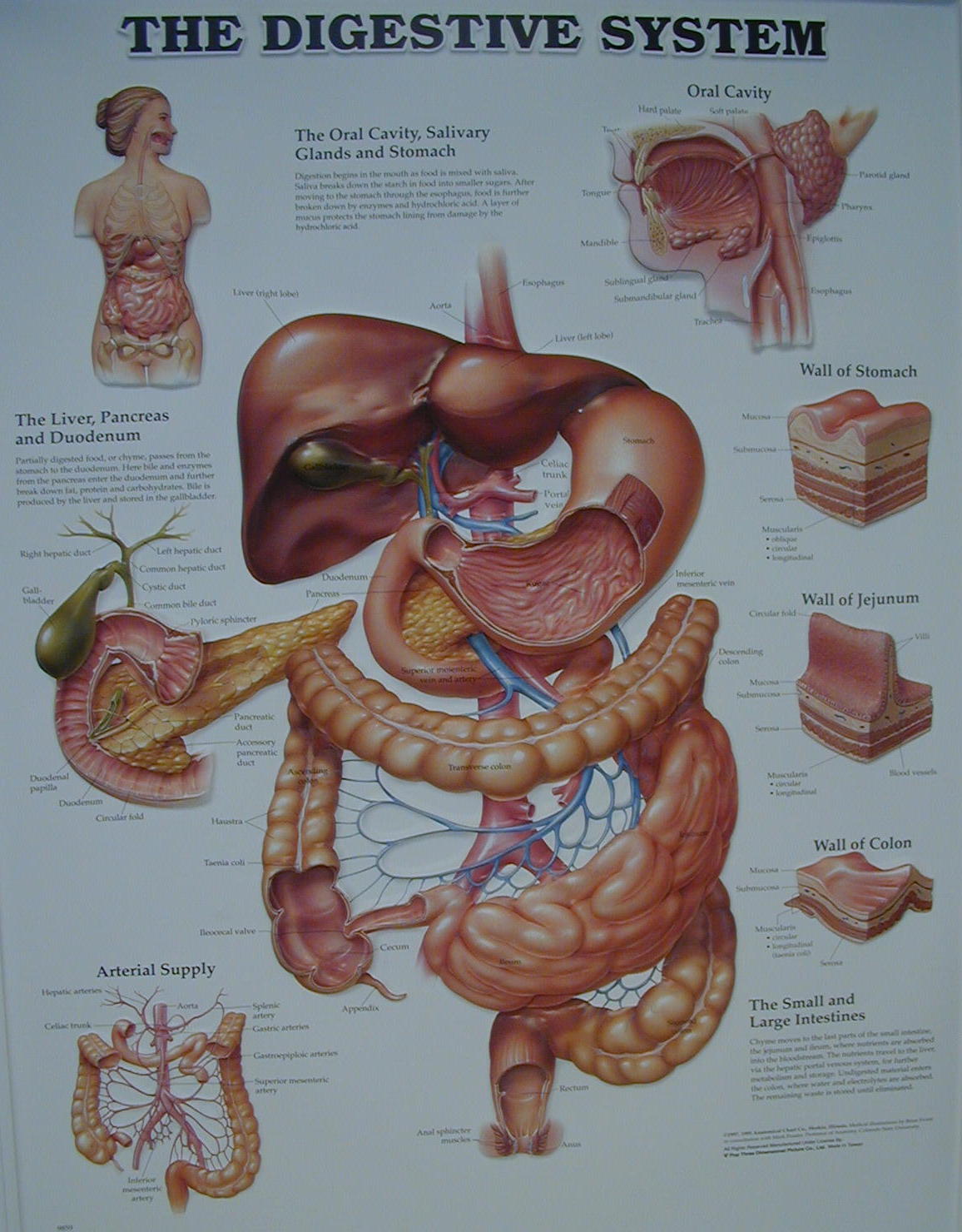
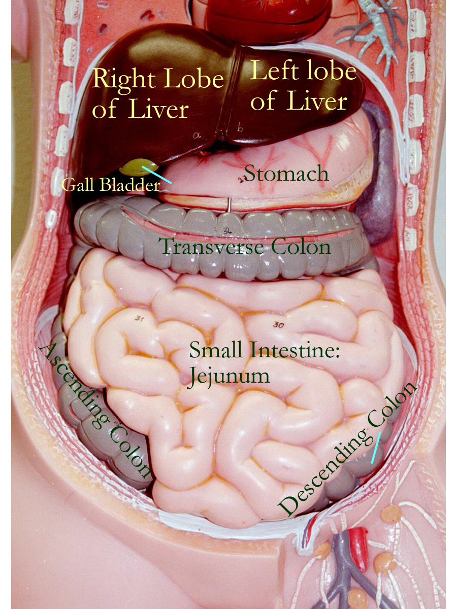
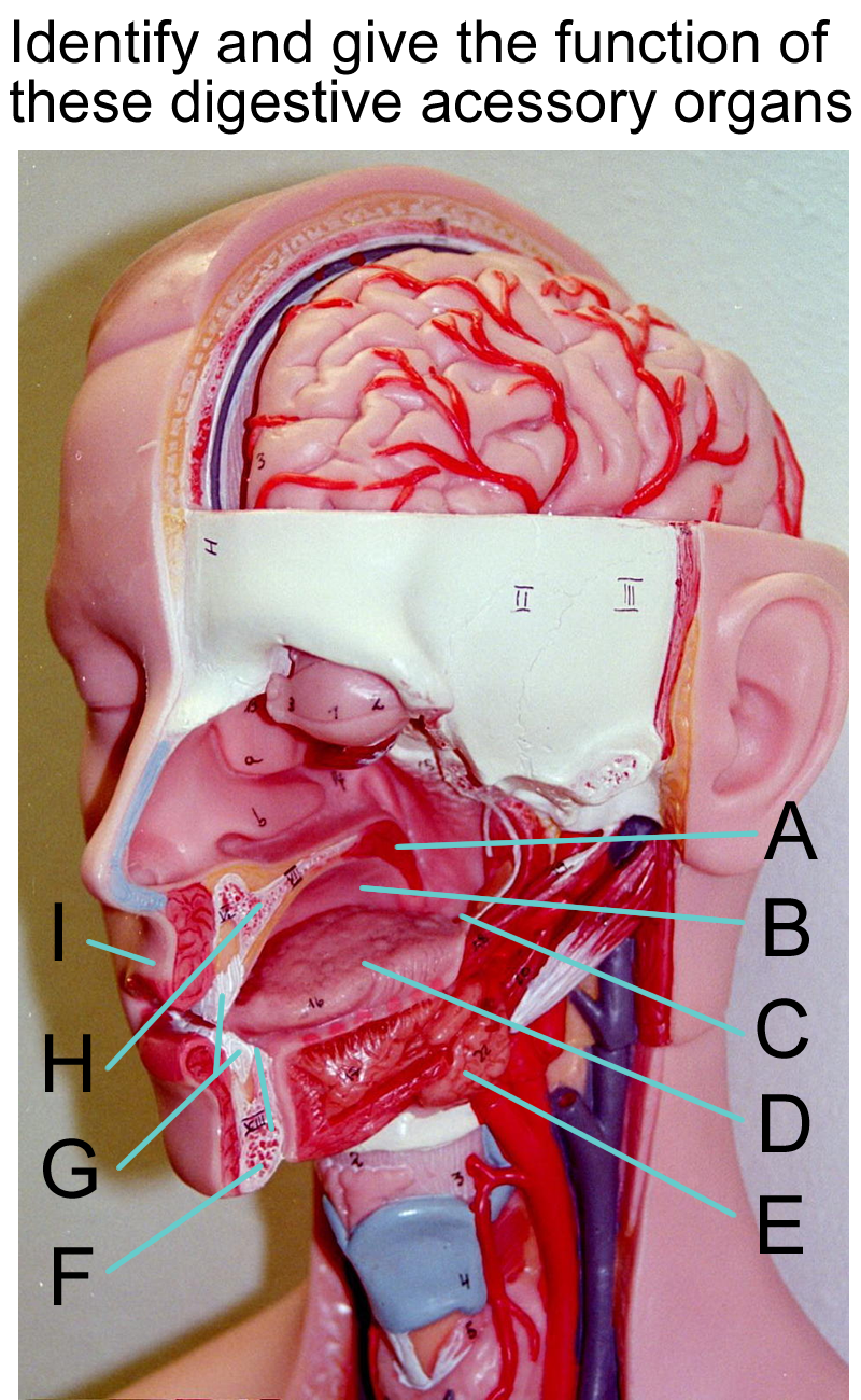
Accessory organs of the gastrointestinal (GI) tract are the second major division of digestive system. These accessory organs aid in digestion and absorption of nutrients for the alimentary canal. Accessory organs are the tongue, teeth, salivary glands, pancreas, liver, and gall bladder.
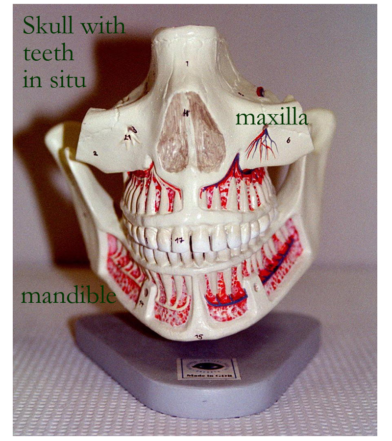
Skeleton with GI models : Anterior, Lateral
Tongue
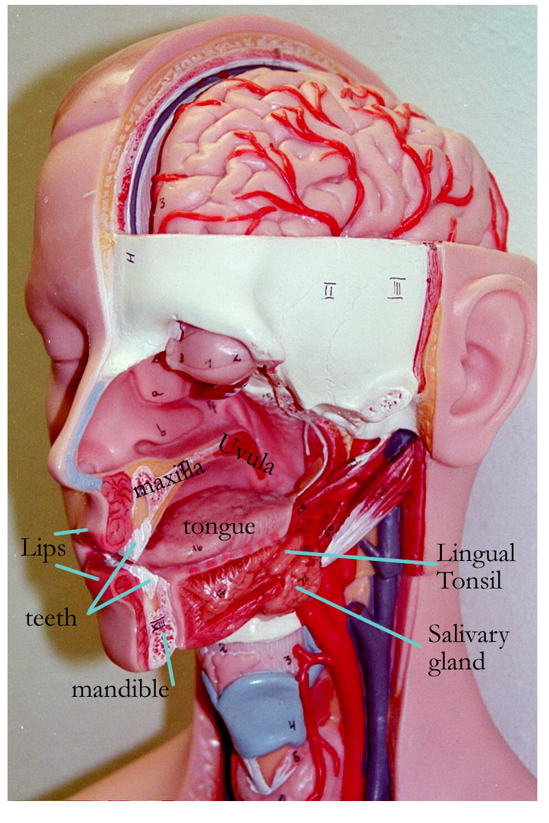
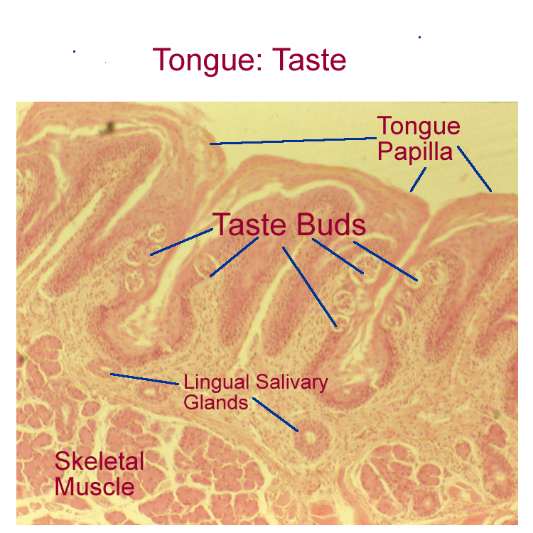
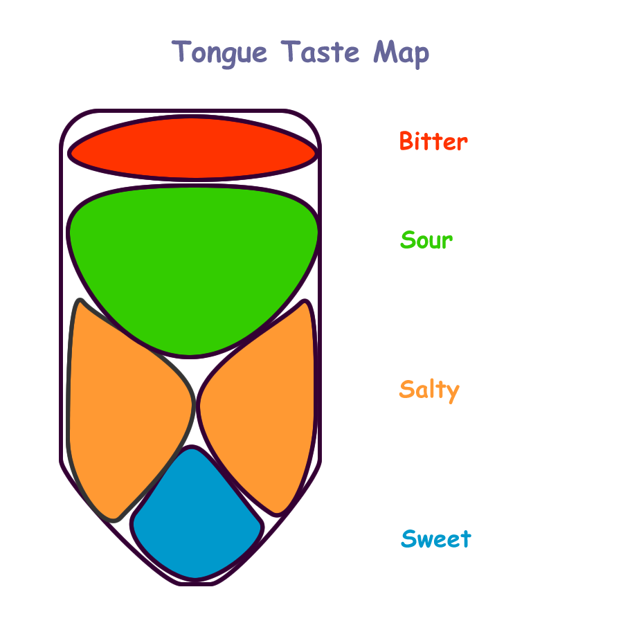
The tongue is composed of intrinsic and extrinsic skeletal muscles in three directional planes and is anchored by attachments to the hyoid bone, mandible, and the connective tissue lingual frenulum. The surface of the tongue is lined by stratified squamous epithelium that forms projections called lingual papillae. Several papillae shapes are recognized: thin, long filiform papillae found in the front of the tongue, mushroom shaped fungiform papillae, and the round flat circumvallate papillae found at the caudal 1/3 of the tongue. In the lateral walls of the papillae are tastebuds linked to cranial nerves. Recall that tastebuds were discussed in detail in the special sensory section. Taste sensations interpreted are sweet, sour, salty, and bitter.
The functions of the tongue are taste, mechanical digestion, and speech.
Teeth
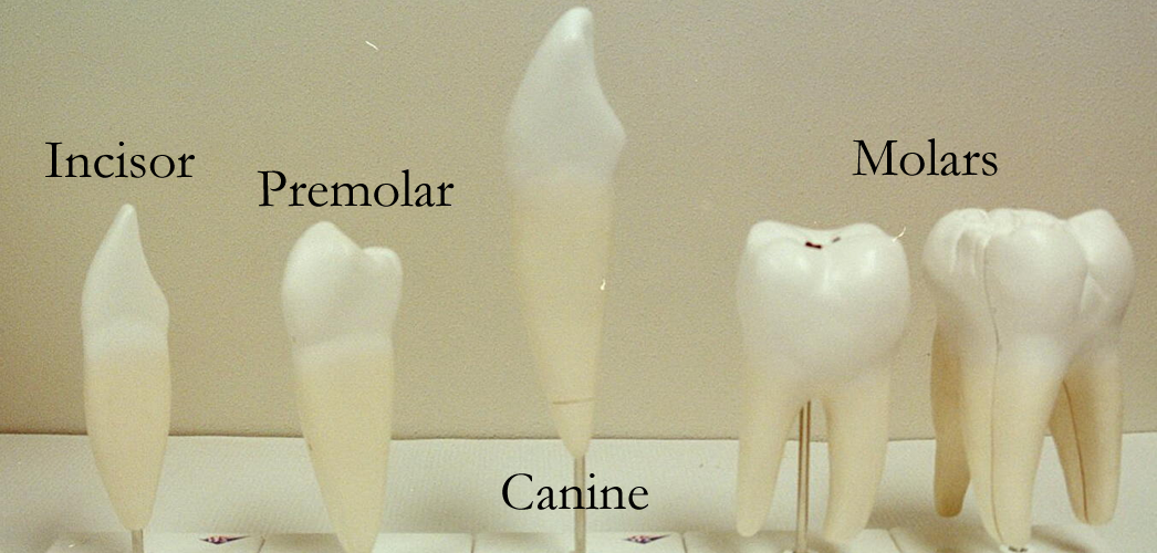
The teeth are housed in the alveolar sockets of the maxillary and mandibular facial bones. They function in mechanical digestion by chewing or mastication as well as in speech of certain
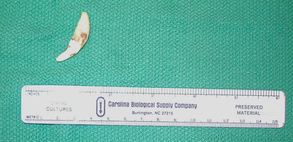
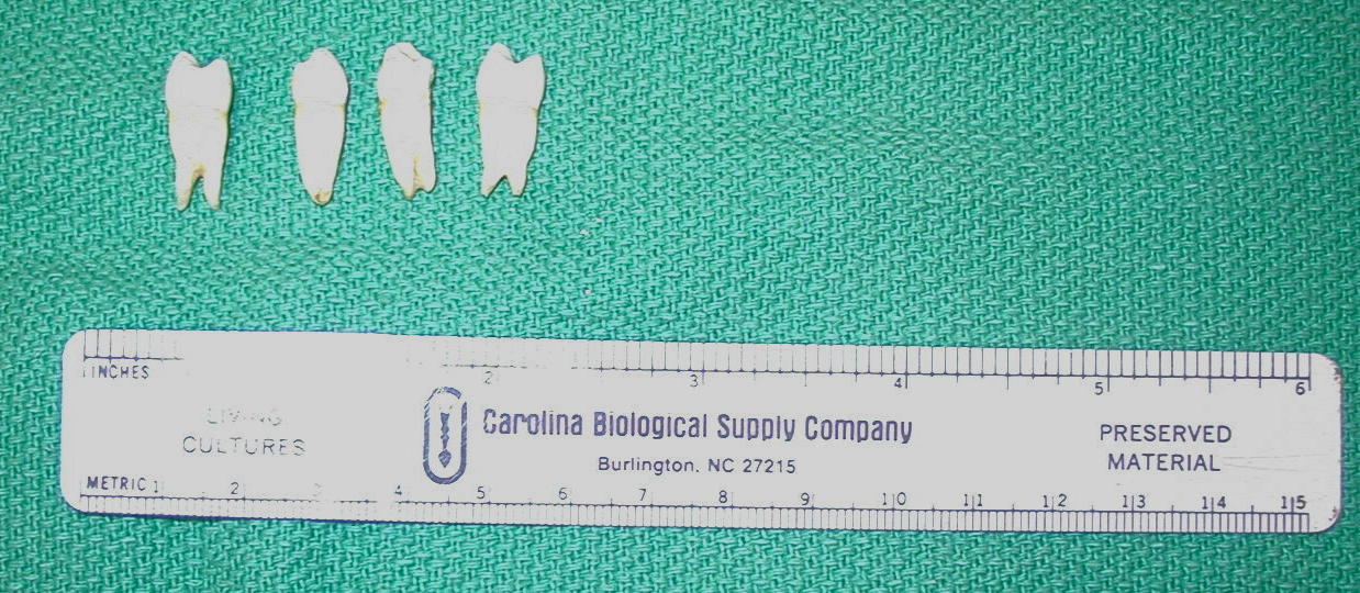
letters and words. In mammals there are two sets of teeth: baby or deciduous and adult or permanent. The dental formula for these sets of teeth is based on tooth types. Incisors are the front set of smaller teeth designed for cutting. Canine teeth are the longer front teeth designed for tearing. The molar groups of teeth are flat and function in grinding. The two types of molars are premolars or bicuspids and molars or tricuspids.
Using the tooth types and location, a dental formula can be constructed. For the teeth in the maxilla or upper arch, half the mouth is used and placed in the “nominator” of the formula. For the teeth in the mandible or lower arch, half the mouth is used and placed in the “denominator” of the formula.
Deciduous Dental Formula 2I 2C 2P x2 = 20
2I 2C 2P x2
Adult Dental Formula 3I 1C 2P 3M x2 = 32
3I 1C 2P 3M x2
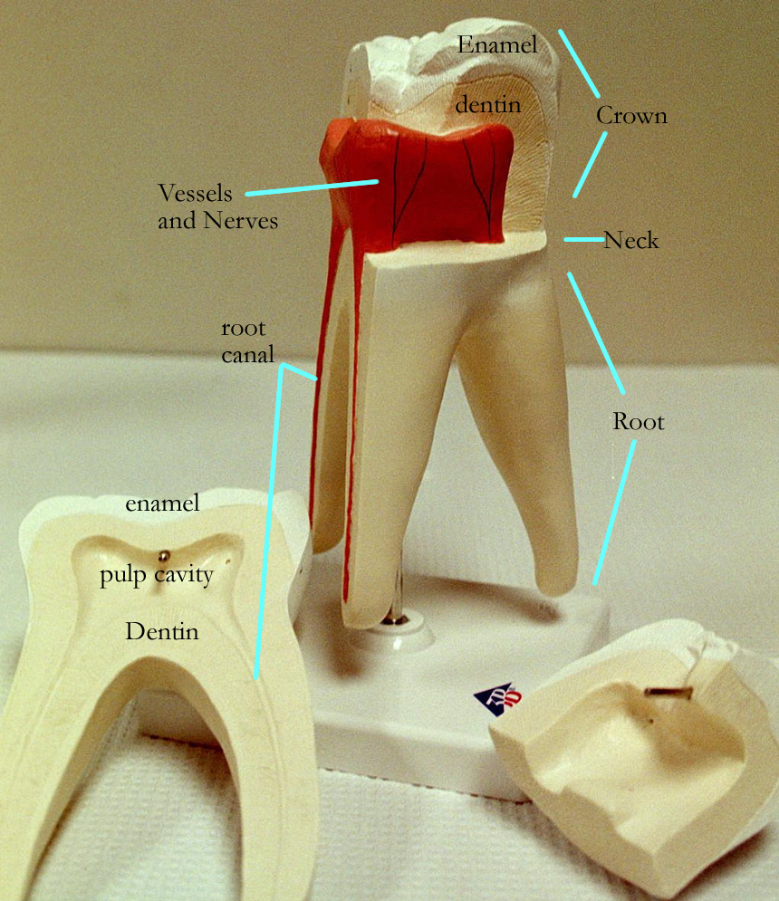
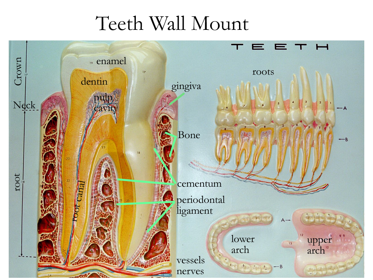
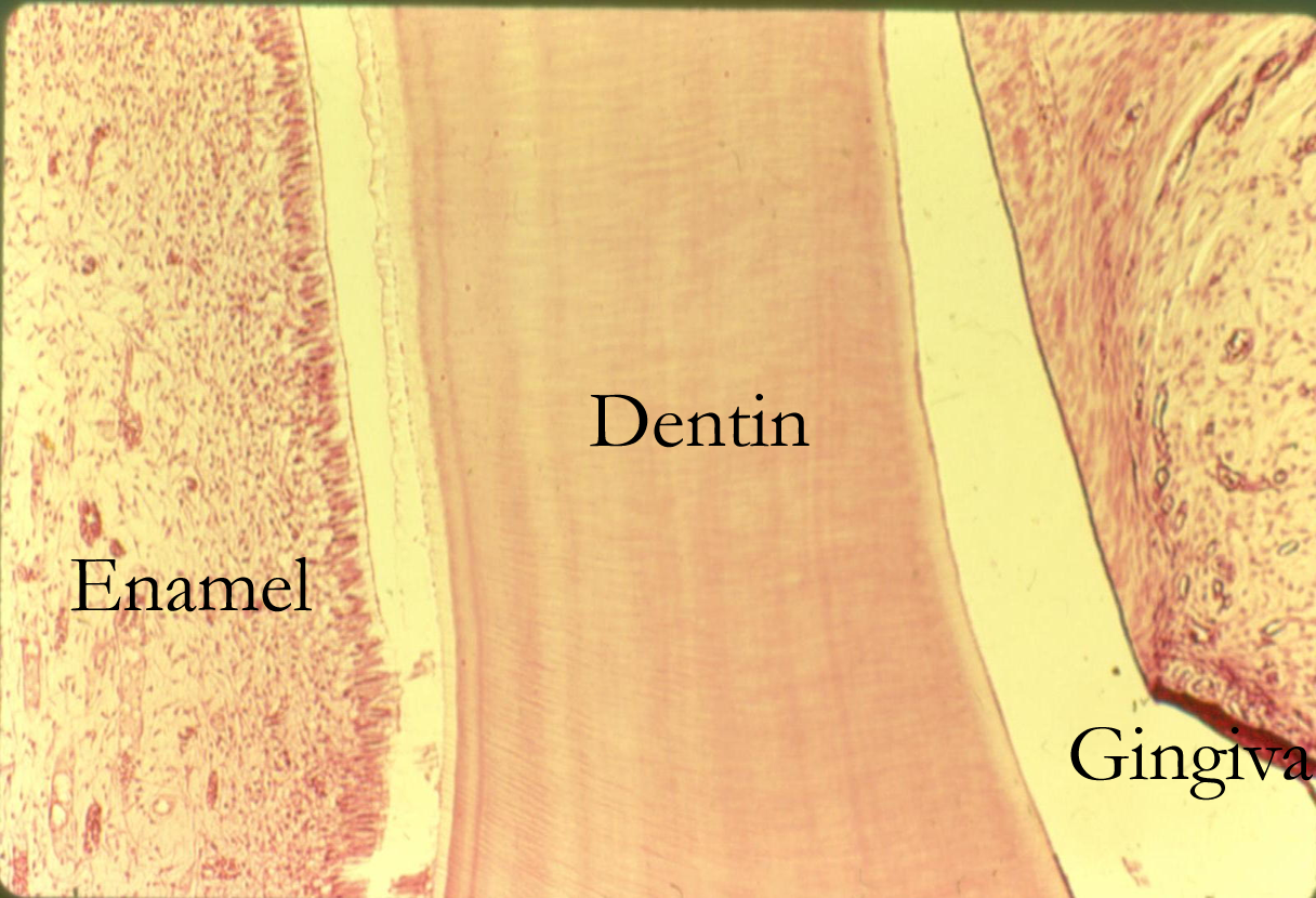
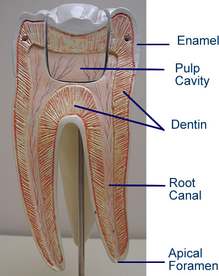
. Gross anatomy of teeth can be divided into three regions:
Crown visible portion of the tooth, dentin covered with enamel
Neck constricted part of the tooth, contains dentin covered by gingival
Root variable number, allows for attachment of teeth and passageway for
vessels and nerves, contains dentin covered by connective tissues.
Root numbers vary from 1, 2, 3 roots depending on tooth size, location, and function. Root dental formulas are also established.
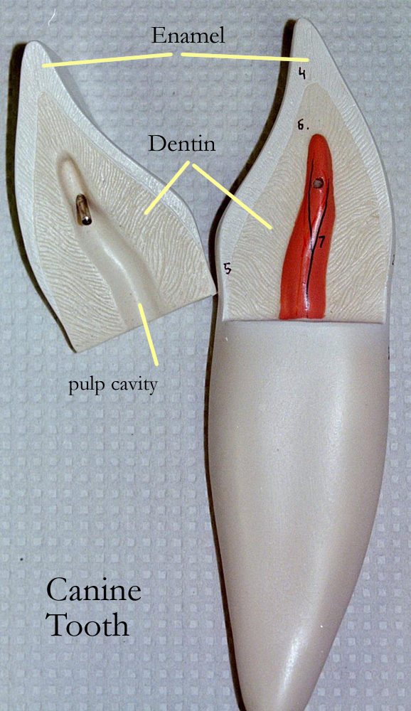
Teeth develop from living cells called odontoblasts which deposit dentin much like osteoblasts deposit bone. Other modified tooth cells secrete enamel, but these cells die after birth. Spaces with in the tooth allow for blood vessels and nerves that supply the teeth. In the crown and neck region there is a space called the pulp cavity and in the root, the root canal. The tooth is anchored in the socket by cementum and the periodonatal ligament.
Tooth Models: Types of Teeth, Gross Anatomy, Interior Anatomy
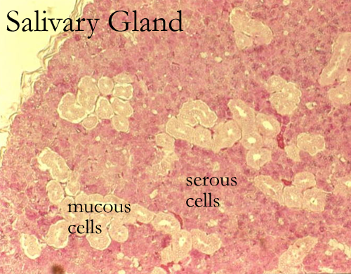
Salivary Glands
Salivary glands are paired on the right and left side in and around the oral cavity.
The intrinsic or buccal salivary glands are found in the lining of the cheeks while the extrinsic salivary glands are found around the oral cavity. The three extrinsic salivary glands are the parotid glands located below the ear, the submandibular glands found under the mandible, and the sublingual glands found under the tongue. Most salivary glands produce on of two types of saliva. The digestive saliva is primarily water with amylase enzymes that start carbohydrate chemical digestion, bicarbonate (HCO3-) for buffering pH, mucus for lubrication, and lysozyme for bacterial destruction. All salivary glands produce digestive saliva. Another nondigestive, thicker saliva is produced during ANS sympathetic activation. The parotid salivary glands can produce both types of saliva.
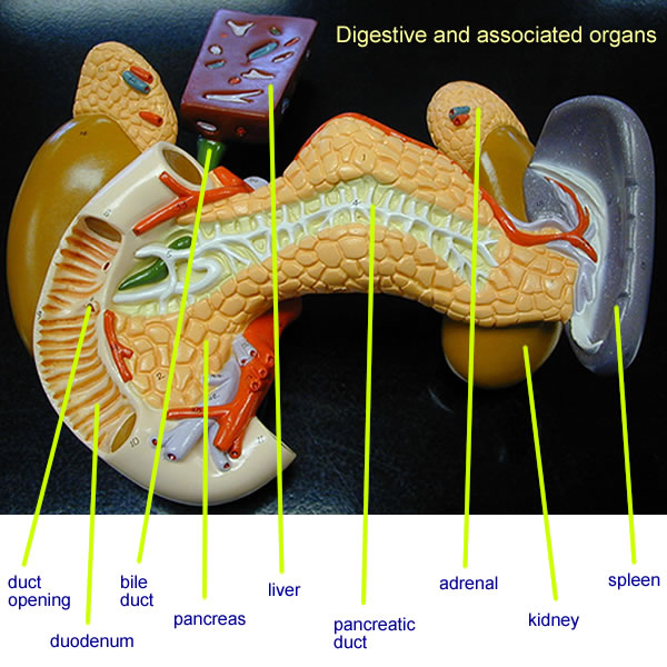
Pancreas
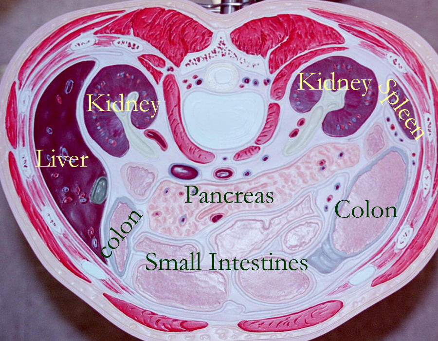
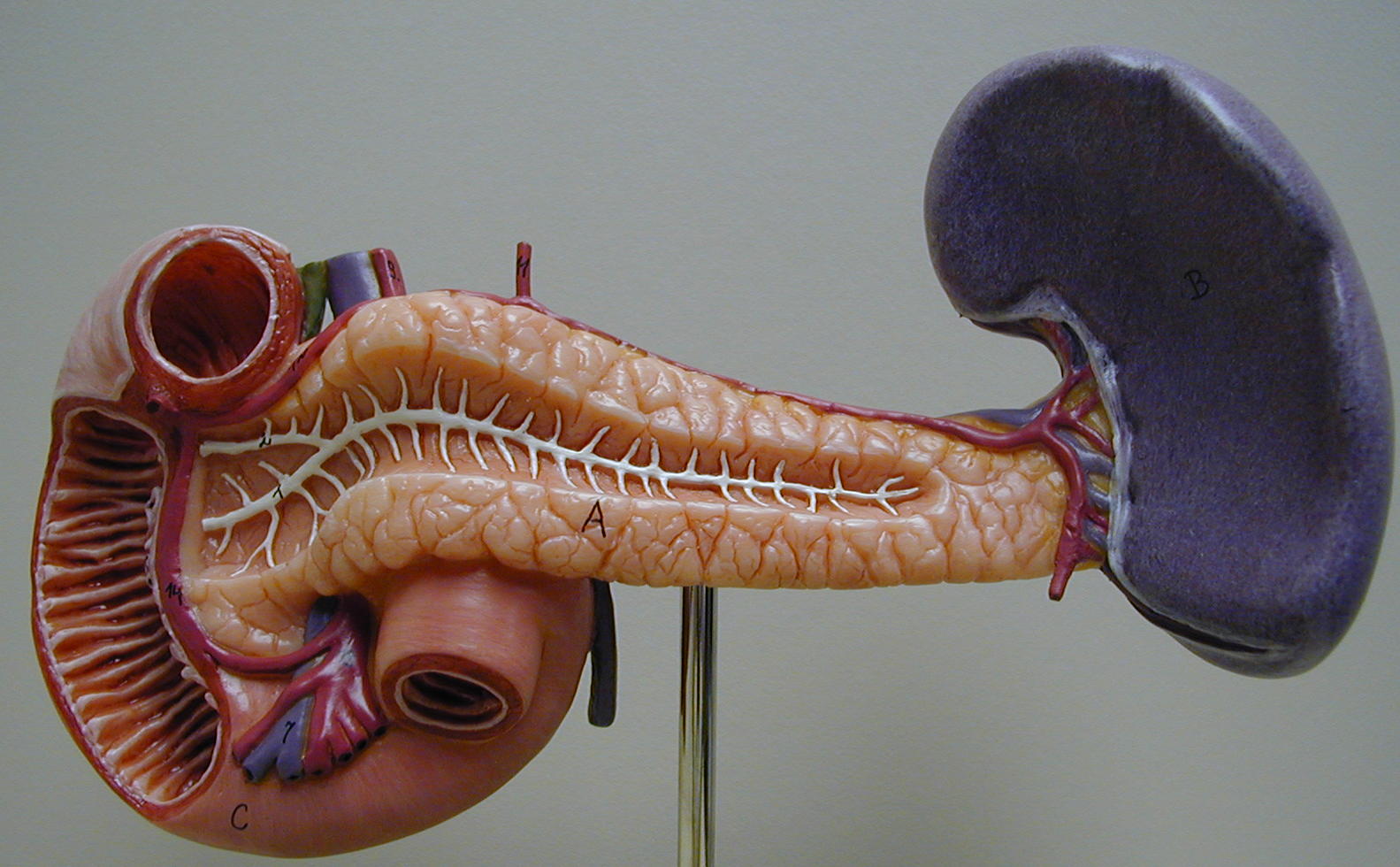
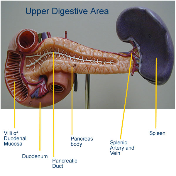
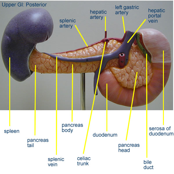
The pancreas is an elongated triangular glandular epithelial organ that sits in the abdominal cavity, inferior to the stomach. Grossly, the pancreas has a head, body, and tail. The epithelial glands found in the pancreas are the endocrine Islet cells and the exocrine acinar cells. The acinar cells of the exocrine pancreas secrete digestive enzymes that are released into the pancreatic duct system that enters the dudodenum. Digestive pancreatic enzymes help in the chemical digestion of carbohydrates with pancreatic amylase, digestion of fats with pancreatic lipase, and digestion of proteins with peptidases and proteases. The pancreas also produces a bicarbonate rich juice to help neutralize the acidic chyme from the stomach. Stimulation of pancreatic secretion is triggered by hormonal secretion from the small intestine. Cholecystakinin (CCK) stimulates release of digestive enzymes and secretin stimulates a bicarbonate rich fluid.
Endocrine functions of the pancreatic Islets were discussed in the endocrine system.
Pancreas with Duodenum: Anterior view labeled, Posterior View labeled
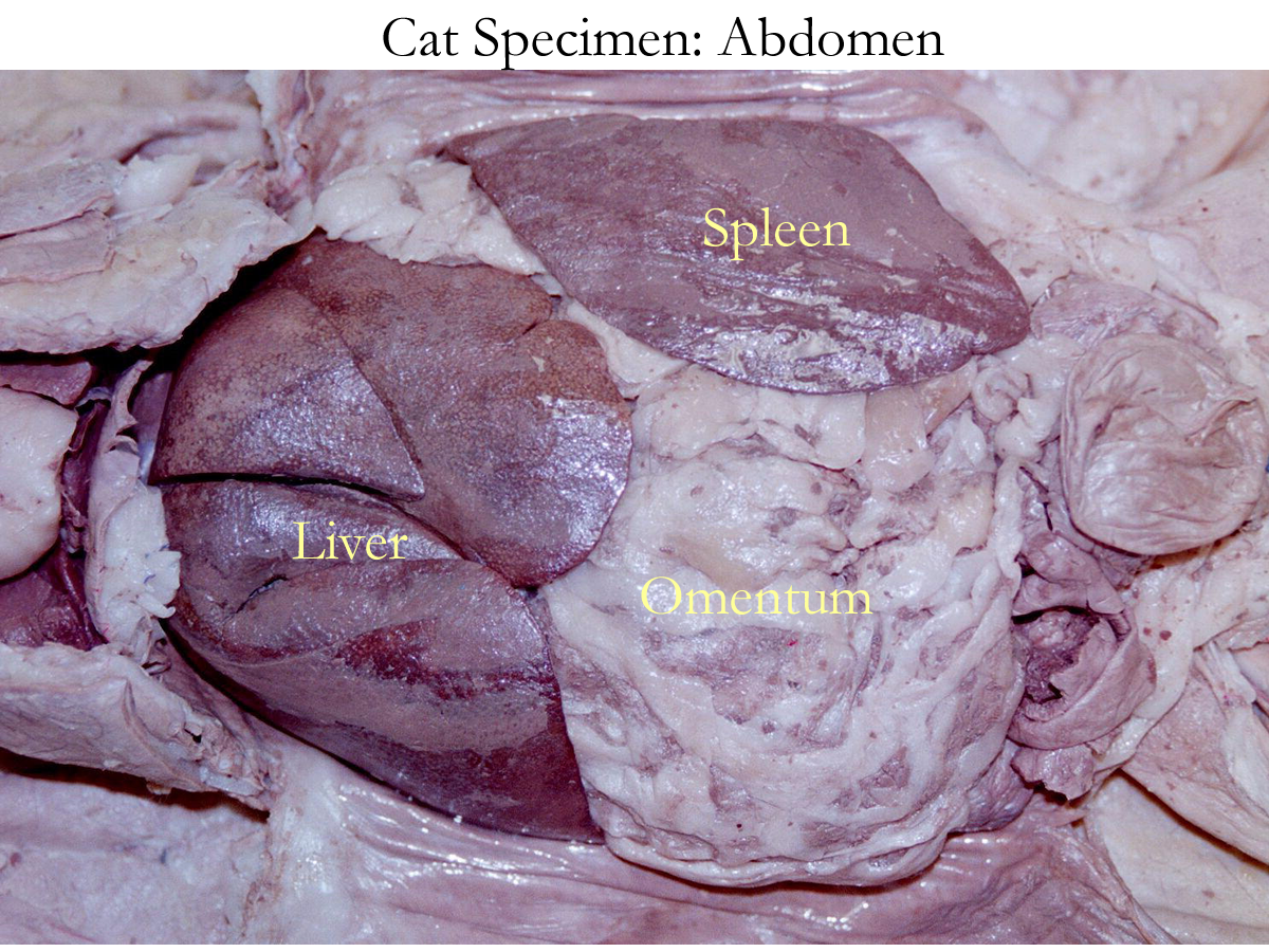
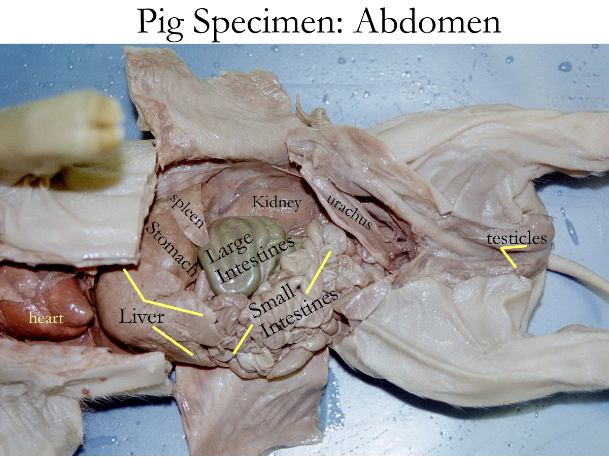
Liver
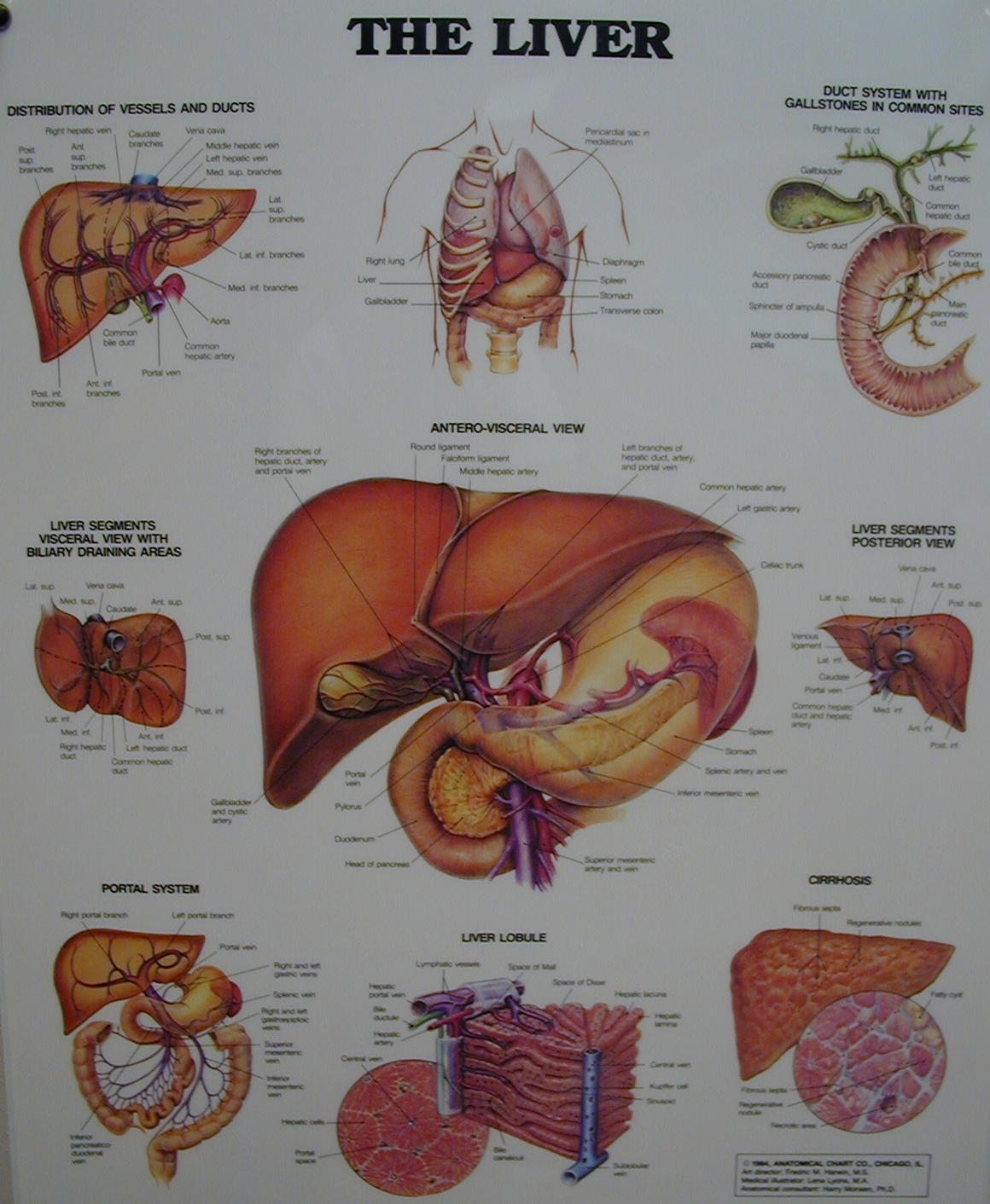
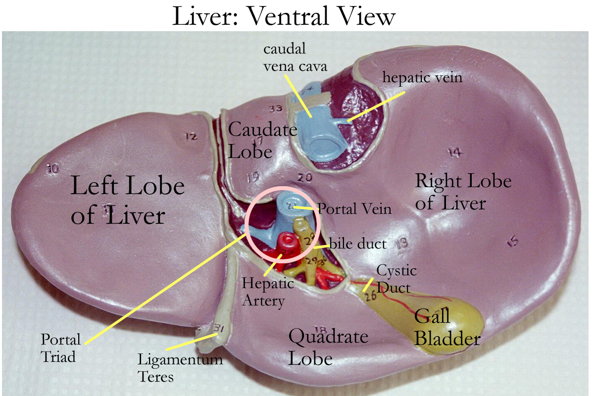
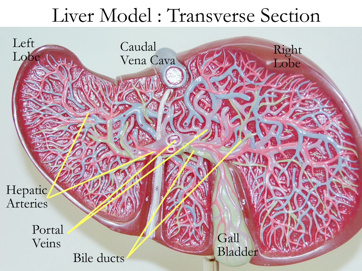
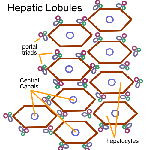
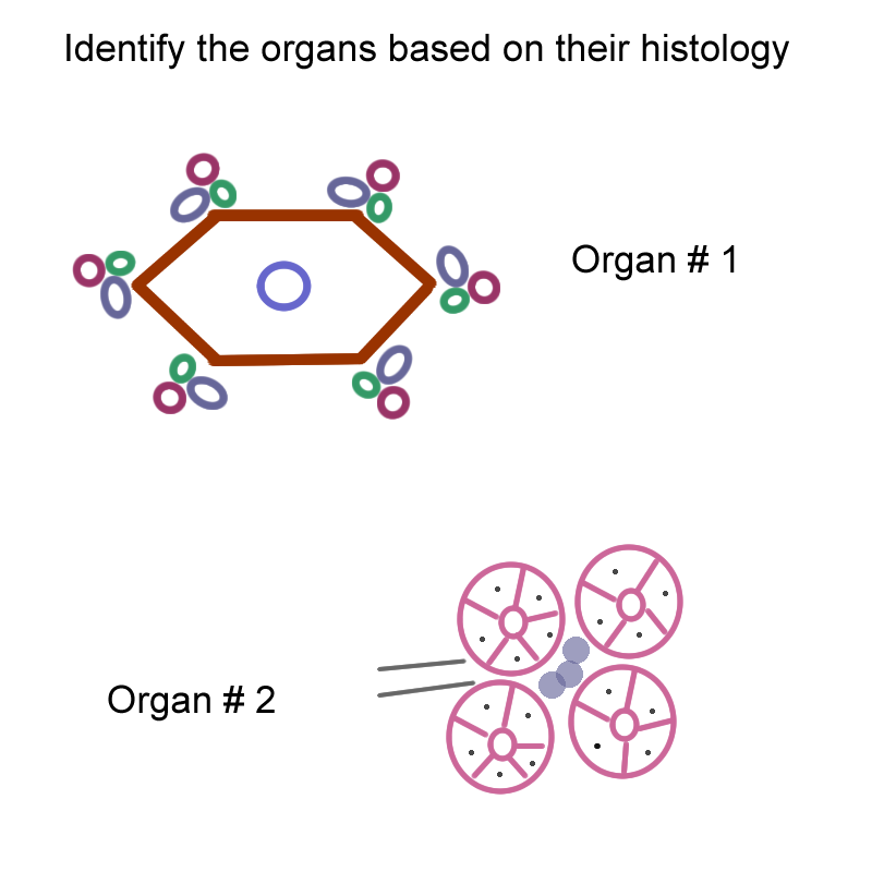
The liver is the largest internal body organs and sits below the diaphragm primarily in the right upper abdominal cavity. The liver is divided into four lobes: two principle lobes, right and left and two smaller lobes, the caudate and quadrate lobes. The liver histologically is glandular cuboidal epithelial cells supported by reticular connective tissue. Functionally, the liver is divided into hepatic lobules by its connective tissue support. The hepatic lobules are made up of these cuboidal epithelial cells called hepatic cells that radiate from central veins. The lobule is hexagonal in shape and at the corners of the hepatic lobule are three structures collectively called the hepatic triad. Each hepatic triad is formed by the hepatic artery, hepatic portal vein, and bile duct. The portal vein brings blood from the digestive organs to the liver for cleansing, the hepatic artery from the aorta is the oxygenated blood that nourishes the liver itself and the bile duct transports bile made by the hepatocytes from the liver to the duct system that connects to the gallbladder.
Functions of the liver are:
Metabolism of carbohydrates, fats, and amino acids
Storage of vitamins (A, D, E, K), minerals (Cu, Fe), and glycogen
Protein synthesis: albumin, clotting proteins, non-essential proteins
Detoxification and filtering of drugs, GI blood, ammonia
Immune defenses due to Kuppfer cells (macrophages in blood capillary sinuses)
Production of bile salts for digestive processes
Bile made in the liver is stored and concentrated in the gall bladder.
Bile is composed of bile salts, water, other salts (Na+, Cl-) and bilirubinBile salts are based on cholesterol and bilirubin is the breakdown product of hemoglobin metabolism. Bile enters the duodenum through the common bile duct derived from the liver and gall bladder.
Bile functions to emulsify fats to enable an increase in surface area of the fat droplets to help lipase enzymes continue chemical digestion of fats.
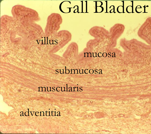
Gall Bladder
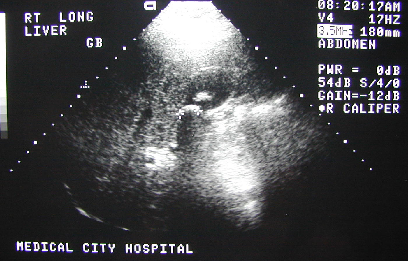
The gall bladder is the bile storage sac located along the undersurface of the right liver lobe. Grossly, the gall bladder has a fundus, body, and neck. The histology consists of a simple columnar mucosal lining with villi that helps in the concentration of bile. When a fatty meal enters the duodenum, CCK is released to stimulate contraction of the smooth muscle in the muscularis layer of the gall bladder in order to push bile into the duodenum. The gall bladder empties first, then bile is released from the liver. Bile is used for fat emulsification to aid in chemical digestion by lipases.
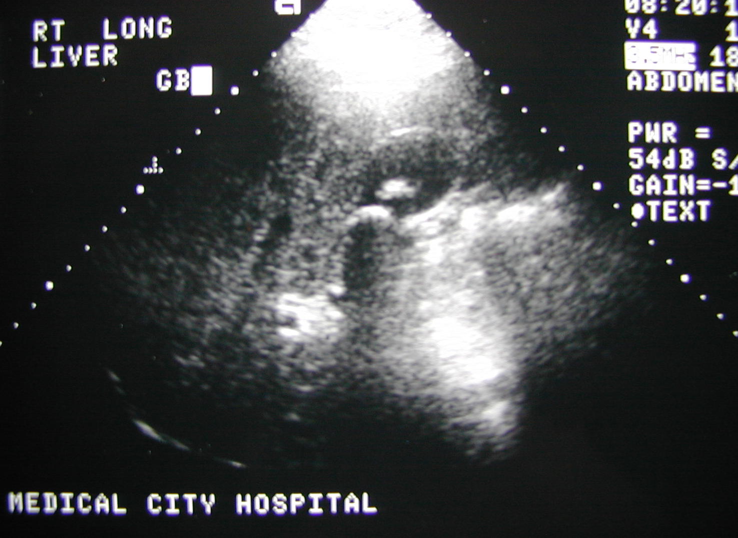
Sonogram of Gall Bladder with Stones
sial/o- salivary gland odont/o, dent/o- teeth
bucc/o cheek gloss/o, lingua tongue
gingival gum pancreat/o- pancreas
chole- bile cholecyst/o- gall bladder
hepat/o- liver -prandial meal
-phylaxis guard steat/o- fat
cholang/o- bile duct ptyal/o- spit
I. ID organs
A Drawing
C Model
D
Specimen
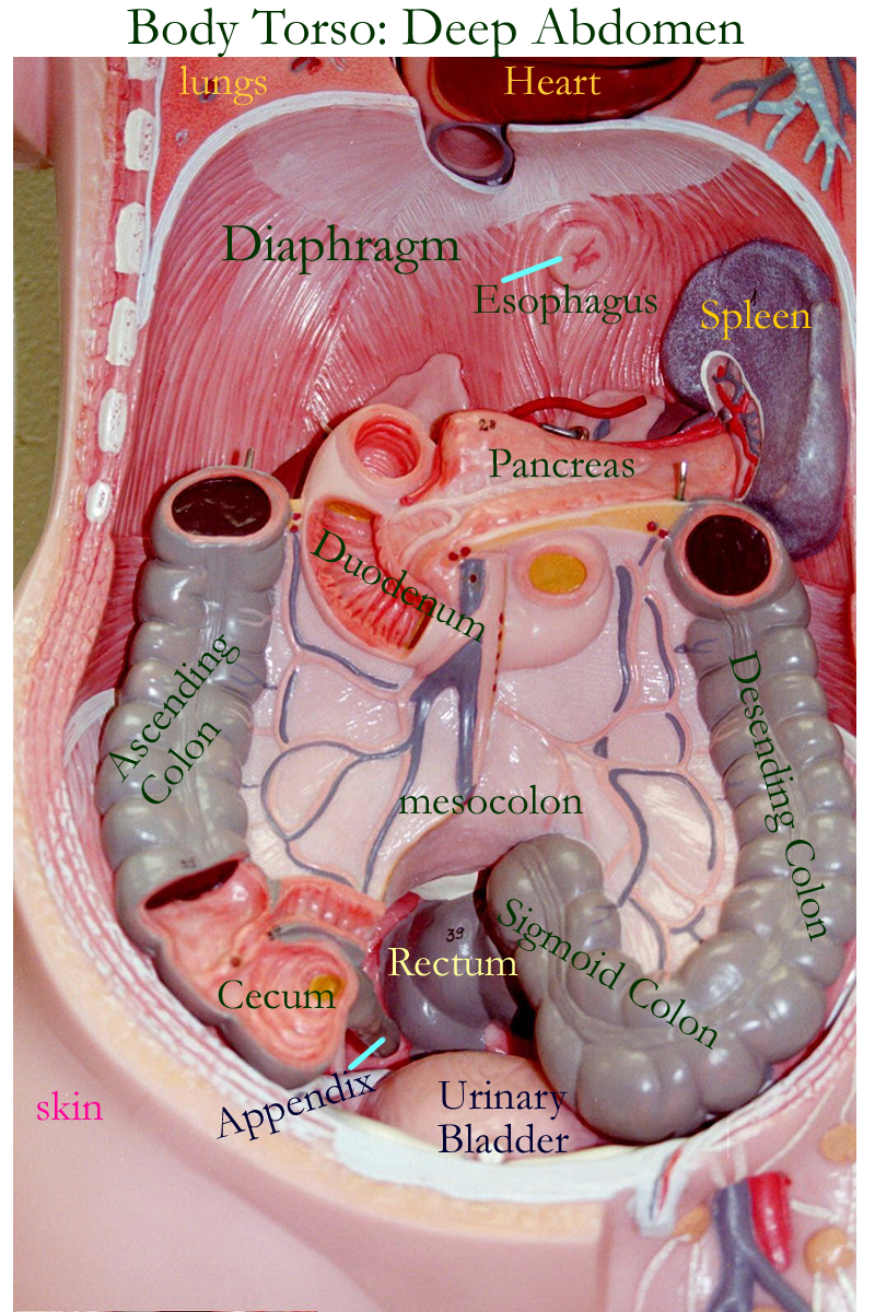
II. Using the 9 segment divisions of the abdominal cavity created
by body planes, name these segments and the digestive organ(s) normally found
in each named segment. Why would this be important to know and understand?
Concept Map: Make
a concept map of the accessory GI structures (gross and histo) anatomy, location
and physiological function. Generally tie these structures to the alimentary
canal discussed in the previous exercise.
Include this map in the LAR lab
report (if selected) as a document insert
or as an
additonal PDF document scan.
Cholelithiasis (Gall stones) Food poisoning
Hepatitis (A, B, C) Dental Caries
Pancreatitis: acute, chronic Cirrhosis
Cancer: hepatic, pancreatic Mumps
Dentist
Dental Hygienist
http://www.nlm.nih.gov/medlineplus/healthtopics.html
http://www.lumen.luc.edu/lumen/meded/histo/frames/histo_frames.html
http://www.gen.umn.edu/faculty_staff/jensen/1135/webanatomy/
http://www.kcmetro.cc.mo.us/maplewoods/Biology/Bio110/Labs.htm
http://calloso.med.mun.ca/%7Etscott/second.htm
http://www.track0.com/canteach/links/linkbodysystems.html
http://www.carr.lib.md.us/schs/science/anatomy/systems.html
http://www.stemnet.nf.ca/CITE/body.htm
http://www.med.virginia.edu/med-ed/phys/practice_board.html
http://www.msms.doe.k12.ms.us/biology/anatomy/digestive/digestive.html
http://arbl.cvmbs.colostate.edu/hbooks/pathphys/digestion/
http://arbl.cvmbs.colostate.edu/hbooks/pathphys/digestion/basics/
http://biology.about.com/library/organs/blpathodigest2.htm
http://members.tripod.com/~rmoskowitz/digestive.html
http://w2.shorecrest.org/MSUS/C-Cruise/htmlpages/anatomy%2Fzoos%2Fgames_links.htm
http://biology.about.com/library/organs/bldigestoverview2.htm
http://www.teaching-biomed.man.ac.uk/mcwilliam/digest.htm
http://www.nlm.nih.gov/medlineplus/digestivediseasesgeneral.html
http://www.niddk.nih.gov/health/digest/pubs/digesyst/newdiges.htm
http://www.medem.com/MedLb/article_detaillb.cfm?article_ID=ZZZ7C4T46JC&sub_cat=338 digestive system
http://www.medem.com/medlb/article_detaillb.cfm?article_ID=ZZZTD0TCGJC&sub_cat=50 teeth
http://www.nlm.nih.gov/medlineplus/digestivesystem.html
http://www.nlm.nih.gov/medlineplus/mouthandteeth.html
1. Name the three extrinsic salivary glands and two functions of saliva.
2. Name the types of teeth and their function.
3. Give the purpose of enamel and the periodontal ligament.
4. Name the major enzymes from the digestive pancreas and their function.
5. Define emulsification.
6. Name four functions of the liver.
7. Give the location and function of the gall bladder.
8. Define enzyme
9. Name the major enzymes from the digestive pancreas and their function
10. Compare and contrast mechanical and chemical digestion.