Biology 2404 A&P Basics Female Reproductive System Dr. Weis
| Objectives | Background | Medical Terms | Activities | Applications | Careers | WWW | Review Questions |
Students should be able to:
* Name, identify, and give the function for the organs related to the female reproductive system:
Primary Sex organ
Duct or Tubular system
External Genitalia
Accessory Organs
* Name the source and feedback control for the female hormones
* Describe the three ovarian phases
* Describe the three uterine phases
* Define oogenesis
* Define related terms
Read related information in the textbook
The female reproductive tract consists of:
Primary Sex Organ ovary
Duct or Tubular system Fallopian tube, uterus, vagina
External Genitalia Vulva
Accessory Organs Mammary Glands, lesser & greater vestibular glands
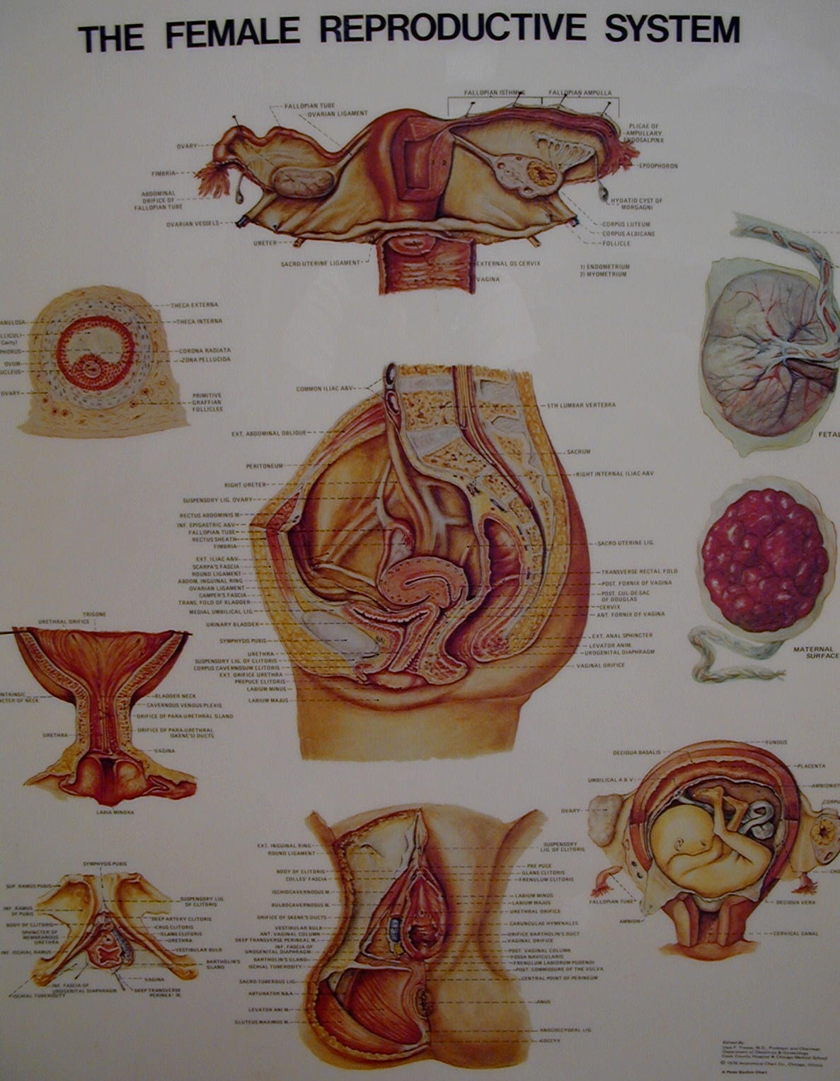
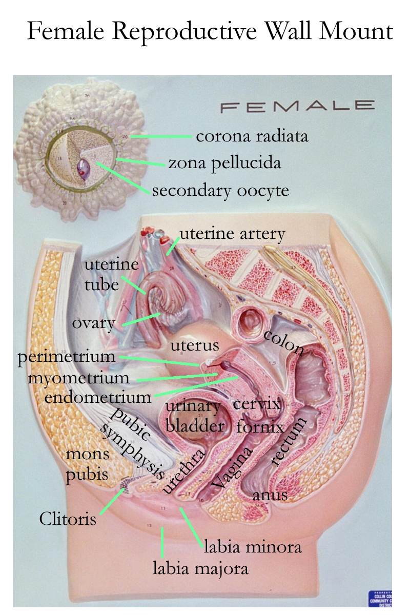
Female repro / Development wall mount
Ovaries
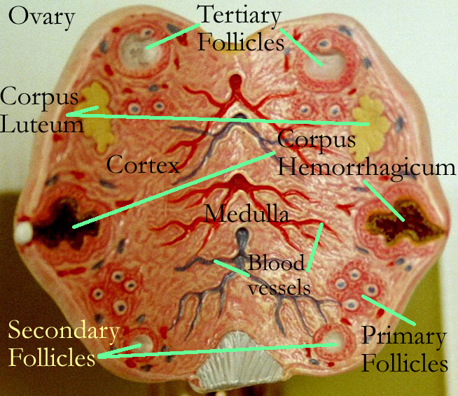
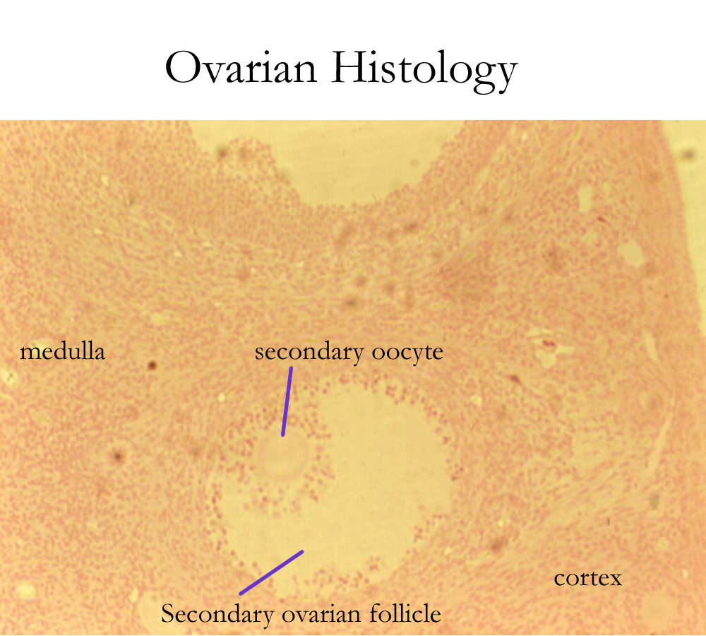
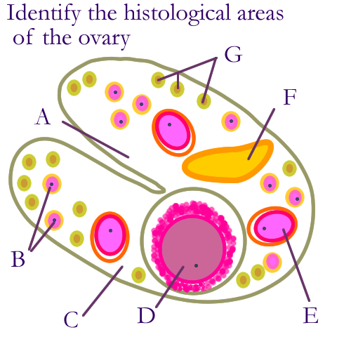
The ovaries are paired oval structures inferior to the kidneys in the mid abdominal region and are suspended by suspensory, ovarian, and broad ligaments. The ovaries are covered by a connective tissue called the tunica albuginea that forms an outer capsule. The ovarian cortex contains ovarian follicles consists of developing immature eggs (oocytes) and surrounding cuboidal cells, thecal cells. In each follicle, the immature eggs undergo oogenesis triggered by the hormones secreted by the thecal cells.
Primordial follicles oogonia precursor cell
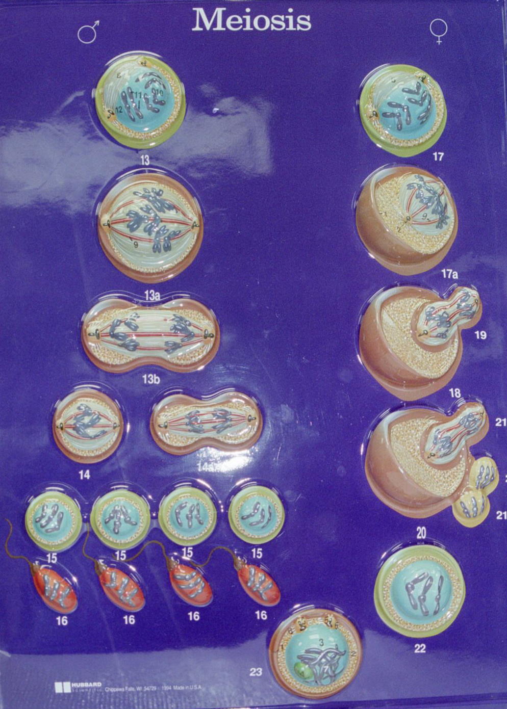
Primary follicles primary oocytes haploid cells, meiosis I
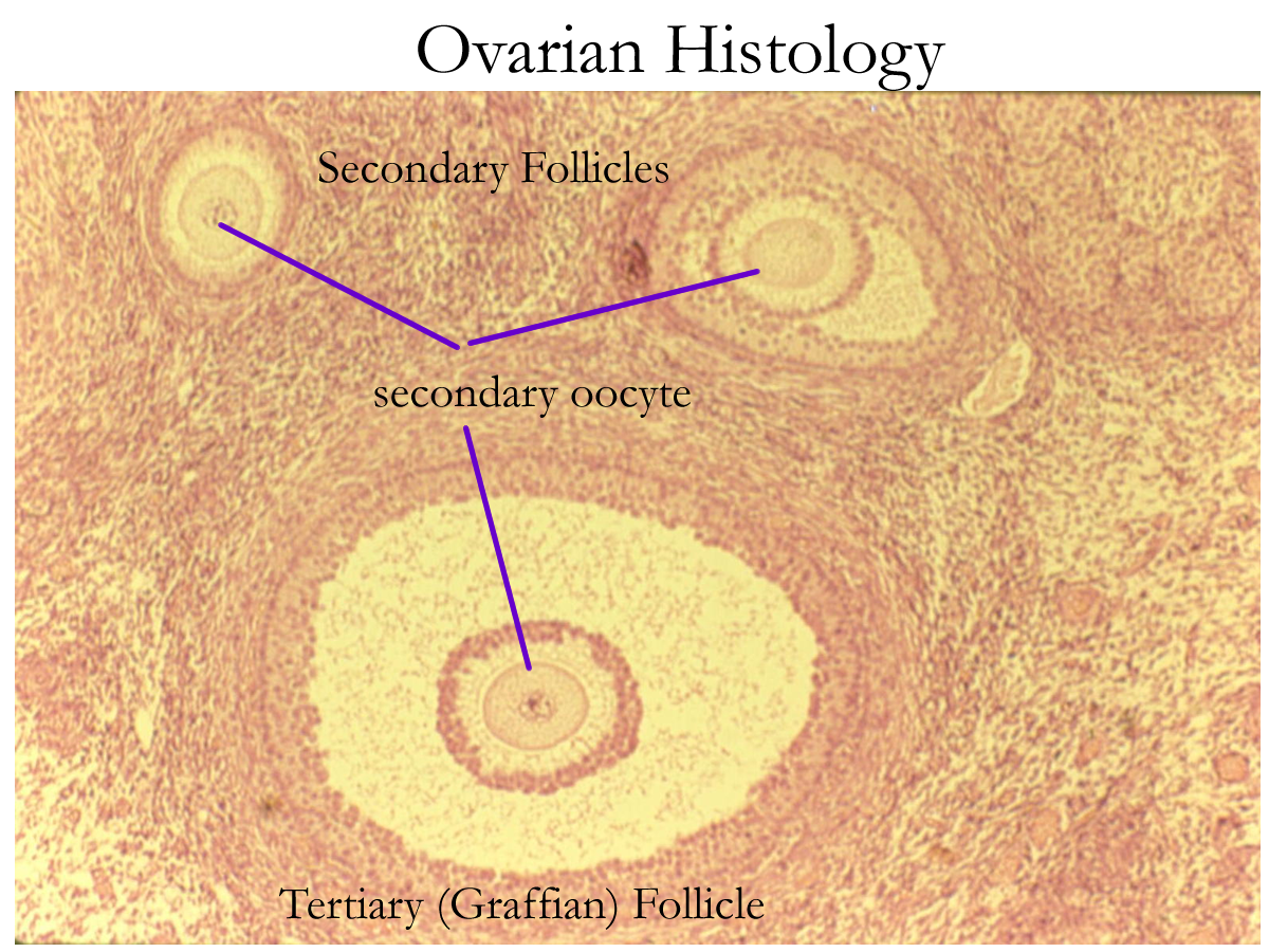
Secondary follicles secondary oocytes haploid cells, meiosis II
Tertiary, Graafian follicles secondary oocytes haploid cells, meiosis II
Atretic follicles deteriorating oocytes no function
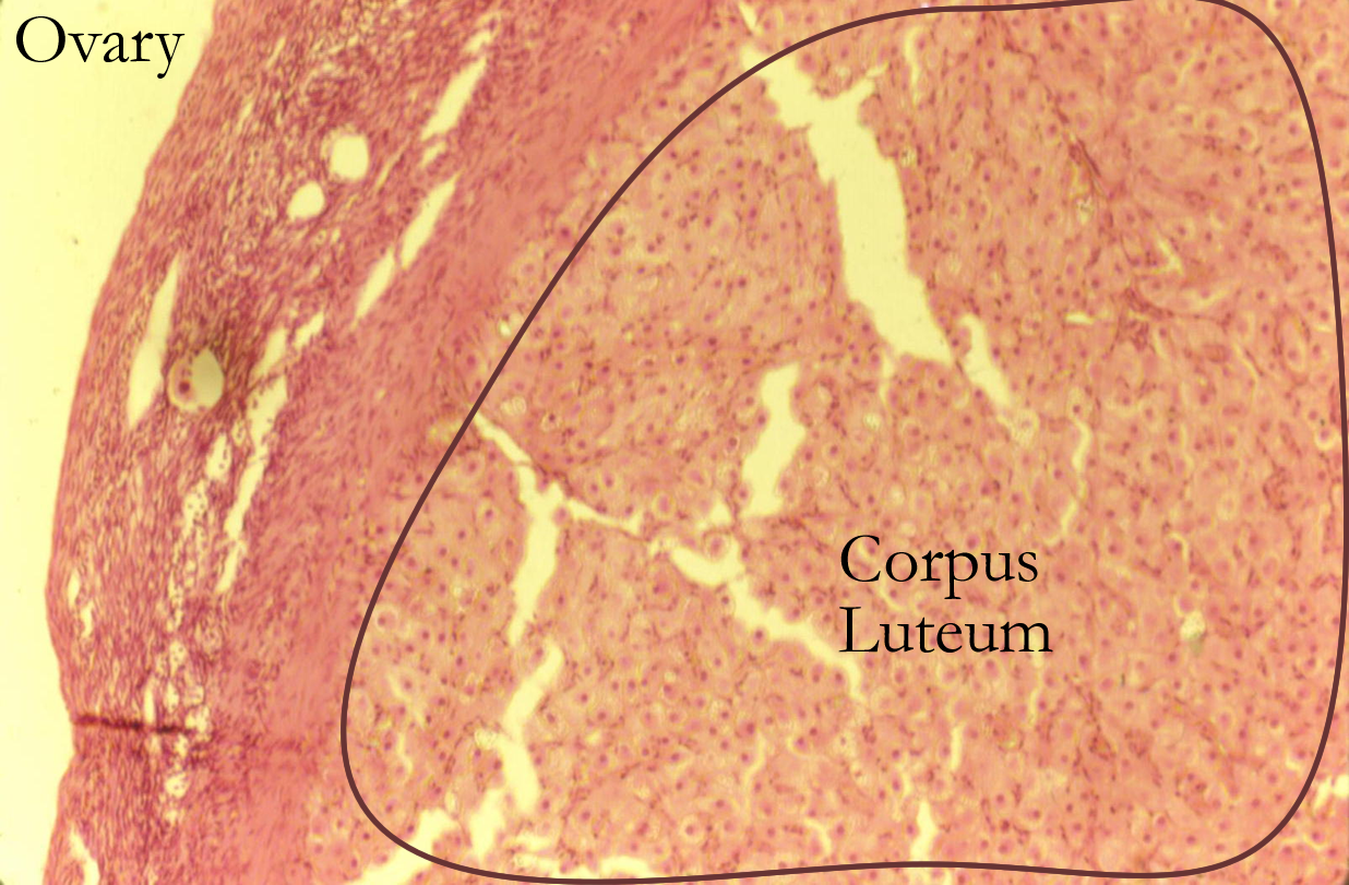
Ovarian Histology: primordial, follicles [primary, secondary, tertiary], CL
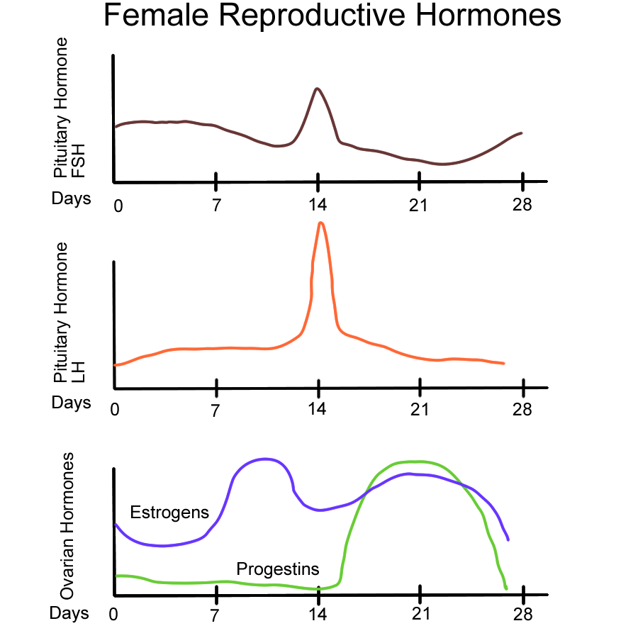
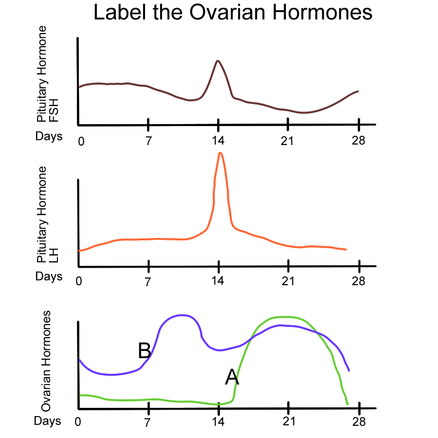
Ovarian Hormones and Cycle
The hormones from the thecal cells that line the follicles are:
DHEA is a hormone precursor to other reproductive hormones that control the ovarian and uterine cycles.
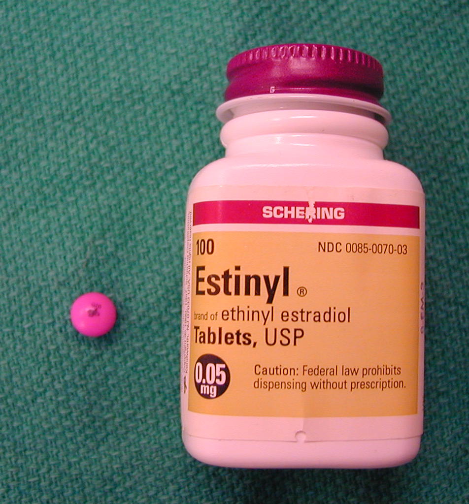
Estrogens estradial, estrone
Progestins progesterone
Androgens testosterone
Estrogens also function in creating secondary sex characteristics: Fat deposition, breast development, hair distribution, skin changes, and bone development
Hormonal control of the ovaries begins in the Hypothalamus with GnRH release triggering response of the AP to release either FSH or LH.
GnRH for FSH ovarian thecal cells reproductive hormones, primarily estrogens
GnRH for LH affects tertiary follicles to causeovulation of secondary oocyte
while the remaining thecal cells form the corpus luteum
In summary, the function of the ovaries is to produce gametes in a process called oogeneis and to produce reproductive hormones in response to homeostatic feedback signals. Hormones control oogenesis and uterine lining changes.
The ovarian cycle is named for the events triggered by hormone response.
Follicular Phase ovarian follicles develop from primordial to Graafian
triggered by estrogen production
Ovulation Graafian follicle ruptures to release secondary oocyte
triggered by AP – LH
Luteal formation of corpus luteum (CL) from leftover thecal cells
function to secrete progestins and estrogens
triggered by LH
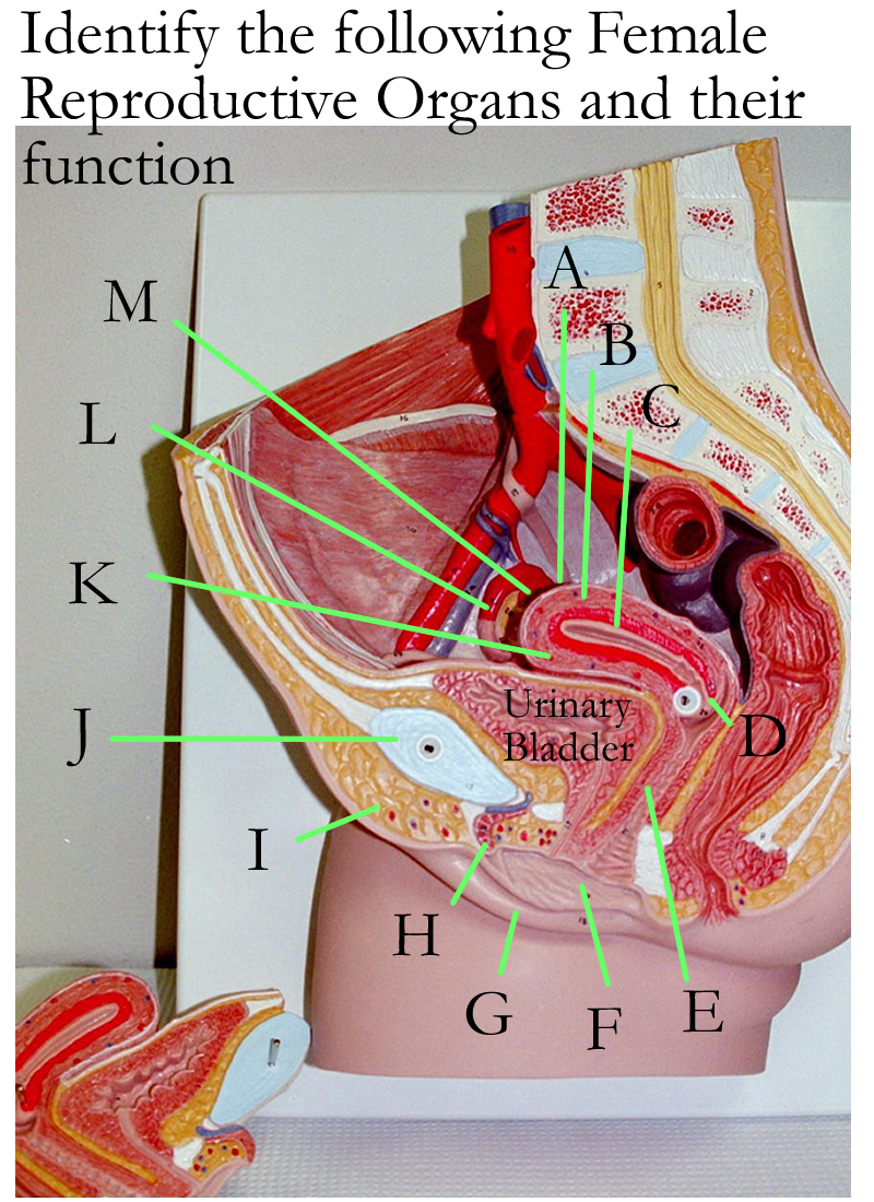
Female reproductive wall mount
Duct System
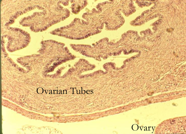
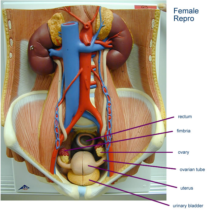
The Fallopian tubes (Salphinx, Uterine Tubes, Oviducts) develop separately from the ovaries. At the lateral end of the tube, there are finger like extensions called fimbria that function to create vacuum like currents to retrieve the ovulated secondary oocytes.
The fimbria is supported by an enlarged trumpet shaped base called the infundibulum.
The infundibulum continues as the expanded portion of the tube called the ampulla and it is where fertilization takes place. As the tube continues, its medial end is attached to the uterus at the isthmus. In summary, the function of the Fallopian tube is in oocyte transport and fertilization.
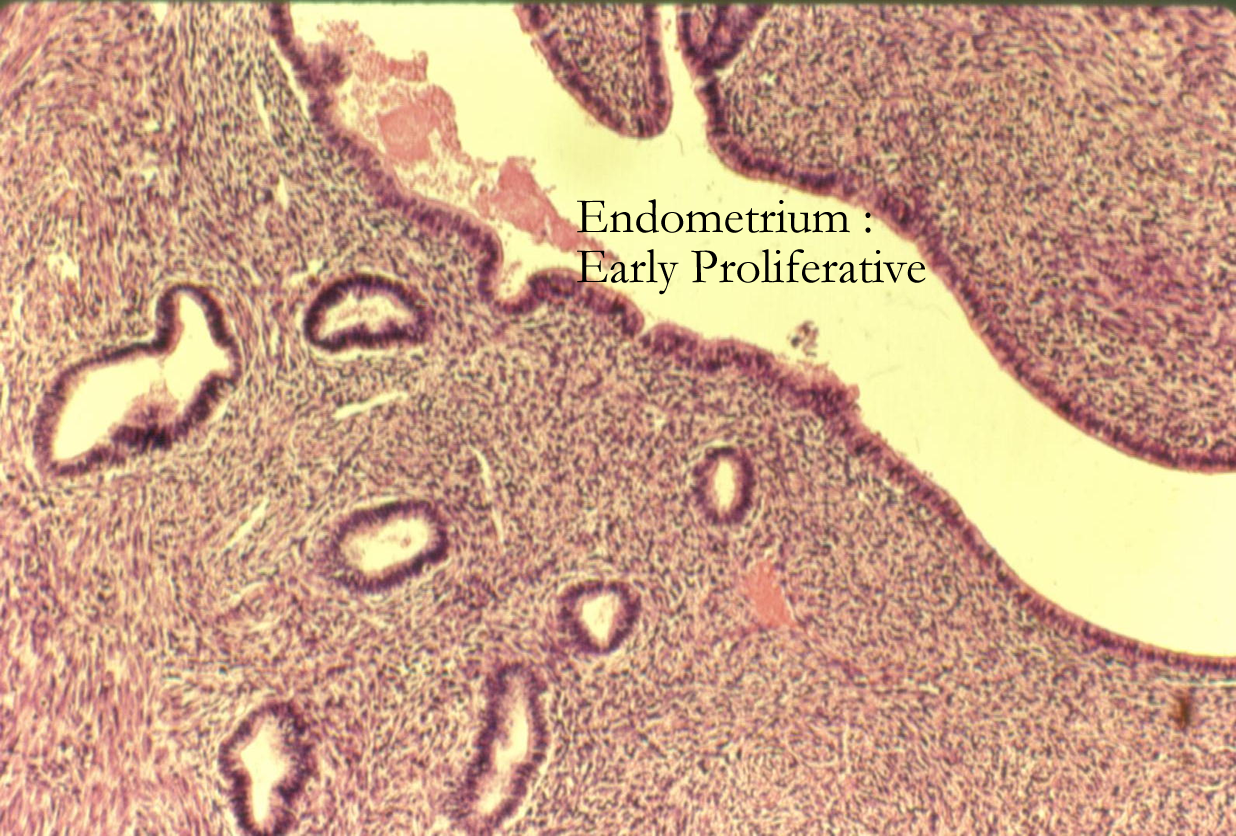
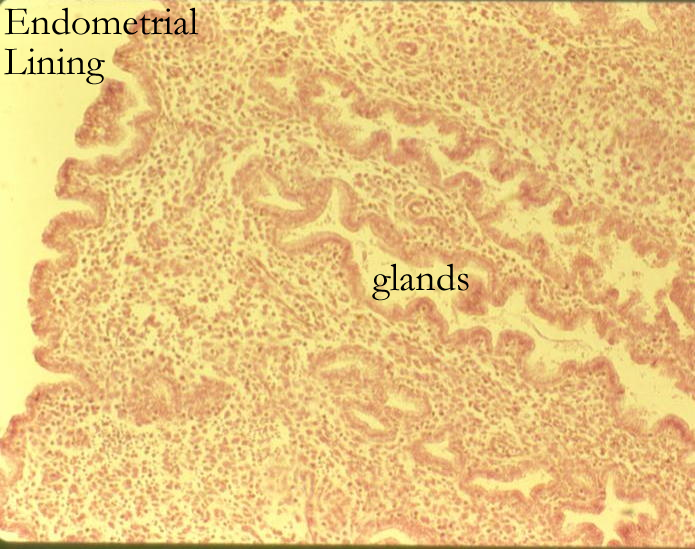
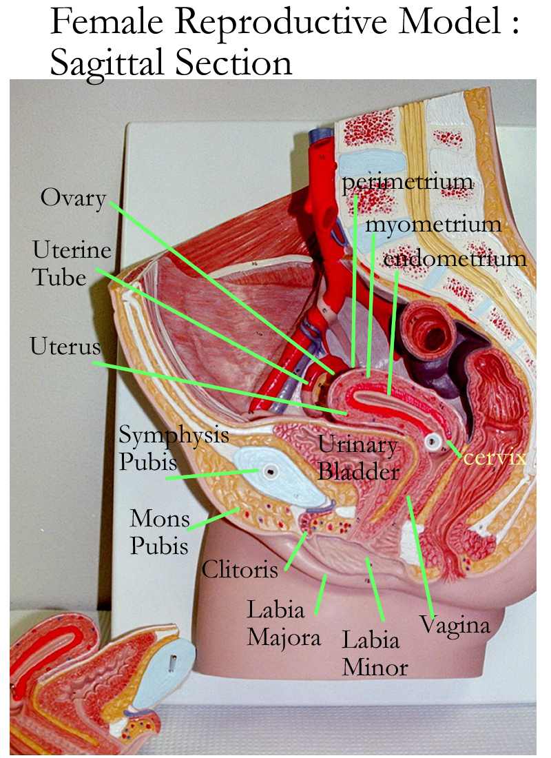
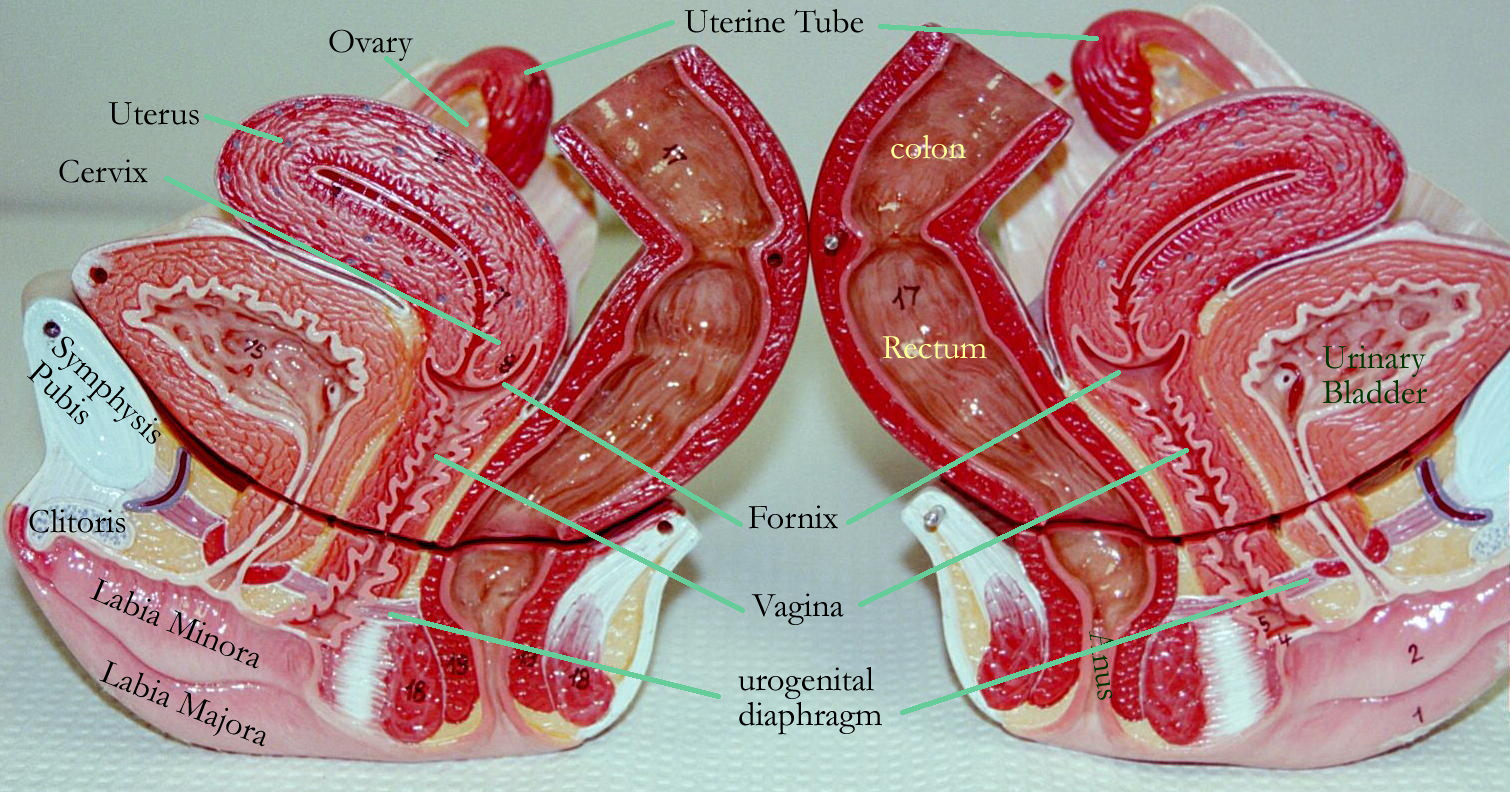
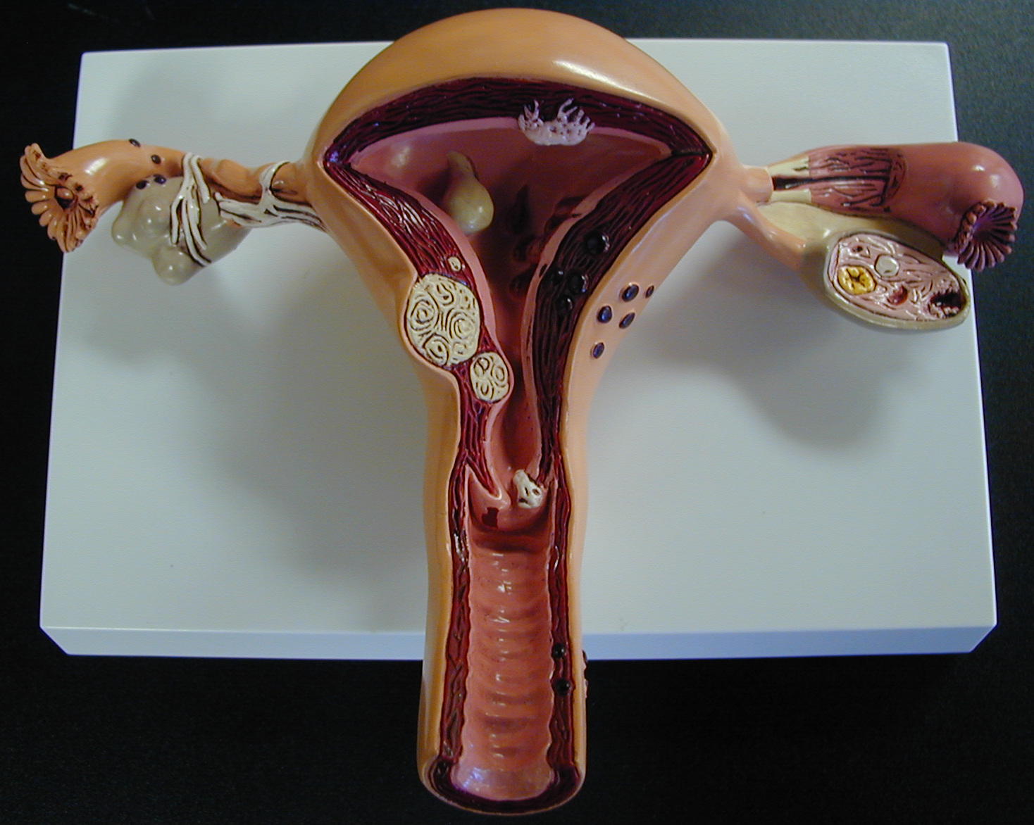
The uterus is a pear shaped structure in the pelvic region of the abdominal cavity suspended by the broad ligament. The external anatomy consists of the fundus, body, and neck or cervix. The internal anatomy of the uterus is composed of three layers. The internal epithelial lining is the endometrium and is a modified simple columnar epithelium in two layers, the basilar layer and the functional layer. The muscle wall is the myometrium and is composed of smooth muscle. The outer layer is a connective tissue layer called the perimetrium formed by the visceral peritoneum.
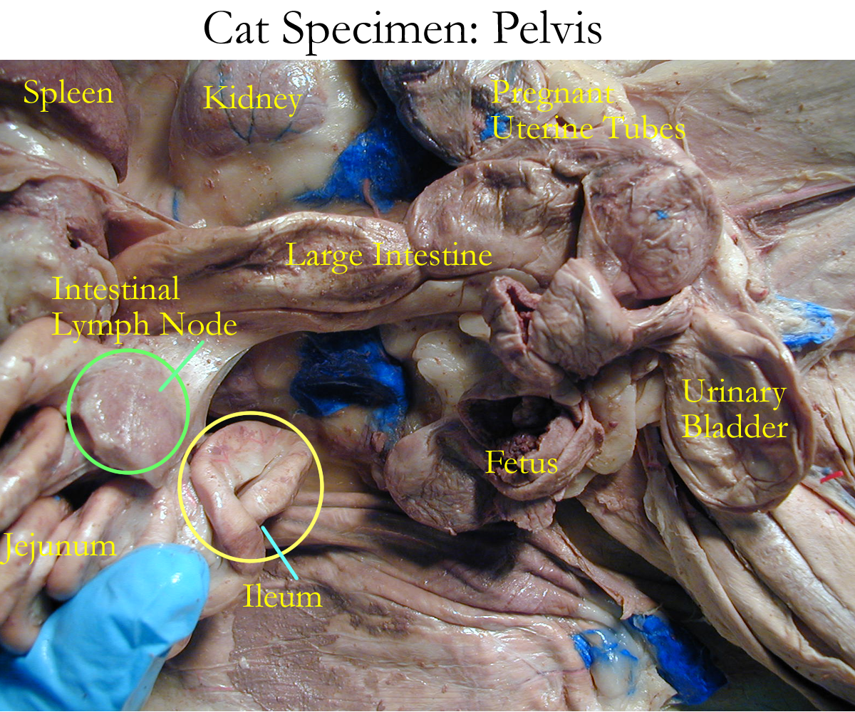
The uterus can change size and shape during pregnancy. The lining of the uterus will undergo several cycles known as the uterine cycle and relates to the hormones produced during the ovarian cycle. The function of the uterus is to provide protection, support, and nutrition to the developing fetus and aid in delivery during labor.
Proliferation (pre ovulatory) basilar layer of endometrium replaces functional layer
triggered by estrogens from the ovary
Secretory (post ovulatory) functional layer secretes nutrients for fertilized egg
triggered by progestins and estrogens from the ovary
Menstrual (post luteal) functional layer of endometrium lost
triggered by a dramatic drop in ovarian hormones
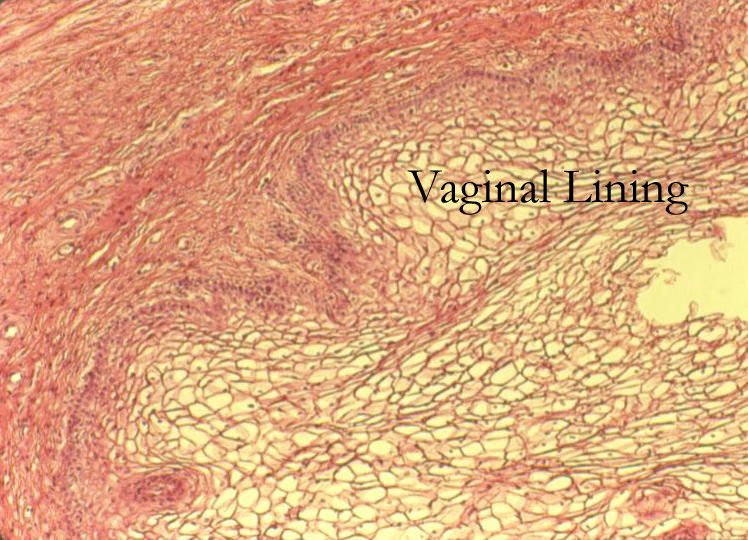
The vagina is a muscular tube that connects the cervix of the uterus at the fornix. The vagina functions as the female organ of copulation, serve as pathway for menstrual flow, and the birth canal. The vaginal lining is stratified squamous and has an acidic pH to prevent overgrowth of bacteria. A thin fold of tissue called the hymen partially closes the distal end of the vagina. The vaginal orifice is the opening of the vagina in the vestibule enclosed by the labia.
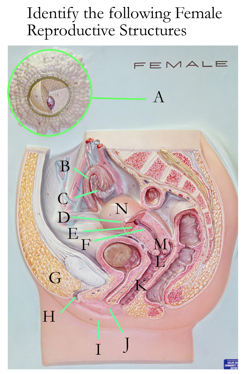
Female reproductive models
Female reproductive wall mount
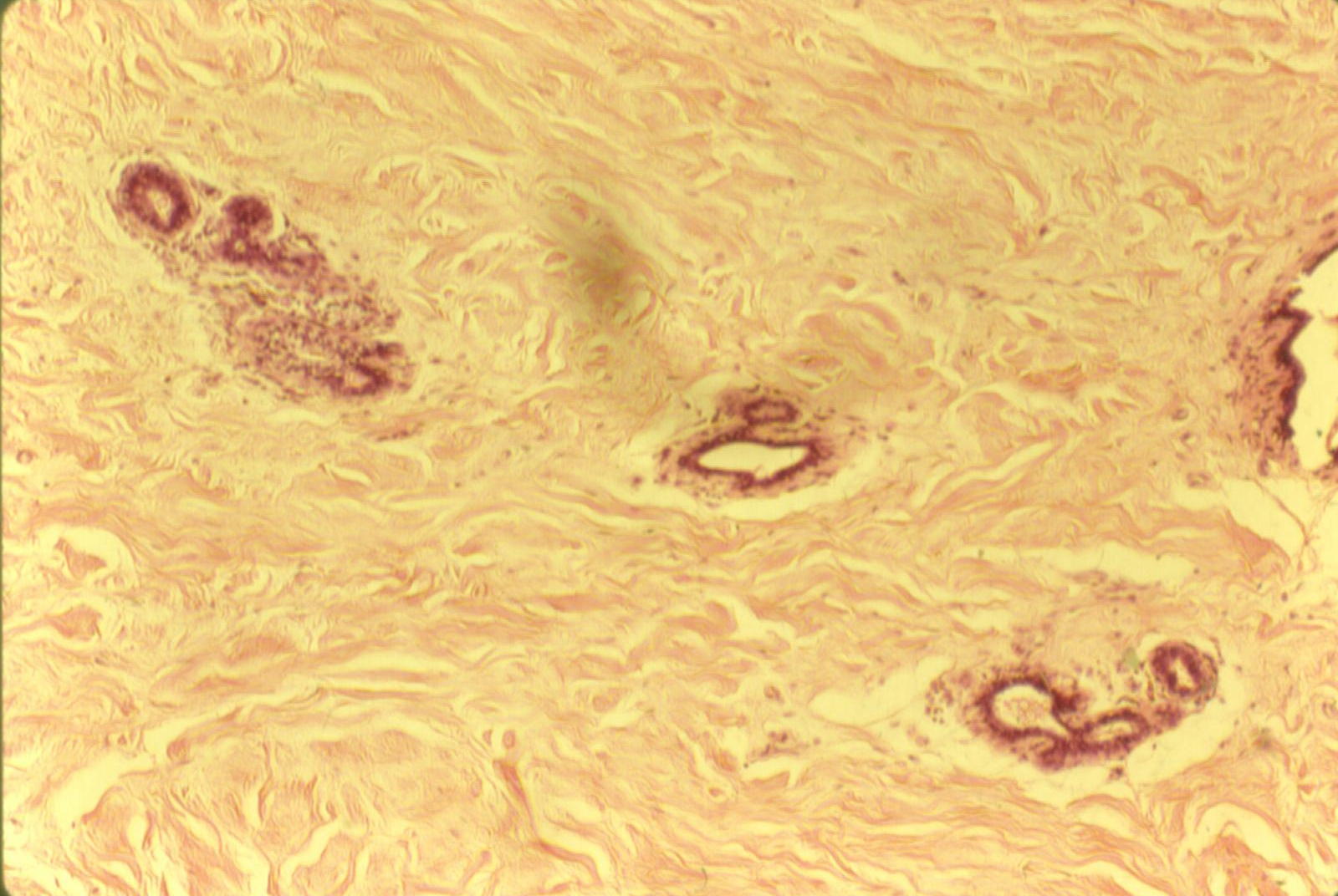
Ovarian and Fallopian tube histology
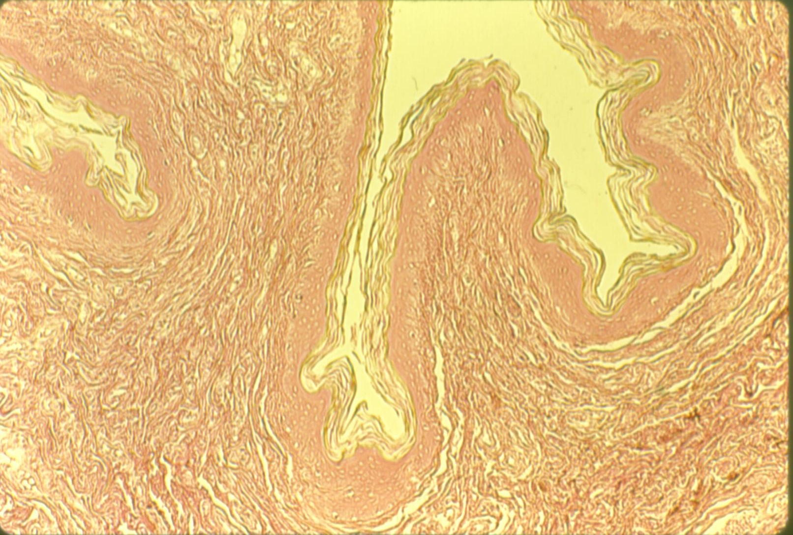
Uterus histology: Proliferative, Premenstrual
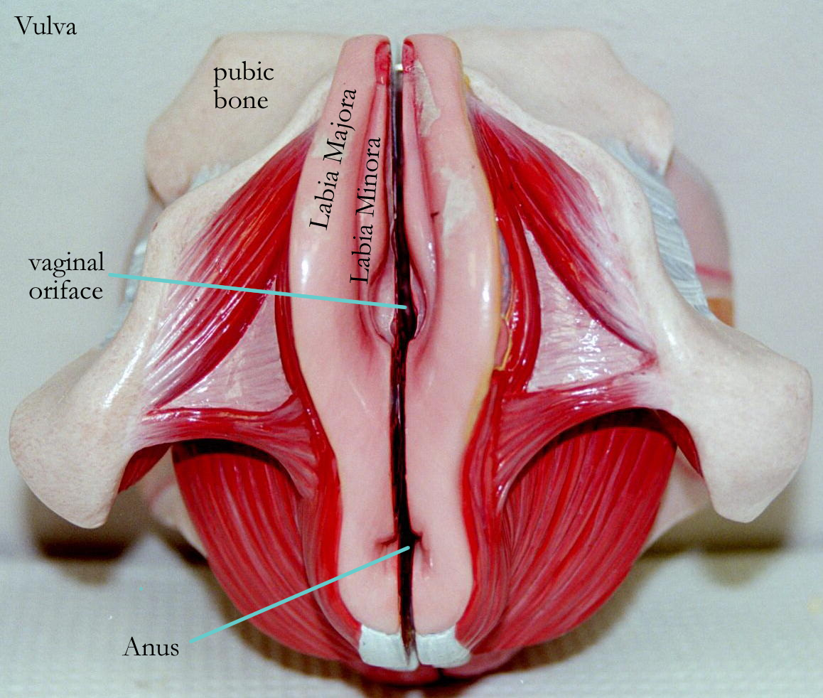
External Genitalia
External Genitalia of the female is collectively termed the vulva or pudendum.
The vulva is comprised of the:
clitoris erectile tissue composed of corpora spongiosum
labia majora analogous to the male scrotum, contains sweat glands
labia minora smaller skin fold that contains sebaceous glands
both labia cover the vagina and urethra to help prevent drying
mons pubis fat over the pelvic symphysis, covered by skin and pubic hair
Accessory Glands
Accessory Organs of the female consist of the:
Lesser vestibular glands, Skene’s glands on either side of the urethral oriface. They are analogous to the male prostate gland and secrete mucus for vaginal lubrication.
Greater vestibular glands, Bartholin’s glands on either side of the vaginal orifice. They are analogous to the male Cowper’s glands and secrete mucus for vaginal lubrication.
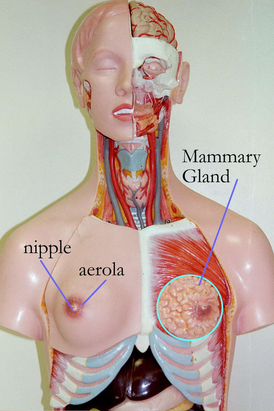
Mammary glands modified sweat glands of the skin that is functional in females
produce milk in response to AP – Prolactin (PRL) hormone
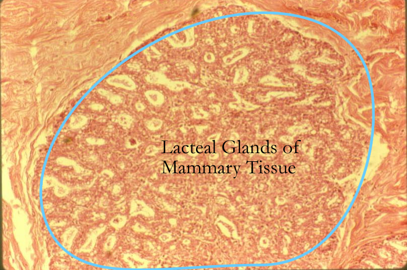
Lactiferous glands are surrounded by fat and supported by
ligaments that anchor the breast tissue to the pectoral muscles
of the chest.
Milk glandular alveoli develop in response to estrogens.
Milk release is under the control of the PP – Oxytocin (OT)
and will travel through lactiferous ducts to the lactiferous sinus and exit at the nipple surrounded by pigmented skin, theareola.
Female Lower pelvis with Urinary System
Mammary Histology: Active, Inactive, Nipple
Problems: Female Reproductive system
-ac pertaining to -algia pain
gyne, gyn/o- women hyster/o- uterus
lapar/o- loin mammilla- breast
mast/o- breast o, oo- egg
-(r)rhea flow, discharge salphing/o- tube
lute- yellow mens- month
ova- egg vagin/o- sheath
cervic/o- cervix colp/o- vagina
episi/o-, vulv/o- vulva galact/o-, lact/o- milk
gynec/o- female uter/o-, metr/o- uterus
labi/o- lip-like oophor/o, ovari/o ovary
ob- against spor- seed
I. ID female reproductive organs
A Drawing
B Models
Concept Map: Make a concept map of the female reproductive system (gross and histo) anatomy, location, and physiological function. Include hormones, source, target, effects. This concept map should be sent as part of the LAR lab report (if selected) as a document insert or as an additional PDF scanned document.
Endometriosis
Uterine Leiomyomas
Fibroids
Mastitis
Uterine Polyps
Ovarian Cysts
Ovarian Cancer
Menorrhagia
Sexually Transmitted Disease (STD)
Gonorrhea, Syphilis, Genital Herpes, Chlamydia, Trichomoniasis, Genital Warts
Pelvic Inflammatory Disease (PID)
Mammography
Cervical Cancer
Breast Cancer
AIDS
Pelvic Inflammatory Disease
Premenstrual Syndrome (PMS)
Amenorrhea
Dysmenorrhea
Menopause
Mammary Dysplasia, Fibrocystic Disease
Gynecologist
http://www.nlm.nih.gov/medlineplus/healthtopics.html
http://www.lumen.luc.edu/lumen/meded/histo/frames/histo_frames.html
http://www.kcmetro.cc.mo.us/maplewoods/Biology/Bio110/Labs.htm
http://calloso.med.mun.ca/%7Etscott/second.htm
http://www.track0.com/canteach/links/linkbodysystems.html
http://www.carr.lib.md.us/schs/science/anatomy/systems.html
http://www.leeds.ac.uk/chb/lectures/anatomy7.html
http://www.med.virginia.edu/med-ed/phys/practice_board.html
http://www.medem.com/MedLb/article_detaillb.cfm?article_ID=ZZZ8QKJ56JC&sub_cat=2 breast disorders
http://www.nlm.nih.gov/medlineplus/pregnancyandreproduction.html
http://www.wcn.org (women's cancer network)
1. Describe the histology of the ovaries and give its functions.
2. Name the functions of the female reproductive ducts:
a) Fallopian tubes
b) Uterus
c) Vagina
3. Give the structure and function of the mammary glands
4. Name the structures of the vulva
5. Define oogenesis
6. Name the three phases of the ovarian cycle
7. Name the three phases of the uterine cycle
8. Name the two hormones that affect the mammary glands
9. Define mitosis
10. Define meiosis