Supplemental Handout for AP 2402 Lab: Hematology Dr. Weis
Introduction:
Hematological
examination and testing of blood provides valuable insight regarding the
health and status of the patient.
This information helps the clinician
in determining a diagnosis, various treatment options, and prognosis for
the patient.
Purpose: The
purpose of this supplemental handout is to provide students with the information
needed to evaluate non-primate blood and gain experience with some of the
various hematological testing methods done in a clinical lab setting.
The
students are directed to their text and lab manual for a more in depth
discussion and presentation on blood.
Objectives:
To safely handle all blood products and lab equipment
To perform specific lab tests on non primate blood
To become familiar with other testing methods for blood
To record and analyze results from specific lab tests
To answer discussion questions at the end of the handout
Precautions: The
emphasis is SAFETY FIRST by use of gloves, cleaning of any spills and disposal
of blood products and
sharps in proper containers.
There is still a small chance that these animals may have contracted some unknown pathogen.
Remember:: SAFETY FIRST
GENERAL PROCEDURE:: Lab tests on Non-primate blood
To Summarize lab procedures you will be doing:
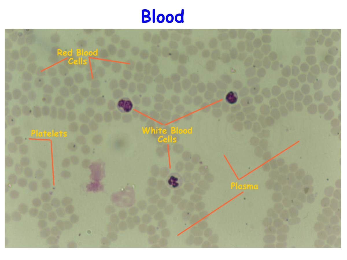
I. Blood smear and stain to observe
a. RBC color, size, shape
b. WEB differential count
c. Platelets appearance and number
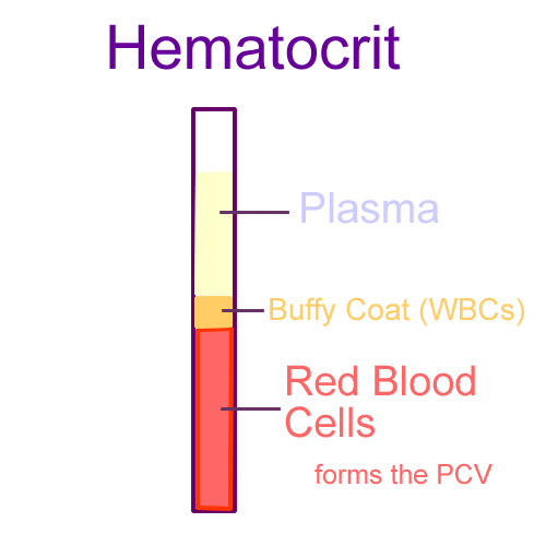
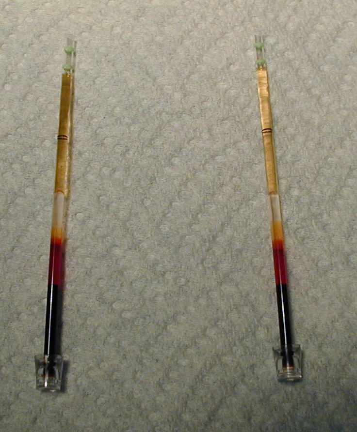
II. Hematocrit (PCV %) and Total protein
III. BLOOD TYPING (ABOD groups)
IV. View other prepared slides: Leukemia, Mononucleosis, Sickle Cell Anemia
Equipment needed:
1. Non-primate blood in proper collection tube:
a. Ruminant (bovine, ovine)
b. Equine
c. Canine
d. Feline
e. Rabbit
There will also be pre-smeared slides to stain for actual use for blood cell observation and differential counts.
2. Slides, 1 box
3. Stain (Diff Quick / or Wrights)
4. Gloves
5. Sharps container and medical biohazard waste bag
6. Lab manual/ Text/ Handout/ Data sheet
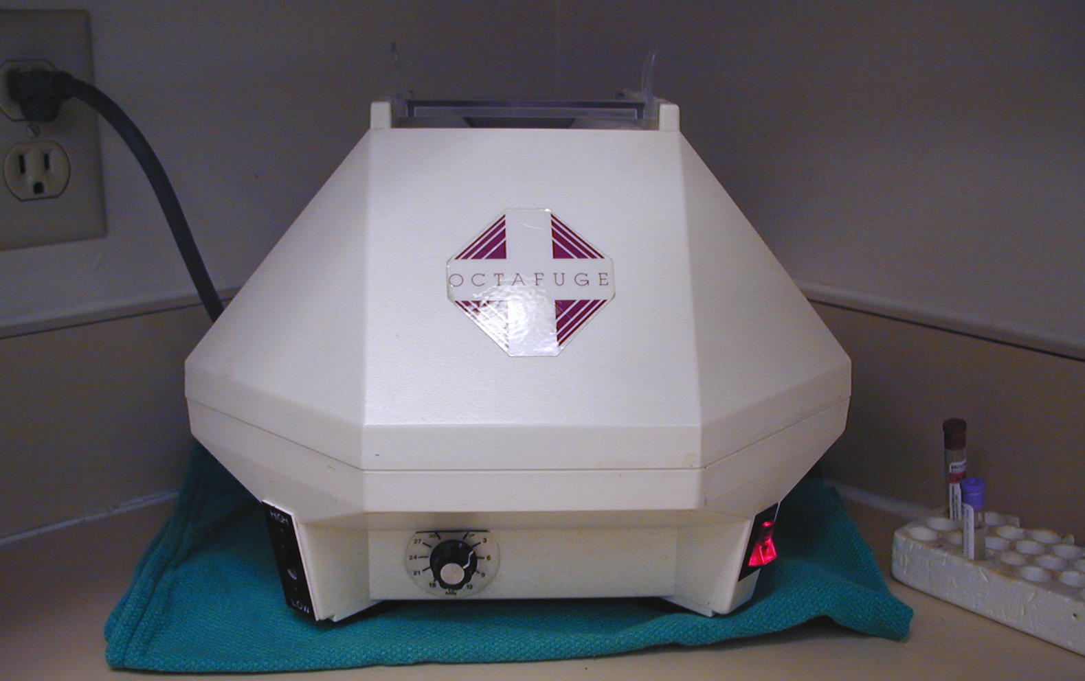
7. Microhematocrit Centrifuge
8. Hematocrit tubes/ clay/ cardreader
9. Clinical refractometer
10. Human ABOD antisera (human antisera test kits)
11. Disposable plastic pipets
12. Toothpicks
13. Grease pen
14. Immersion oil
15. Hemacytometer
16. Unopette Reservoir System for RBC and WBC
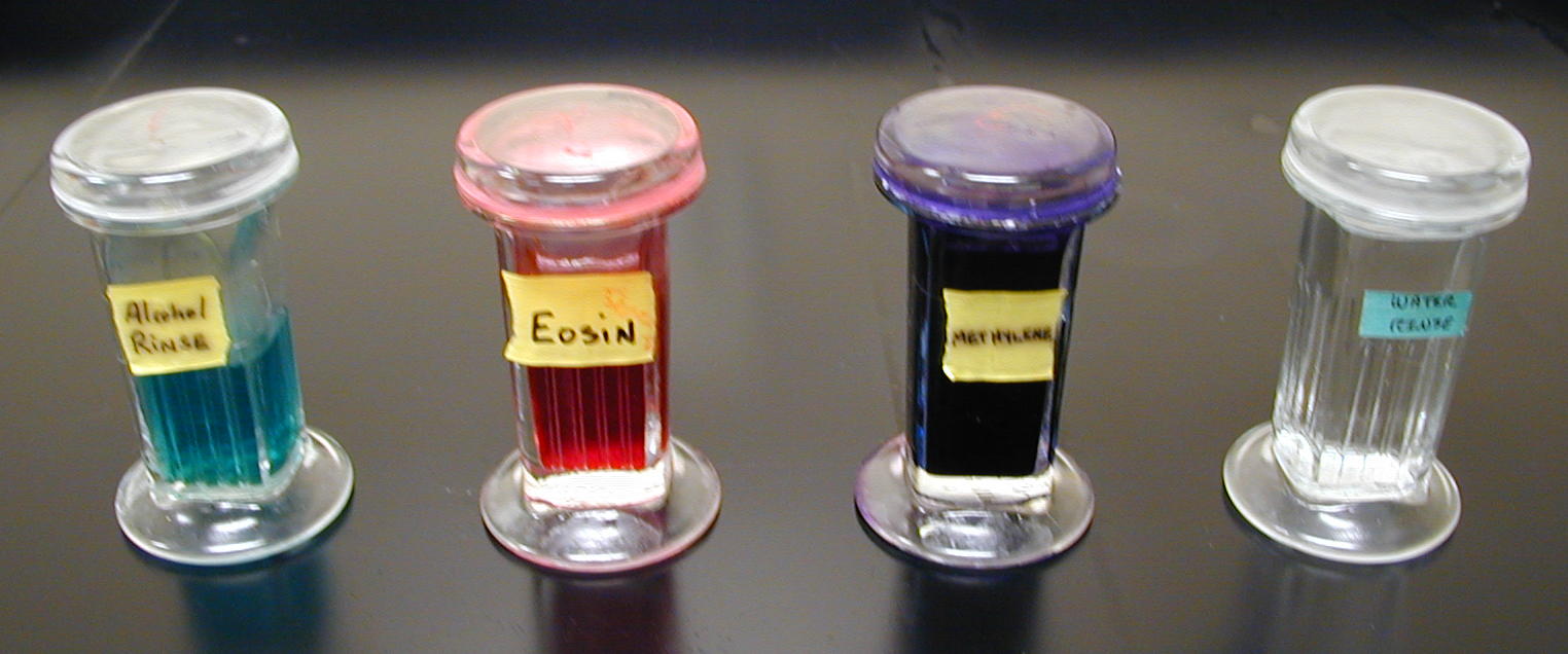
To use diff quick stain:
Dip dried slide into each copland jar 10 times at 1 second intervals
Allow excess stain to drain off before switching to next jar.
The first jar will be a light blue alcohol wash
The second jar will be a red eosin stain
The third jar will be a dark blue methylene blue stain
The fourth jar will be dH20 for rinse.
Please keep the lids on these stains at all times
Use forceps to dip the slides or you will have stained fingers
PCV
ABO and Rh blood typing
Observation
of agglutination reactions with human ABO and anti D sera in different
animal species has been observed.
Since the epitope for
the ABO antigens consists of an oligosaccharide with four sugar components,
the results can be explained due to similarities in these small saccharide chains rather than to the more variable peptide
sequences of the antigens.
Results
of positive agglutination reactions using animal blood and human antisera have
not widely been published.
From classroom observation, these are possible
results * :
Canines.....B+
Rabbits.....B+
Goats.......A+
Bovine......O+
Equine......O+
Sheep.......O-
Feline.......A or B
* remember that non-primate blood types are actually classified differently than human ABOD groups.
Canine blood types are called dog erythrocyte antigen 1.1 (DEA 1.1) which can be positive or negative (smilar to human Rh types). Feline blood types include groups assigned type A, type B, and the rare type AB. Both dogs and cats develop alloantibodies IgG and IgM like humans, so that blood typing experiments using human antisera can demonstrate agglutination reactions.
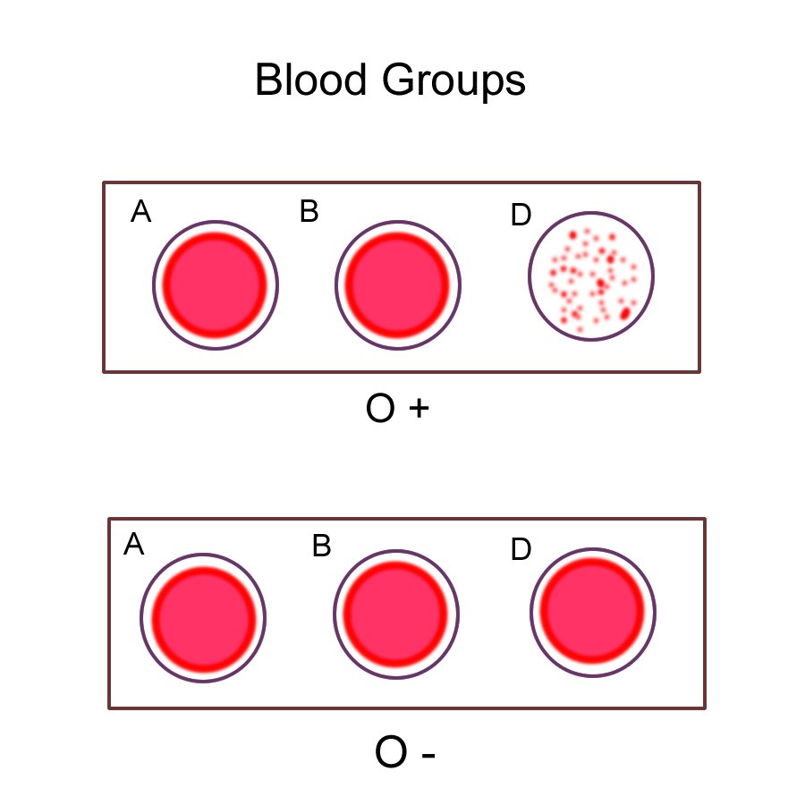
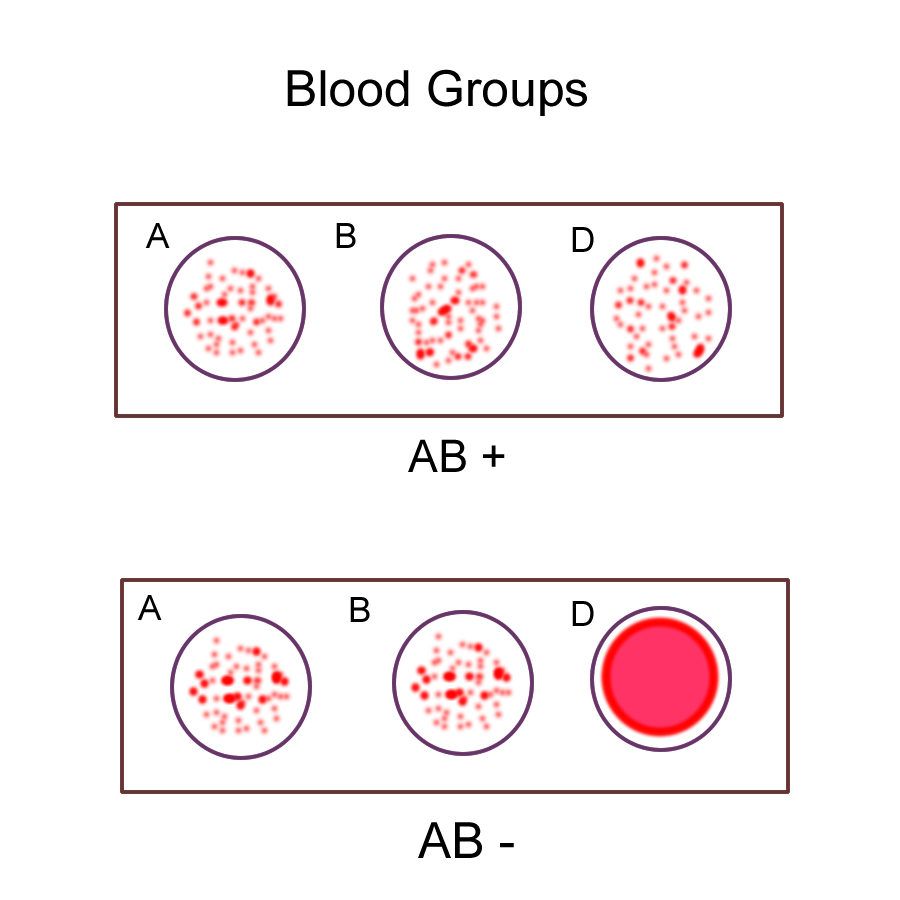
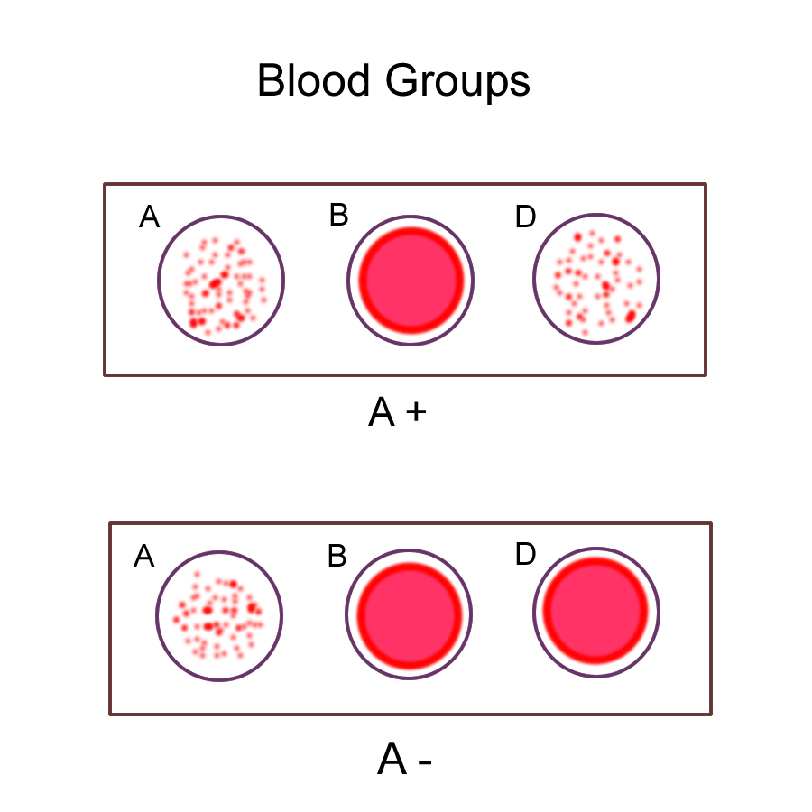
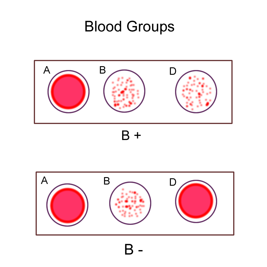
Mark a card or slide with the appropriate species and perform the blood typing as directed.
A
positive reaction will appear as small dark spots at the margin of the
mixing area.
The reaction takes approximately 5 minutes. If the blood
remains a homogenous pool, then you can assume that the antigen is absent.
ABOD reactions
Additional Laboratory Report Questions
1. Compare and contrast mononucleosis to agranulocytic leukemia.
2. If a person with blood type A marries a person with blood type B, what are all the possible blood types for their offspring.
3. What is a transfusion reaction and why does this occur ? What are the possible consequences ?
REFERENCES
Tortora, Gerard J., Tallitsch, Robert B. Laboratory Exercises in Anatomy and Physiology, 4th ed., New York, Macmillan Publishing Company, 1993.
Evans, David L., Evans, Henriette. 1994. Using livestock blood in A&P Labs, HAPS convention handout, May 1995
Page, T.W. 1994. Blood substitues for the physiology laboratory.
HAPS News Feb., 1994
Watkins, W.M. 1966. Blood group substances in the ABO system : the genes control the arrangement of sugar residues that determine group specificity. Science 152:172.
Duncan, J. Robert, Prasse, Keith. Veterinary Laboratory Medicine
Clinical Pathology, 2nd ed., Ames, Iowa State University Press, 1986.