Microscopy and Cytology Review Experiment
I. Using the proper microscope technique, view a prepared slide of squamous
epithelium from a cheek smear.
Draw and label what you see in the space provided:
II. One of each lab partners will make a cheek smear wet mount to view and
identify major cellular components.
- Gently scrape the inside of your mouth with the broad end of a toothpick
- Stir the scrapings in a drop of water on a microscope slide and place a
cover slip on it
- Observe the specimen under 4x and 10x objective power
- Stain the slide with one drop of methylene blue placed at the edge of the
cover slip
- Draw the stain across the specimen by placing a tissue or paper towel at
the opposite edge of the cover slip from the original drop of stain
- Observe again with the microscope and idenity the following basic cell parts
using the 40x objective lens.
- Nucleus
- Nucleolus
- Cytoplasm
- Plasma membrane
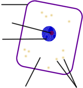
III. Identify the labeled parts on the cheek cell drawing below and give their
unique function: