Microbiology Lecture Notes
Prokaryotic Cell Structure & Function
Dr.
Weis
Prokaryotic Cell Structure
Size
~0.2µm -2µm diameter
2-8 µm length
Single-celled, if aggregates form groups
Shape: Rod (bacilli), round (cocci), spiral (helix), pleomorphic
Rod variations: long, slender, square, oval ends
Number: single, double, chain
Divide along their short axis, so there is
no aggregate groupings
Round variations: oval
Number: single, double, x4 (gaffkya), cube, cluster, chain, x8 (sarcina)
Divisions create groupings as stated
above
Spiral variations: curved, corkscrew, helical, comma
Pleomorphic: Archaea bactera
Variations: square, star
Cell Wall Appendages
1) Glycocalyx
All bacteria secrete some type of glycocalyx to protext them from drying.
Polysaccharide polymer +/- peptides or polypeptides outside the cell wall
NOTE: If only contain sugars, then called Extracellular polysachharide
Can be:
Organized and thick = Capsule, firmly attached
Unorganized and thin = Slime layer, loosely attached
Regular structured, outer viscous thin layer = S-layer
The glycocalyx will form a biofilm to
help surround bacterial colonies. The negative charge of this glycocalyx structure helps repel the WBCs of
the immune system.
2) Flagella
Length 10µm - 20µm
Not very thick, need special stain to view
Lacks the "9 + 2" microtubular structure
seen in Eukaryotes
Function: motility
Arrangement:
One (1) = monotrichous, at one pole
At both ends = Amphitrichous, single at each pole
Tuft = lophotrichous, two or more flagella at one or both poles
Entire = peritrichous, over the entire surface
If an endoflagella, it will create the axial filament that causes a corkscrew shape such as those of the Spirochetes.
3 parts
a) Filament with flagellin protein wound and intertwines as helix to form a hollow core, normally not surrounded by a sheath. Because of the hollow core, it does not flex. Flagellar (H) protein functions as an antigen. Hollow core allows for repair if flagellum is cut off. Repair happens at the tip, not at the base.
b) Hook – flexible coupling that is an "L" shaped attachment of the filament to basal body, allows filament to rotate.
c) Basal Body- 35 different proteins arranged in rings that anchor structure by way of a central rod in cell wall and cell membrane.
Minimum: 2 in plasma membrane. Can have two in cell wall.
G(-) have 4 rings (2 outer and 2 inner); G(+) have
2 rings
Function: Provide motility by rotation due to ATP driven proton pump
Two proteins in the flagellar motor: MotA and MotB form a proton channel through the cytoplasmic membrane. The rotation is driven by the proton gradient created by the electron transport system as protons accumulate in the space between the cell membrane and cell wall and then return to the cytoplasm via the channel. Half of all known bacteria are motile.
Clockwise moves called tumbles, no real forward progress but can move away from a repellant [ (-) taxis]
Counterclockwise moves called runs, forward progress and moves towards an attractant [(+) taxis]
Moves organism to food source, enables organism to avoid phagocytosis
by immune system.
3) Axial Filaments – Seen in Spirochetes, also called endoflagella because they spiral around the cell in between the outer membrane and cell wall. Similar to prokaryotic flagella and are attached to basal motors at either end of the cell.
Filaments attached at opposite ends move relative to one another to
create a spiral motion rotating the cell like a corkscrew.
4) Fimbriae and Pili
Pili are thin protein tubes made of pilin that originate from the cytoplasmic membrane. The pilus shaft is composed of pilin and at the end is an adhesive tip that corresponds to the glycoprotein or glycolipids of the host cell. The adhesive tip can be switched in order to adhere and pull towards different types of cells. Can be used in conjugation, a type of asexual reproduction.
Fimbriae are short, nonmotile fibers and found at the poles or along the entire surface. Used primarily for adherence. Fimbria can also be used in the glycocalyx to help form biofilms.
Two types of Pili:
a) Short attachment "pili" [fimbria], numerous
Can anchor to tissues via adhesion proteins
b) Long conjugation pili or sex pili, one or two
Primarily used for transfer of DNA
Movement of prokaryotes is fairly random in a homogenous environment.
Can become specific based on environmental stimuli such as
Movement toward a stimulus is positive taxis, movement away is negative taxis.
For movements in a coordinated run, the flagella are rotating counterclockwise.
In an uncoordinated/random or separate movements, flagella are rotating clockwise creating a tumble like motion.
Chemoreceptors are located in the cell membrane or periplasm of the bacterium which bind the attractants or repellants
Cell Wall
Most all bacteria have a semi rigid wall which surrounds the plasma membrane.
Mycoplasmas do not have a cell wall and their cell membrane has pumps to maintain proper osmotic pressure. Their cell membranes contain different sterols that help in stabilization.
Function : to maintain shape, protect, and prevents organism from rupturing
Structure of Cell Wall :
Peptidoglycan of Eubacteria contain muramic acid (M)
[Archaea bacteria do not have muramic acid]
Composed of repeating & interlocking amino+disaccharides (NAG = N-acetylglucosamine and NAM = N-acetylmuramic acid) forming a carbohydrate backbone monomer [glycan part] linked by a ß 1-4 bond with an additional short tetrapolypeptide (4 AA) side chain [peptido part] that attachs to the NAM polymer.
The 4 AA side chain contains L-Alanine, D-Glutamate, mes-diaminopimelic acid
(DPA), and D-alanine. Therefore, a single peptidoglycan unit
is composed of
In order for bacteria to increase the size following binary fission, the
links in the peptidoglycan must be broken so that
new polymers can be added and linked (transglycolation) and the tetrapolypeptide crosslinks resealed.
Bacterial
enzymes are involved such as:
Interference with this process will result in a weak cell wall and lysis of the bacterium.
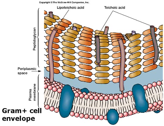
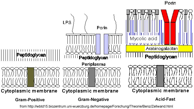
G(+) bacteria have simple cell walls composed of many layers of peptidoglycan which form 60%-90% of their cell wall, up to 50% of the cell weight to create a 25-80nm thick structure that is heavily crosslinked. Teichoic acids are also found, composed of polymers of glycerol or ribitol joined by phosphate groups. Amino acids such as D-alanine are attached. Teichoic acid is also linked to muramic acid and helps link various layers of the peptidoglycan mesh together.
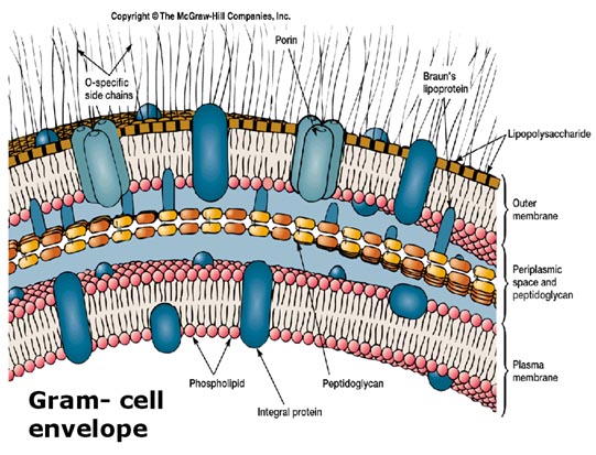
G(-) bacteria have complex cell walls containing one or a few layers of peptidoglycan to create a 3nm thick structure, which makes up 10%-20% of the cell wall and only intermediately cross linked. No teichoic acids are found. The peptidoglycan layer sits in a space containing periplasm fluid between the cell membrane and the outer membrane which allows for H2O, nutrients, and protein transport. The outer membrane of the G(-) cell wall contains similar structures to the cell membrane in addition to lipoproteins and lipopolysaccharides. Porin proteins are also found that act as a channel through the outer phospholipid membrane. These channel proteins allow the passage of nutrients, vitamins, and viral attachments. The lipopolysaccharide [LPS] in the outer membrane consists of an O-polysaccharide that is antigenic (creates an immune response), a core structure, and a Lipid A, which is considered to be an endotoxin. Lipopolysaccharides [LPS] can also impede the entry of drugs into the cells and therefore, create more resistance to antibiotics.
Additional importance is placed on the capability of antibiotics such as
penicillin to prevent the construction of peptidoglycan in G(+)
bacteria. Lysozyme produced in an immune response
(in tears) can disrupt the linkage between the carbohydrates (
Atypical Cell Walls
No cell walls or very little material is present in
A) Mycoplasmas: plasma membrane has sterols, no cell wall
B) Archaea: cell wall has pseudomurein
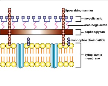
C) Acid Fast organisms have a glycolipid outer membrane (instead of phospholipids) composed of mycolic acid, arabinogalactan-lipid complex, and lipoarabinomannan. They do have a thin peptidoglycan layer and a phospholipids cell (plasma) membrane (much like the G – bacteria).
Comparison of G(+) and G(-) and Acid Fast cell walls
Cell (Plasma) Membrane
Lies internal to the cell wall and encloses the cytoplasm
7nm thick
Phospholipid bilayer (hydrophobic and hydrophilic regions)
Proteins:
Peripheral: to provide attachment sites for chromosomes, pili, flagella
Integral: Active Transport via permeases; Electron transport system, ATP synthase
Lacks sterols seen in Eukaryotic cells, but has sterol-like substance called a hapanoid that helps stabilize the membrane.
Selectively permeable:
Water, Gases, Lipid soluble molecules can diffuse
Ions use pores
Water soluble molecules need carrier proteins or transporters
+/- pigments for photosynthesis
Function: Anchors DNA during replication and separation during division
Site of Energy production (electron transport system)
Peptidoglycan synthesis
Protein synthesis for cell membrane
Contains the bases of flagella
Waste removal
Involved in the formation of endospores
Membrane transport
a) Active (ATP or proton) : Used for Glucose and AA
a. Antiporter system: moves molecules in opposite directions
b. Symporter system: moves two molecules the same direction
c. Group Translocation: chemical is altered as it is transported
to help trap it inside the cell. [Glucose -> Glucose-6
Phosphate]
b) Passive
a. Simple Diffusion (lipid soluble)
b. Facilitated diffusion (AA, nitrogenous) called uniporter
c. Osmosis (water)
Contain extracelullar enzymes that can break down substances to be transported.
Can envaginate (fold in) to create structures necessary for aerobic metabolism or to create thylakoid membranes the contain chlorophyll for photosynthesis. Other envaginations called mesosomes are artifacts created during preparation processes used to observe bacteria.
Importance of the cell wall in chemotherapy:
Certain antibiotics (AB) and disinfectants/antiseptics can alter the cell membrane.
Terminology
Cell envelope = cell membrane + cell wall +/- glycocalyx
Cytoplasm = Cytosol + Organelles
Cytosol
- 80% water
- proteins (primarily enzymes)
- Carbohyrates
- Lipids
- Salts / Ions
Nuclear Area (Nucleoid)
a) Single (haploid) unit of long, supercoiled, continuous closed circular (two ends covalently bonded together) double stranded DNA containing the bacterial chromosome (genome), attached to the plasma (cell) membrane.
It contains the genetic material of the bacteria.
Very little protein is associated with bacterial DNA, but there are histone-like proteins (HU, IHF, H-NS) bonded to the DNA to create about 50 domains which help make it more compact. An enzyme, DNA gyrase (topoisomerase II) helps to supercoil each domain to help cause a winding and twisting of the DNA around itself creating a compact size.
Other DNA topoisomerase enzymes are essential in unwinding, replicating, and rewinding the circular, supercoiled DNA.
REMEMBER : NO MITOSIS STAGES, NO MEIOSIS (only one chromosome) !!
The bacterial chromosome is generally 1000µm long and frequently contains as many as 3500 genes.
Bacterial DNA replication is bi-directional and leads to the replicated DNA strand created at the replication forks appearing like a Greek Letter, called a theta structure.
Protein synthesis occurs much like it does in Eukaryotes:
Genes located along the DNA are transcribed into RNA that undergo translation (mRNA, tRNA, rRNA). Recall that the DNA base sequences determine which proteins / enzymes an organism can synthesize which therefore determines the type of chemical reactions that can occur.
NOTE: Some chemotherapeutic agents can inhibit normal nucleic acid replication.
b) Small circular rings of DNA called plasmids that contain supplemental genes:
a. Not essential for growth, are capable of autonomous replication
b. Double stranded, helical (some may have a linear form)
c. Can have anywhere from 1-700 copies of a plasmid in a cell
d. Associated with plasma membrane proteins
e. Replicate independently from bacterial chromosomes
f. Contain genes for toxins, AB resistance, other enzymes : examples include
R-plasmids in certain bacteria have the genes that code for production of sex pili and antibiotic resistance. Exchange plasmids during a process called conjugation
Other plasmids can code for exotoxins or endotoxins
c) Transponons (transposable elements or jumping genes)
a. Small pieces of DNA (1-12 genes long) that encode enzymes that move the gene from one DNA location to another.
b. May be found as part of the nucleoid or in plasmids
c. Contains genes coding for antibiotic resistance
d. Uses transpoases, enzymes that cut and reseal the DNA during the exchange, so they can cut themselves out and insert themselves into another nucleoid or plasmid
e. Specific transponons called Integrons that carry multiple gene clusters that can integrate and accumulate as this unit can move from one piece of DNA to another
Metachromic Granules
i. Volutin – reserve of PO4= for ATP
ii. Can help in ID of certain bacteria, such as Diptheria
Polysaccharided granules
i. Glycogen- store carbon and energy reserve
ii. Starch
Lipid – storage
Sulfer – store sulfur
Carboxysomes – enzymes for use of CO2 as carbon source
Gas vacuoles – buoyancy
Magnetosomes – iron magnetite to help in orientation
Poly-beta-hydroxyalkanoate (PHA) – plastic like polymer that functions as a carbon and energy storage.
Highly durable/ resistant dehydrated cells formed internal to plasma membrane.
Little or no metabolism occurs
Created by specialized vegetative cells when nutrients are depleted and are formed to help certain bacteria survive extreme environmental conditions.
Primarily seen with G(+) bacteria.
Formation called sporogenesis or sporulation.
Process: Vegetative cells are exposed to harsh environmental conditions. Cells begin sporulation by replicating their DNA and creating a septal wall that closes off the replicated DNA. This structure is called a forespore. The membrane layers can synthesize additional peptidoglycan layers to form a series of thick protein coats called the spore coat, a keratin like protein. The original cell disintegrates (lysis) and the spore is then released. Sporulation takes around 15 hours.
Arrangement of spores can help in classifying organisms:
Central/middle
Subterminal (near the end)
Terminal (at the end)
The final spore structure contains
Resistant outer coats (cortex, spore coat, exosporium)
Core: Nucleiod region with some ribosomes, RNA, enzymes
It can remain dormant for many years and survive the harshest environments, even if using
Chemicals
Heat / Drying
Radiation
Boiling
Reversal to vegetative state is by germination and the breaking down of the protective layers which will occur once proper environmental conditions return.
Spores require special stain to view and are the most resistant structures.
Examples of bacterial genuses that forms spores are Bacillus and Clostridium.
Anthrax Bacillus anthracis
Tetanus Clostridium tetani
Botulism Clostridium botulinum
Gas Gangrene Clostridium perfringes