Biology 2404 A&P Basics Lab Exercise 5 Tissues & Membranes Dr. Weis
| Objectives | Background | Medical Terms | Activities | Applications | Careers | WWW | Review Questions |
Students should be able to
* give the four major tissue groups and their function
* give examples of each major tissue type, their location, and function
* name the five types of membranes and their function
* give definitions of related terms
* know the meaning of related word parts and medical terms
Anatomy and Physiology Background
Read related textbook information
Tissues comprise the next level of organization since they are formed by cells with similar structure and function. There are four major tissue groups, each of which combines to form the organs and their systems. The major tissue groups and their general functions are:
1) Epithelial function as linings or coverings
2) Connective functions to connect and support
3) Muscle functions to provide movement
4) Nervous functions to communicate and control
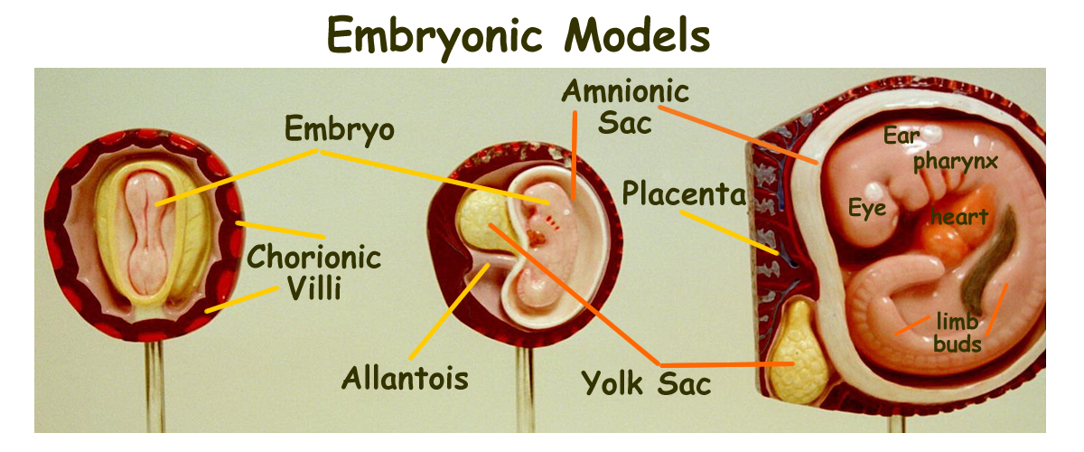
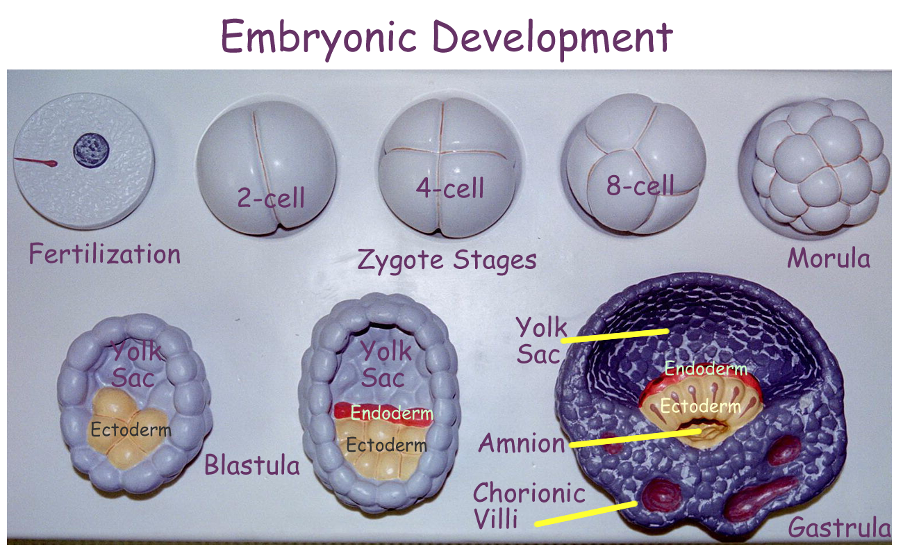
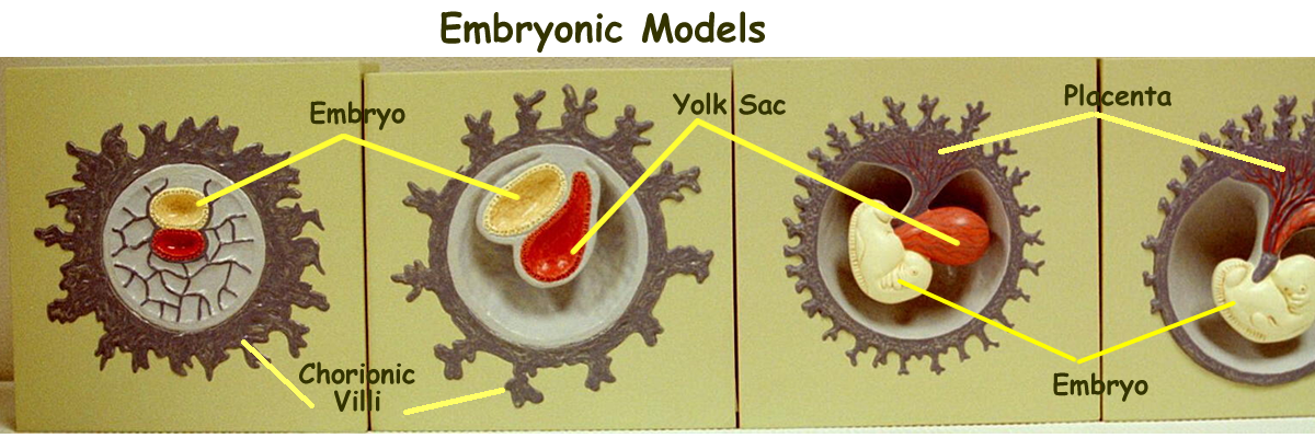
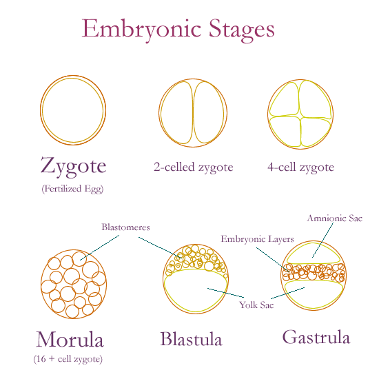
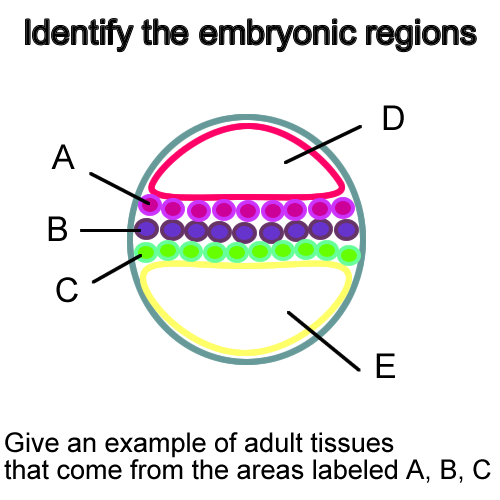
Adult tissues are derived from embryonic tissues that develop from a fertilized egg called the zygote. Mitotic cell divisions create blastomeres (also known as stem cells) that migrate to various areas once the zygote morula enters the uterus. Fluid from the female reproductive tract helps to push these blastomeres into regions. The outer cells become part of the fetal placenta while the inner group of cells forms the embryo.
Three layers of blastomeres create the embryonic tissues that help to form the adult tissues and organs. The embryonic tissue layers are from outside to inside as the:
Ectoderm
Mesoderm
Endoderm
The ectoderm gives rise to all nervous tissue and the epithelium of the skin.
The mesoderm gives rise to all connective and muscle tissues.
The endoderm gives rise to most of the epithelial tissues and glands.
Embryology Models : Embryonic, Development, Extra-embryonic membranes
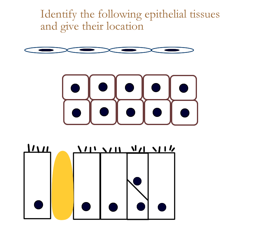
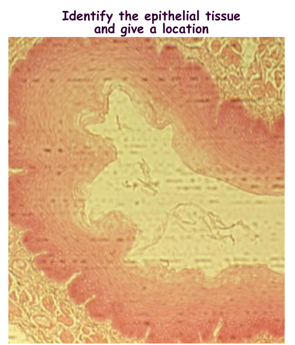
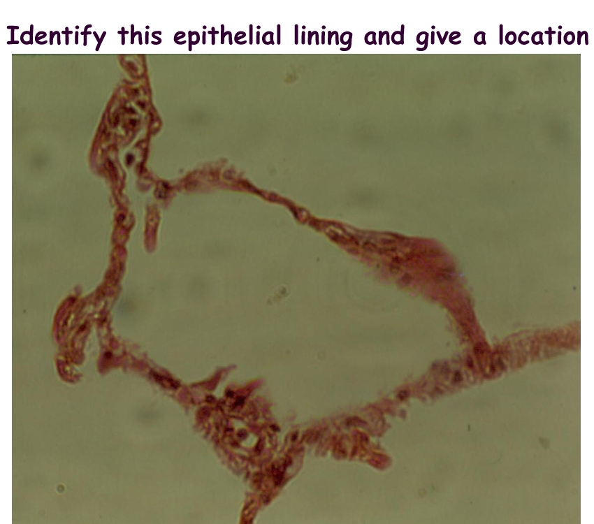
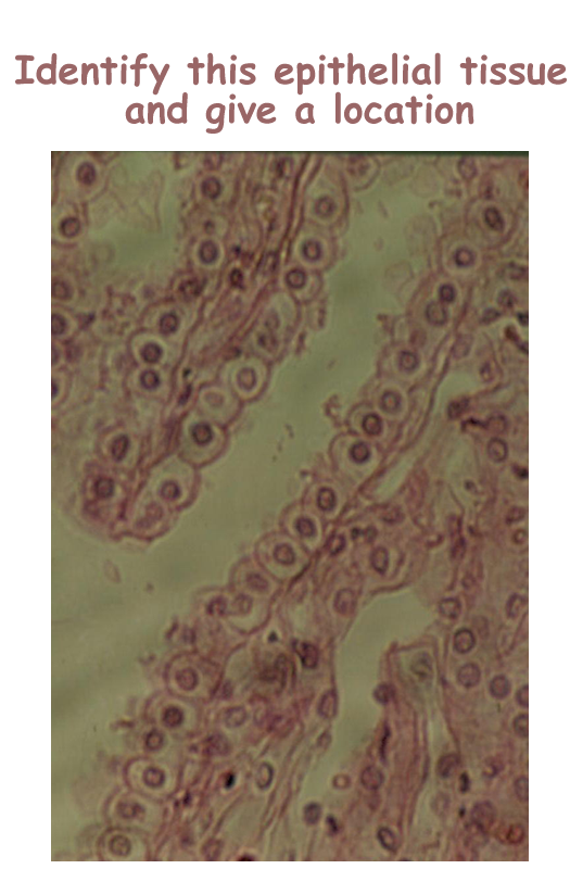
Epithelial Tissues
Two major types of epithelial tissues are those that form membrane linings and those that are specialized as secretory glands. Epithelial tissues are named based on their structure and in turn their structure dictates their function and hence their location.
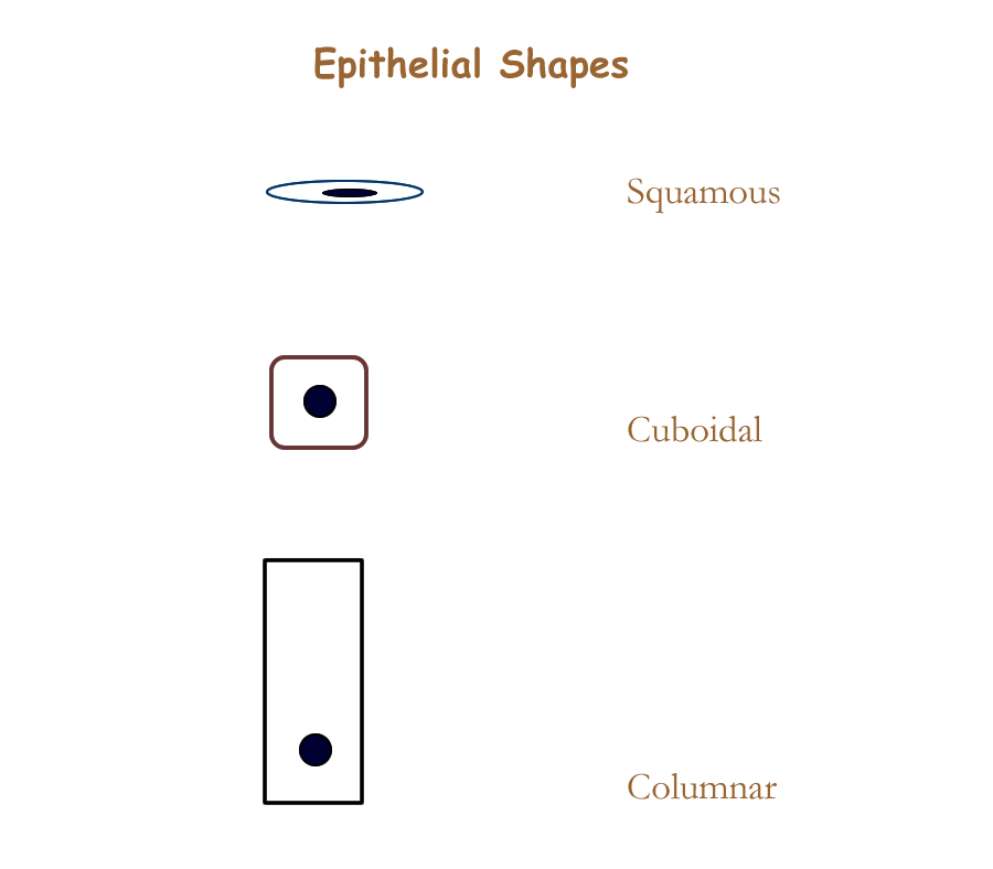
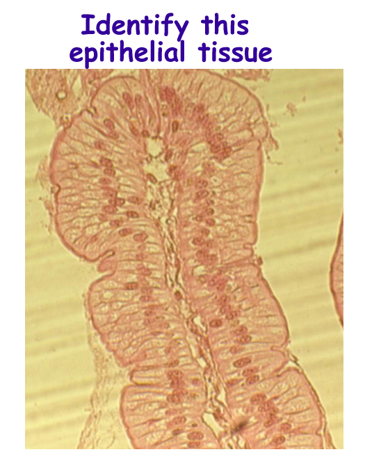
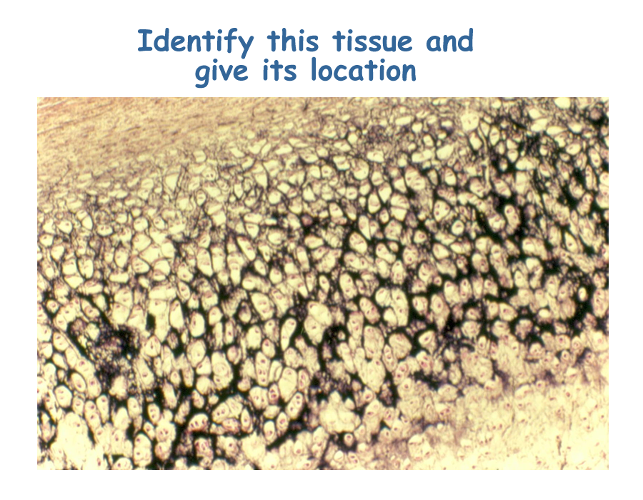
Epithelial tissues that form membrane linings are named based on the shape of the cell and the number of layers. There are 8 types of epithelial linings, six regular and two that are modified versions of a few of the other six. The cell shapes come in three types:
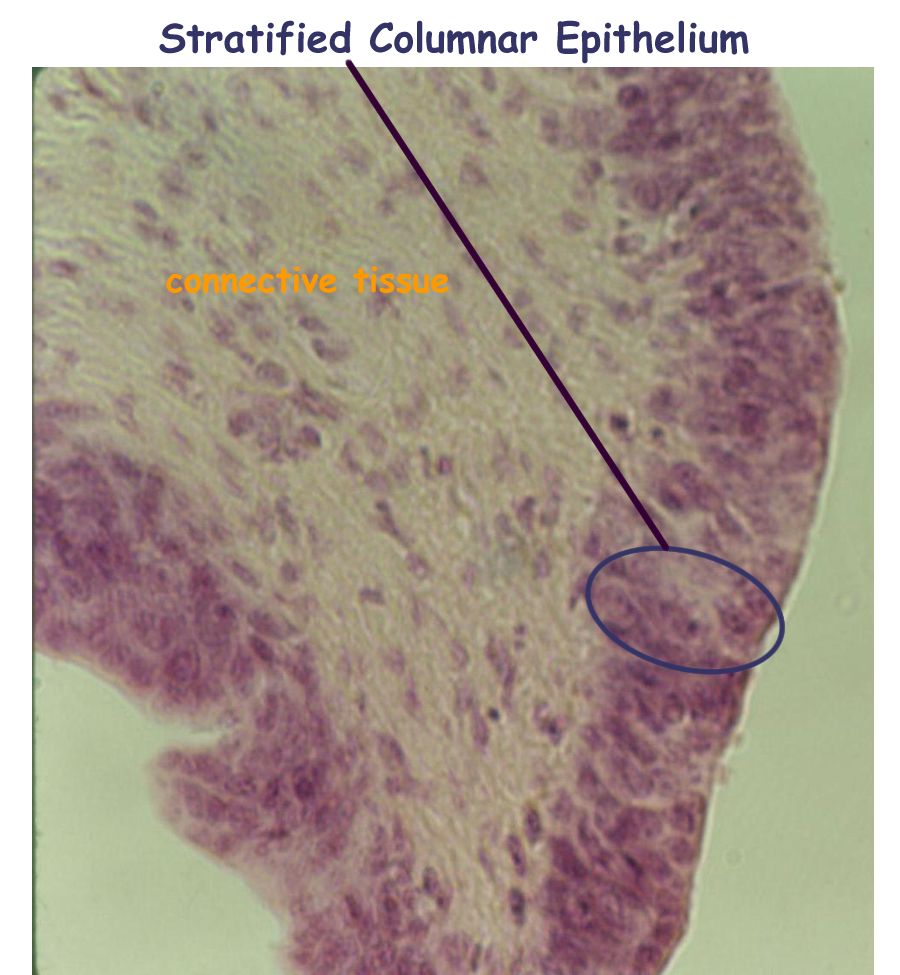
flat or squamous, square or cuboidal, and tall or columnar. The number of layers is either one single layer called simple or more than one layer called stratified.
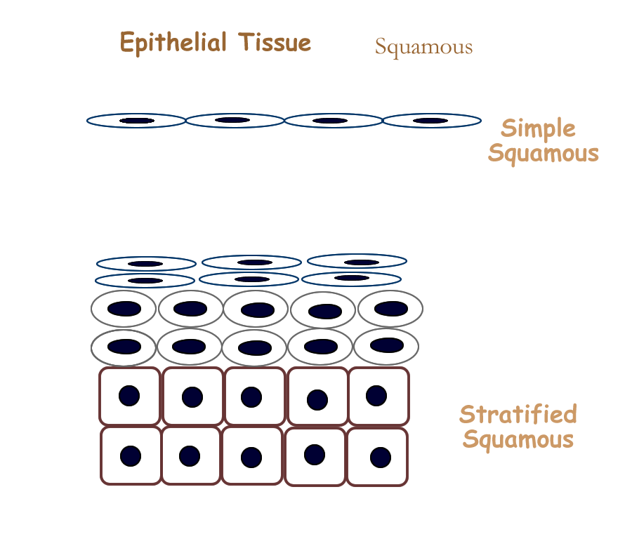
If you combine the layer with the shape you end up with the six regular types of epithelium. For example, a single layer of flat cells would be called simple squamous.
Since it is very thin and delicate, it wouldn’t be found on surfaces that need a lot of protection, like the skin. It is better suited for diffusion and so this simple squamous lining is part of the cardiovascular system. It lines the heart and blood vessels.
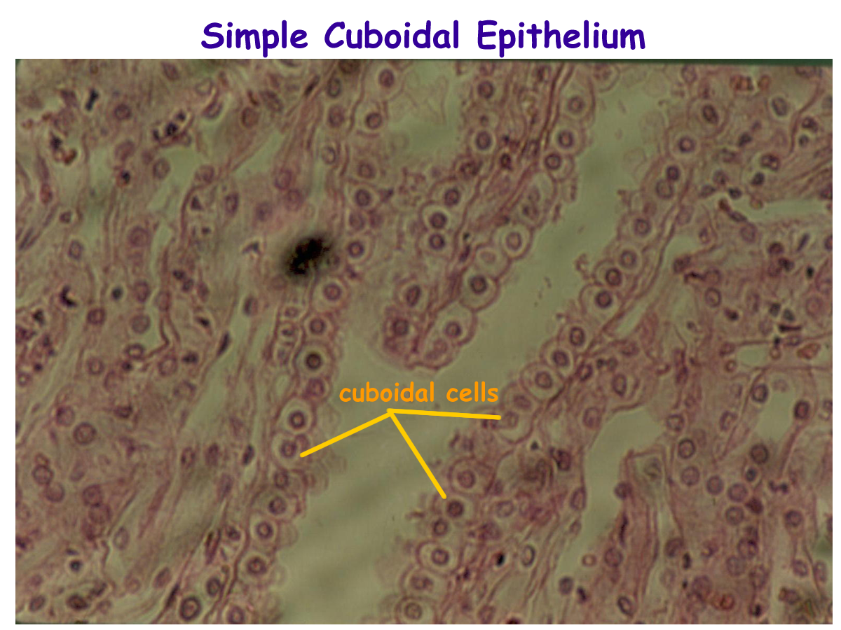
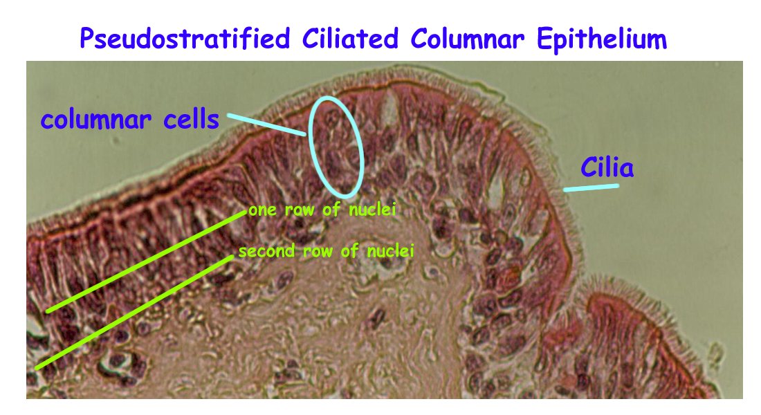
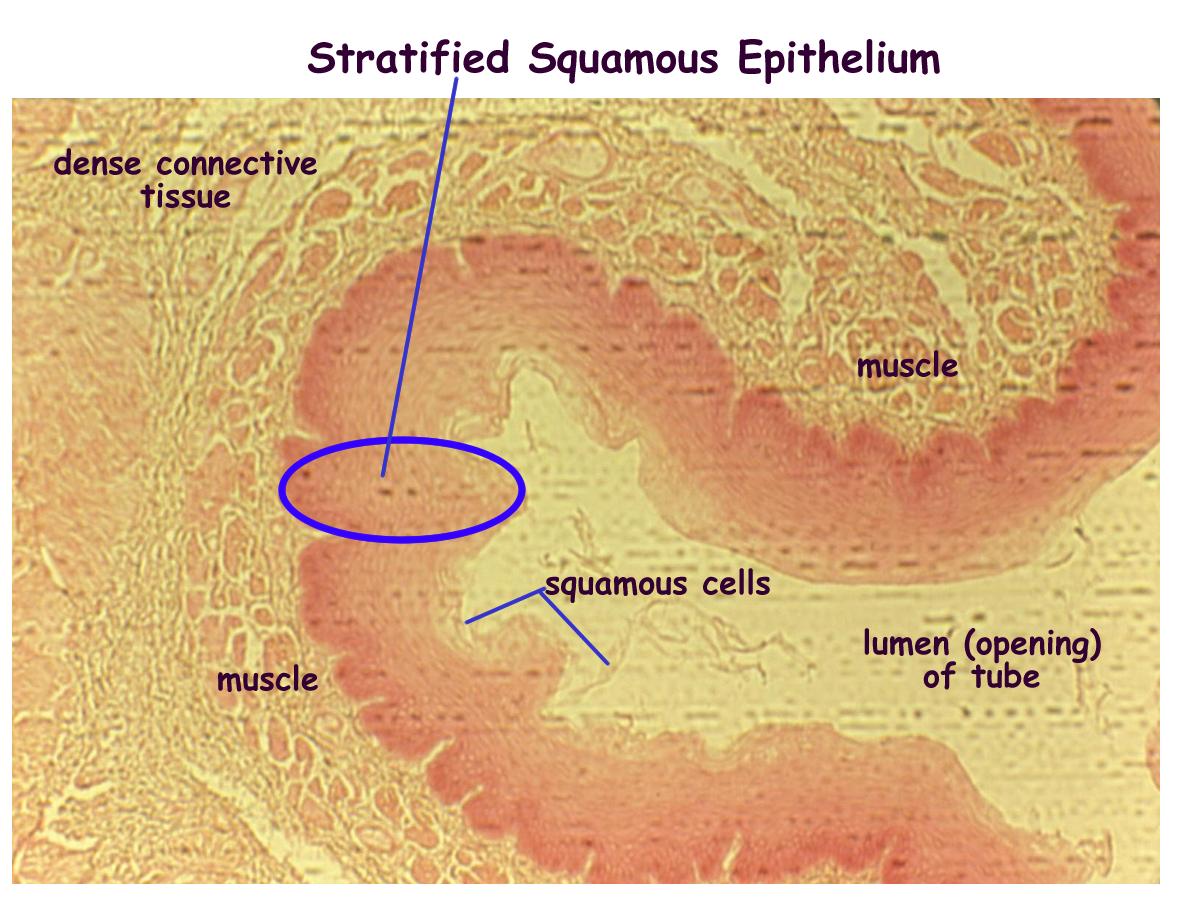
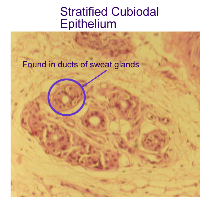
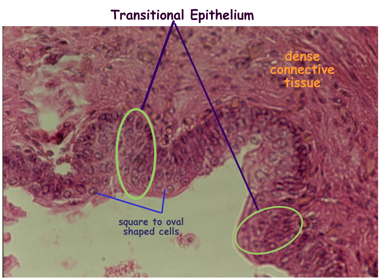
The six expected epithelial layers, their description and function are
Simple Squamous one layer of flat cells lines cardiovascular
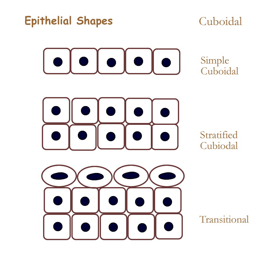
Simple Cuboidal one layer of square cells most common type of lining, found in kidney tubules
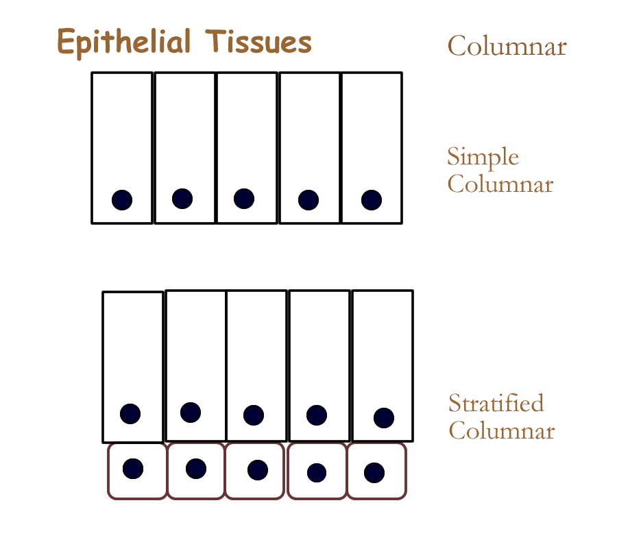
Simple Columnar one layer of tall cells lines most of digestive tube
Stratified Squamous many layers, the last is flat forms the epidermis
Stratified Cuboidal two layers, the last is square found in glands
Stratified Columnar several layers, the last is tall found in reproductive tubes
The other two epithelial tissues are based on the stratified cuboidal and the stratified columnar linings.
Transitional modified stratified cuboidal lines most of urinary
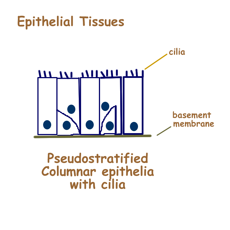
Pseudostratified columnar modified stratified columnar lines upper respiratory
Epithelial tissues are attached to and supported by a noncellular, protein rich layer called the basement membrane. The basement membrane comes from secretions of epithelial tissues and connective tissues that form glue like adhesive which joins the two tissues together. This is needed because blood supply for all epithelial tissues comes from the lower connective tissue structures. The process provides exchange by diffusion and allows the nutrients across and the removal of wastes from epithelial tissues.
Healthy epithelial tissues regenerate regularly anywhere from 3 days to 30 days.
The epithelial tissue that form glands are based on simple cuboidal and stratified cuboidal tissues. Their function is to secrete. Some of the other epithelial membrane linings can secrete, but only in certain regions. Epithelial glandular primary glandular function is to secrete.
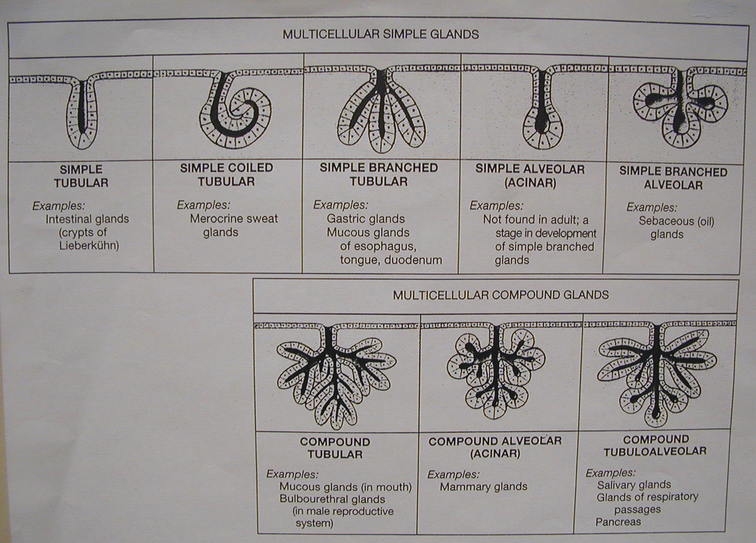
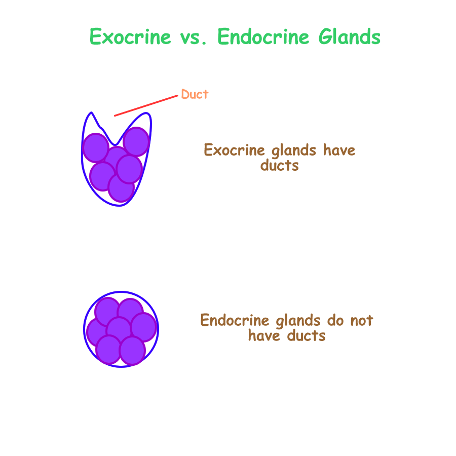
Their classification is also based on structure and varies from the linings. Glands are classified into two major groups, those that have ducts and those that do not. Ducts are passageways for the secretion to travel. If a gland does not have a duct, then the blood becomes the means for secretory travel.
The two major epithelial glands are:
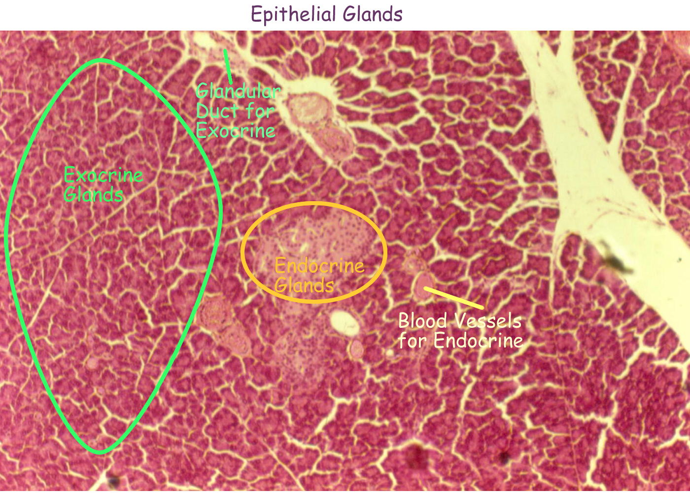
Exocrine have a duct or tubule as part of their structure
Endocrine do not have a duct or tubule as part of their structure
Glandular cells are cuboidal in shape, so that is not needed in the name.
Exocrine glands are classified based on number of cuboidal cells and number of ducts.
Sometimes specific exocrine glands are classified based on their secretion type.
Endocrine glands are classified based on their secretion or location, since the number of cells will always be multiple and the duct number will be zero. Endocrine tissues will be discussed in a later chapter and exercise.
Exocrine Gland classification
Number of cells single or unicellular and many or multicellular
Number of ducts single or simple and many or compound
Shape of glandular system tubular, coiled, acinar, alveolar, mixed
So we can combine the structural elements to come up with our exocrine classifications.
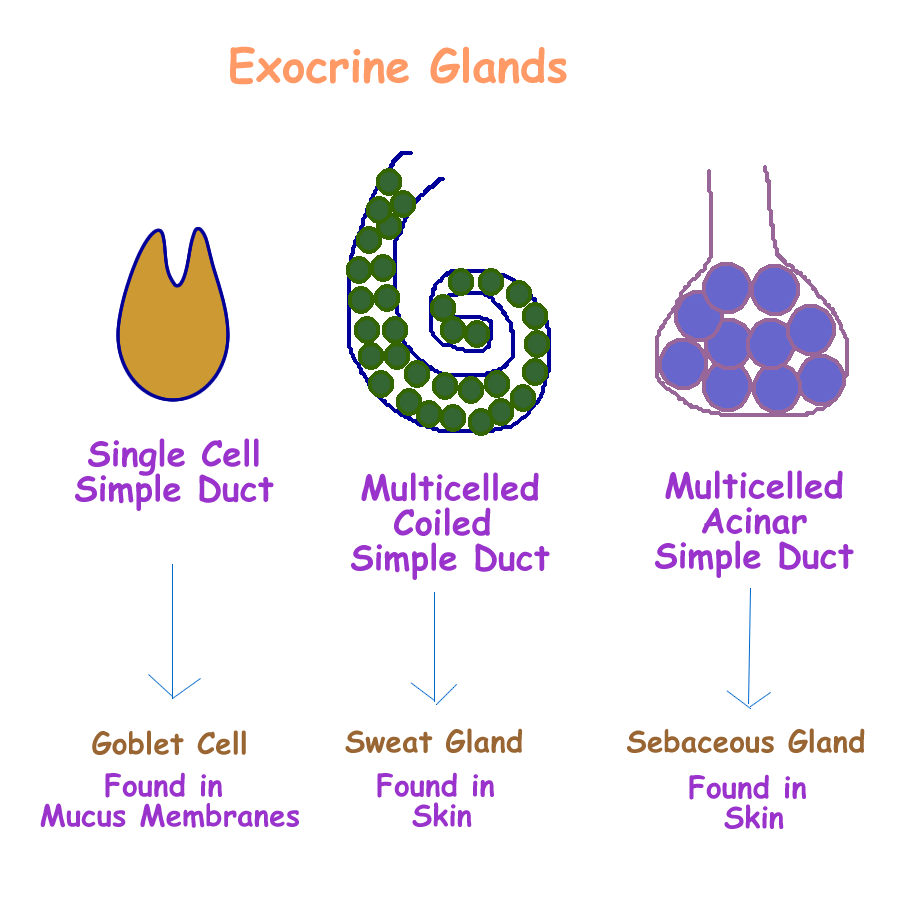
Simple Unicellular one duct from one cell
Simple Multicellular one duct from many cells
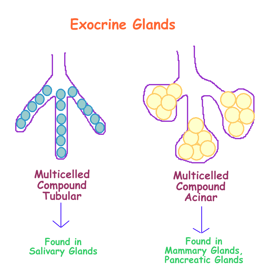
Compound Multicellular many ducts from many cells
As you can see, there is no compound unicellular, as one cell can not have multiple ducts.
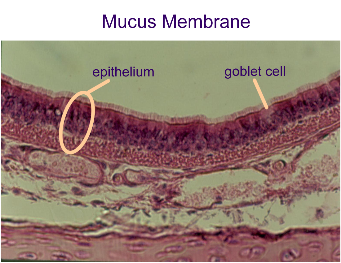
Simple Unicellular glands have another name. They are called goblet cells.
Their secretion is called mucin and when this mixes with water, it becomes mucus.
These cells are found in the linings that form mucous membranes, the linings of open body cavities. Examples of these goblet cells can be found in the digestive and respiratory linings.
Simple Multicellular glands are found throughout the body and are usually named for the secretion produced. Gastric glands secrete gastric juices, sweat glands secrete sweat, and sebaceous glands secrete sebum.
Compound multicellular are found in specialized areas, such as mammary glands for milk production, the pancreas for digestive enzymes, and the salivary glands for saliva.
Drawings of Glands : Exocrine Simple Duct System, Exocrine Complex Duct System
Histology of Glandular Epithelium
Drawings of Epithelial Tissues
Exocrine Glands verses Endocrine Glands
Histology of Epithelial Tissues
Histology of Membranes
Synovial (specimen)
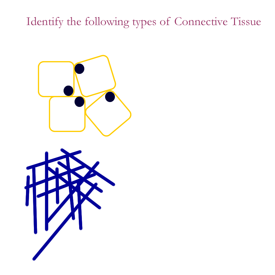
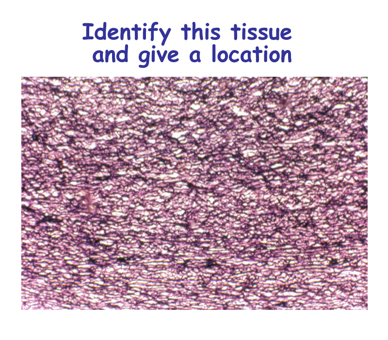
Connective Tissues
Connective tissues from a large group of tissues that are found throughout the body.
All connective tissues are comprised of three parts:
specialized cell(s)
fiber(s)
a fluid called the ground substance
The cells of connective tissues usually secrete or make the fiber and fluid.
Sometimes the fluid and fiber are combined to form the matrix.
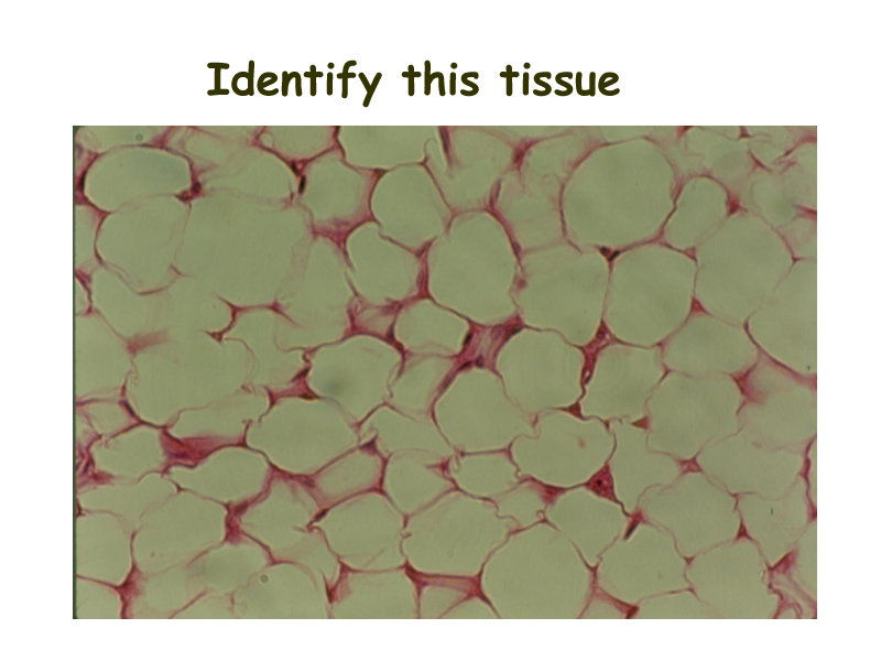
Connective tissues can be classified into several major groups and subgroups:
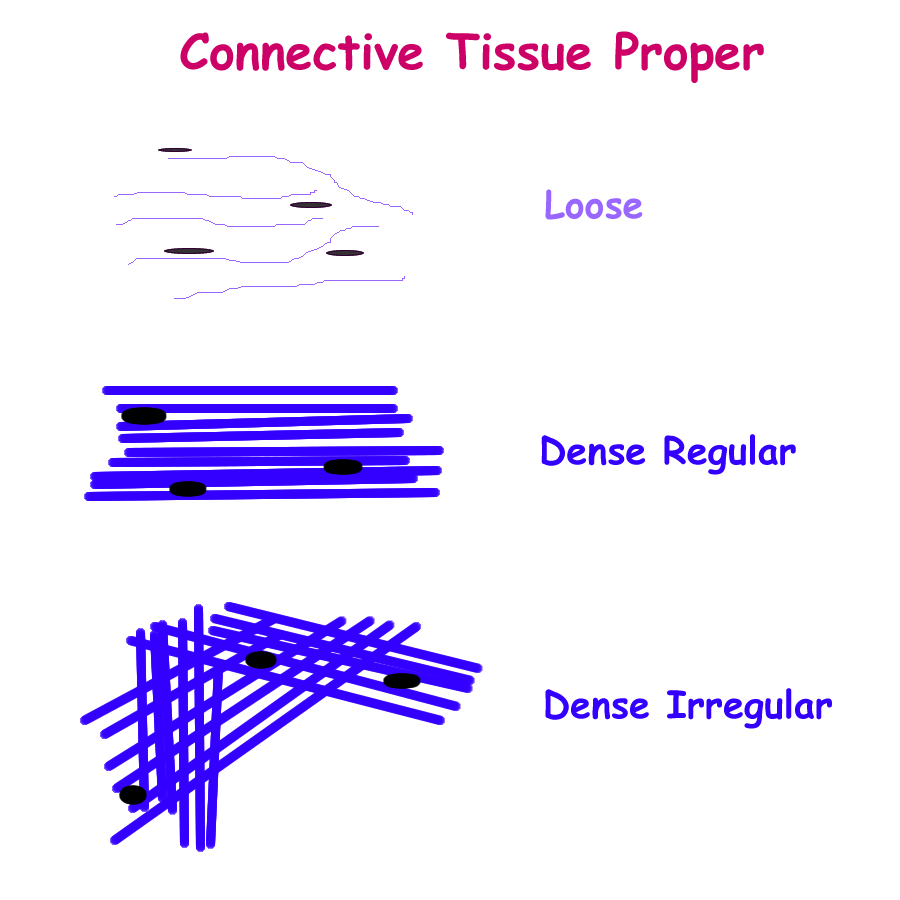
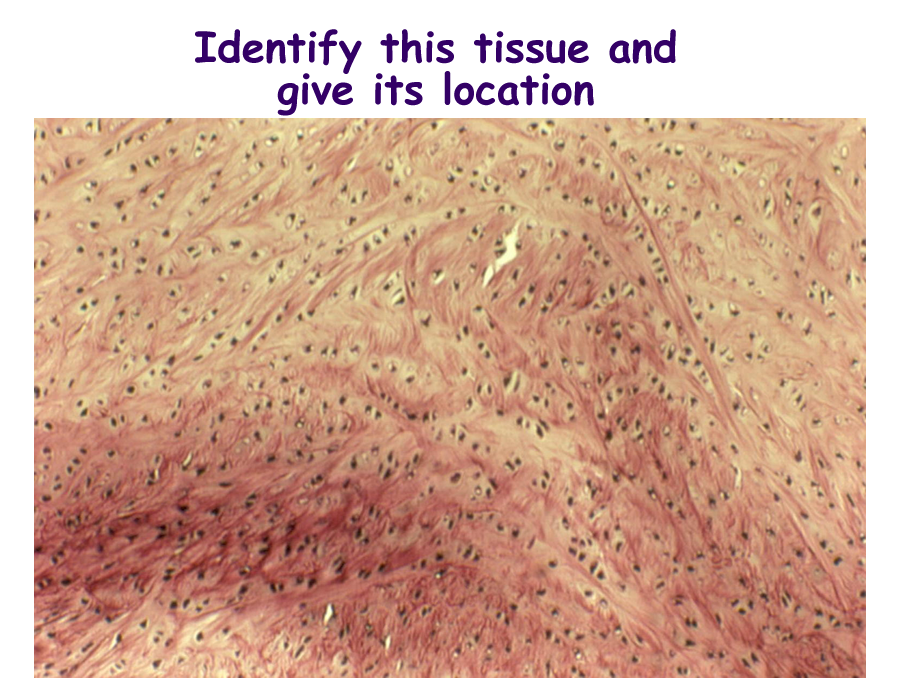
* Connective Tissue Proper
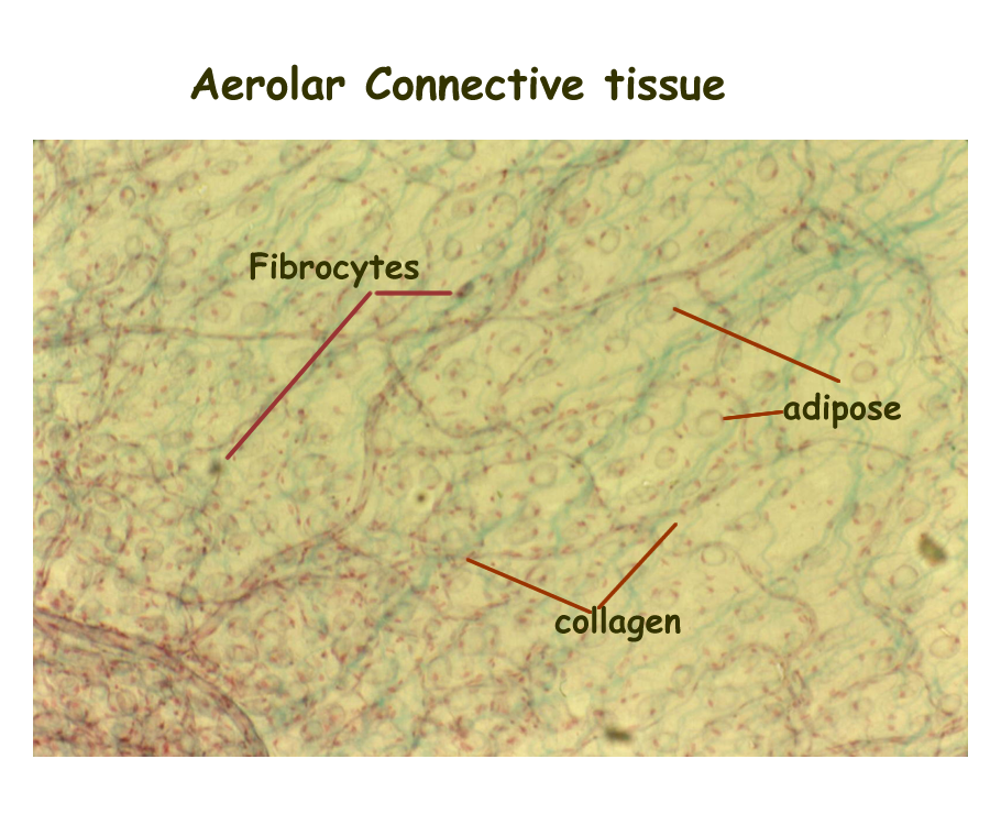
- Loose or aerolar
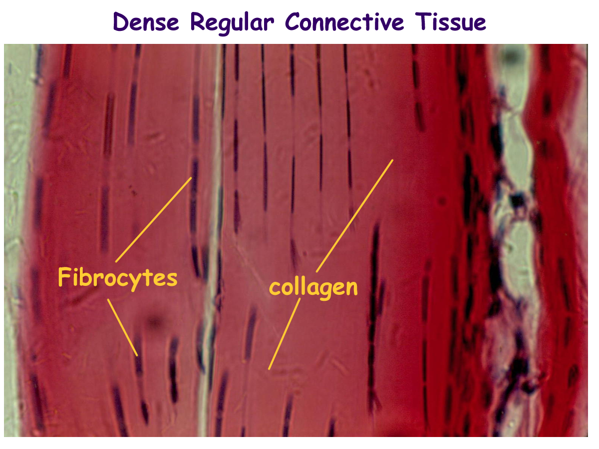
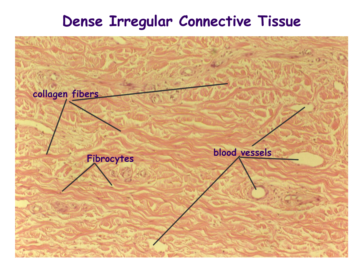
- Dense: regular and irregular
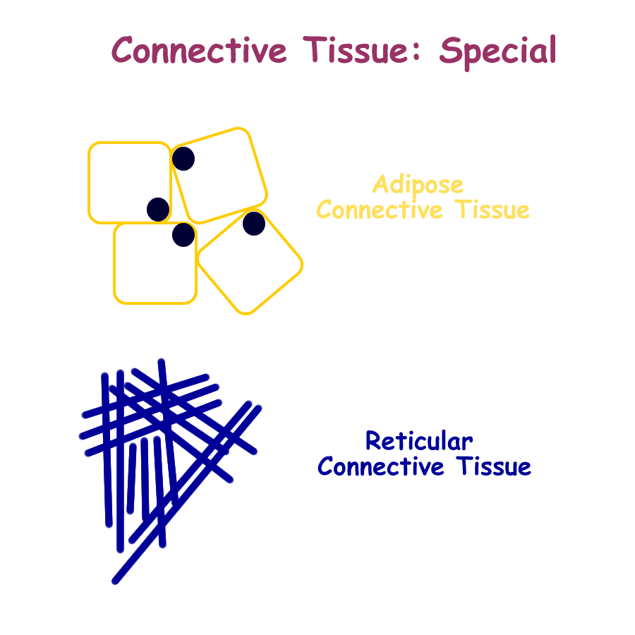
- Specialized: adipose, reticular, pigmented
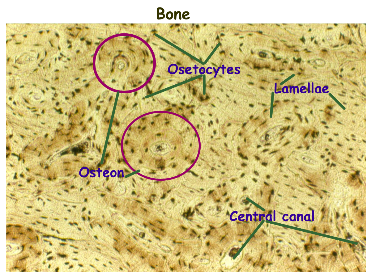
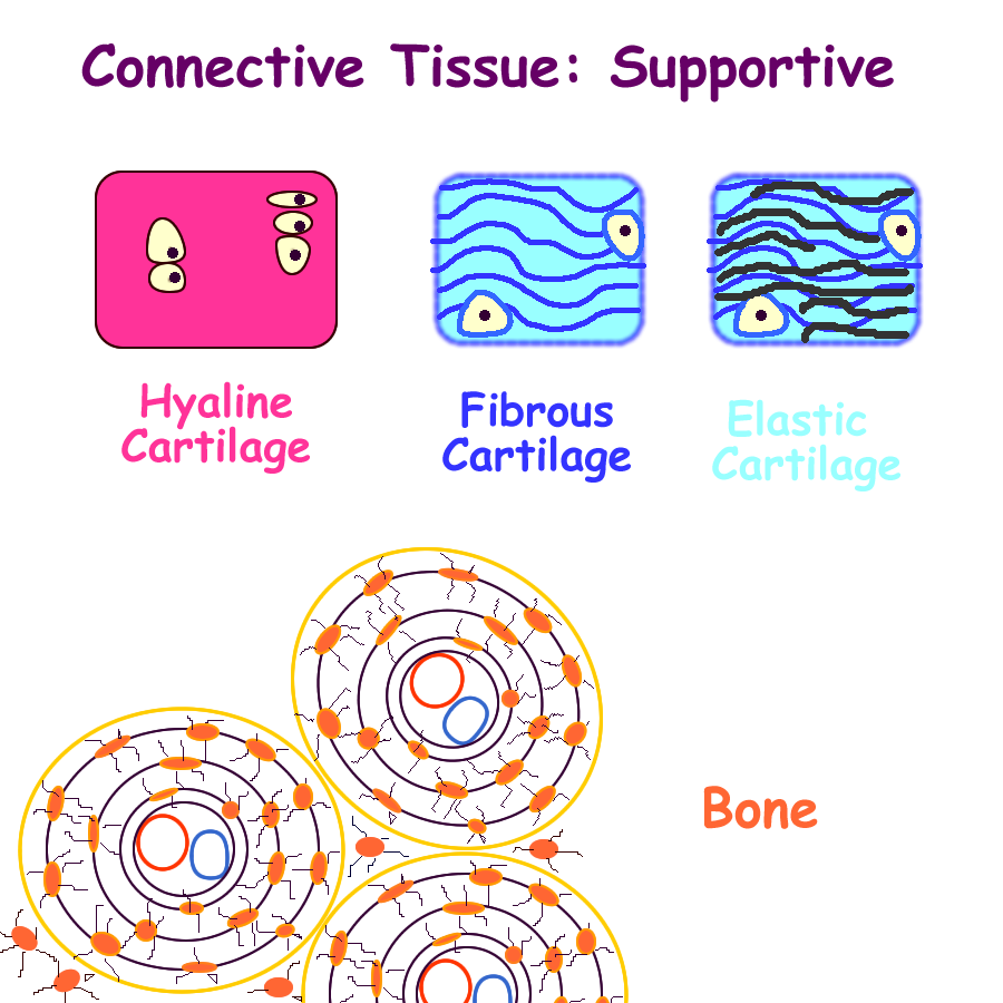
* Supportive
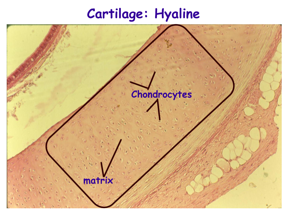
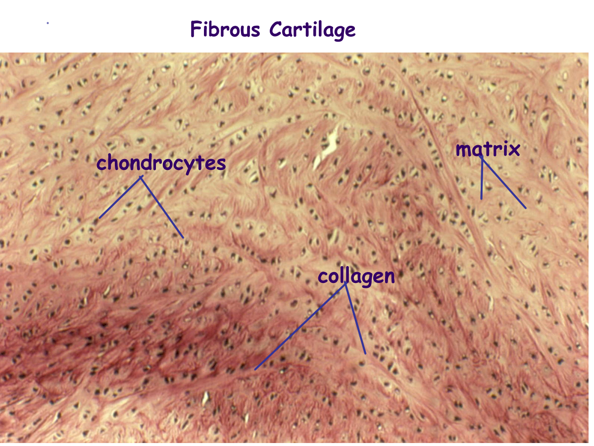
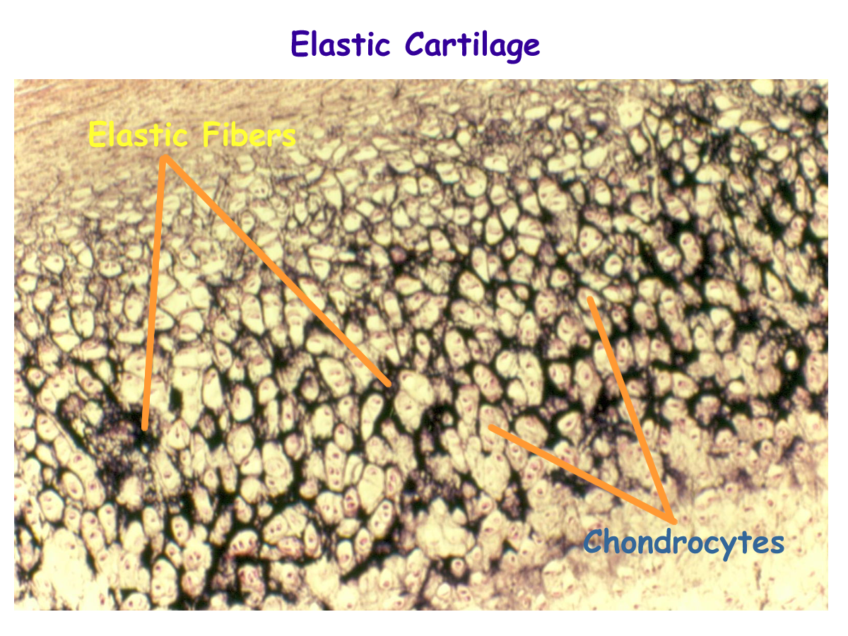
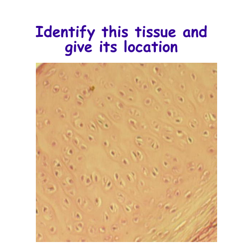
- Cartilage: hyaline, fibrous, elastic
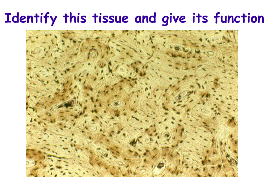
- Bone
- Dentin
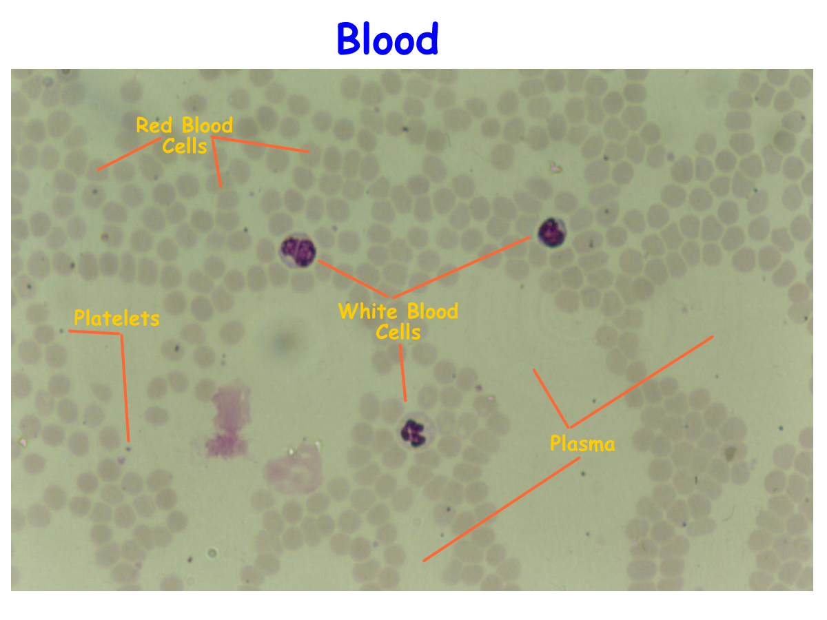
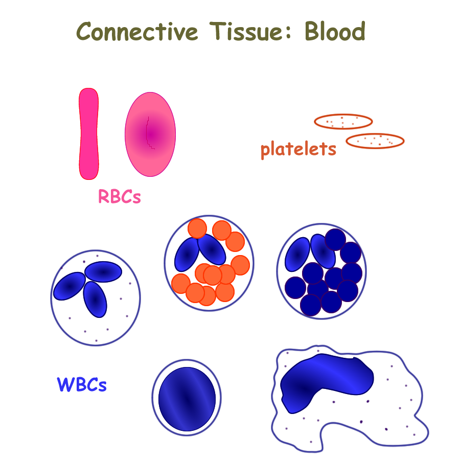
* Blood
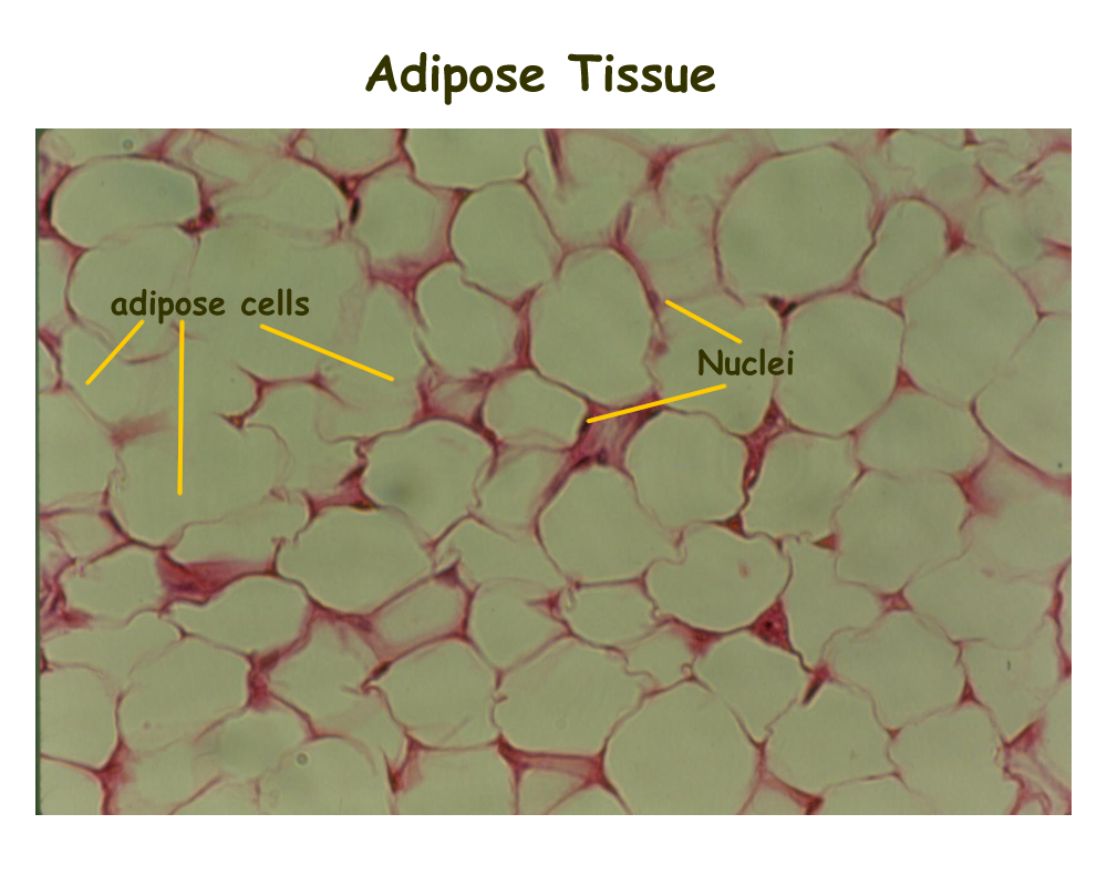
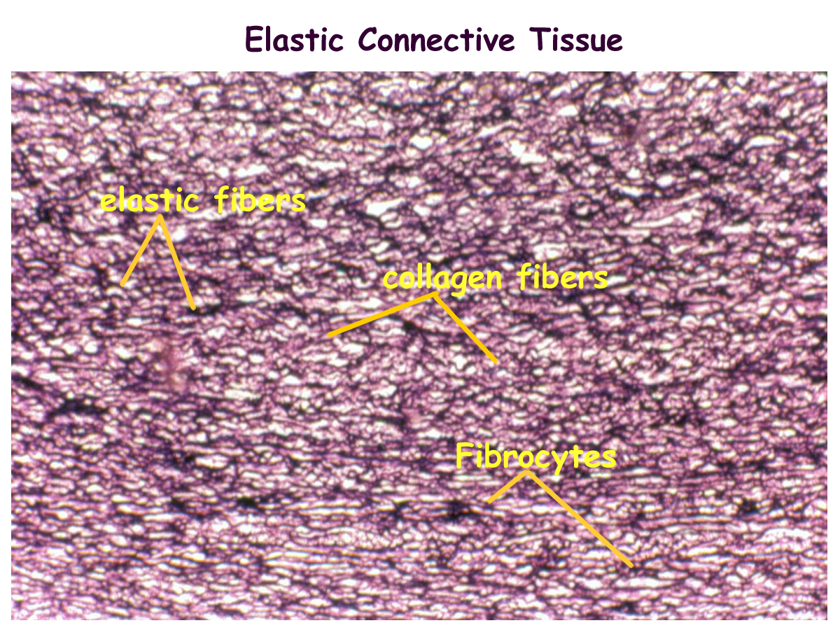
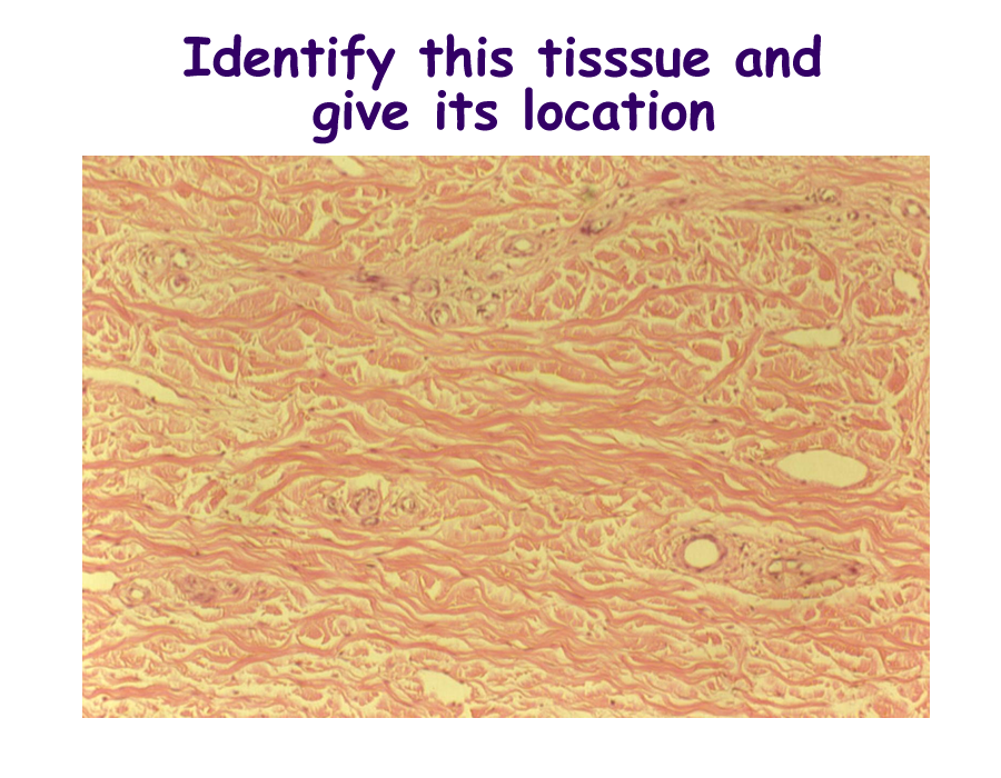
In the connective tissue proper group, the main cell is the fibroblast. In the case of adipose or fat, the main cell is the adipocyte. The major fiber is collagen.
Elastin can also be found in connective tissue and provides stretch when needed.
Loose or aerolar tissue provides a filler to prevent spaces between tissues.
Dense regular tissue form tendons and ligaments and dense irregular is found in the dermis of the skin.
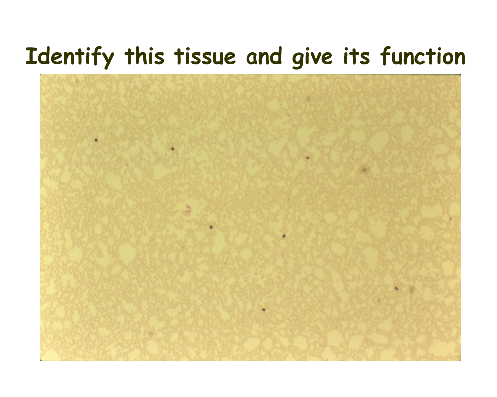
Adipose tissue is found in the subcutaneous region and around most internal body organs.
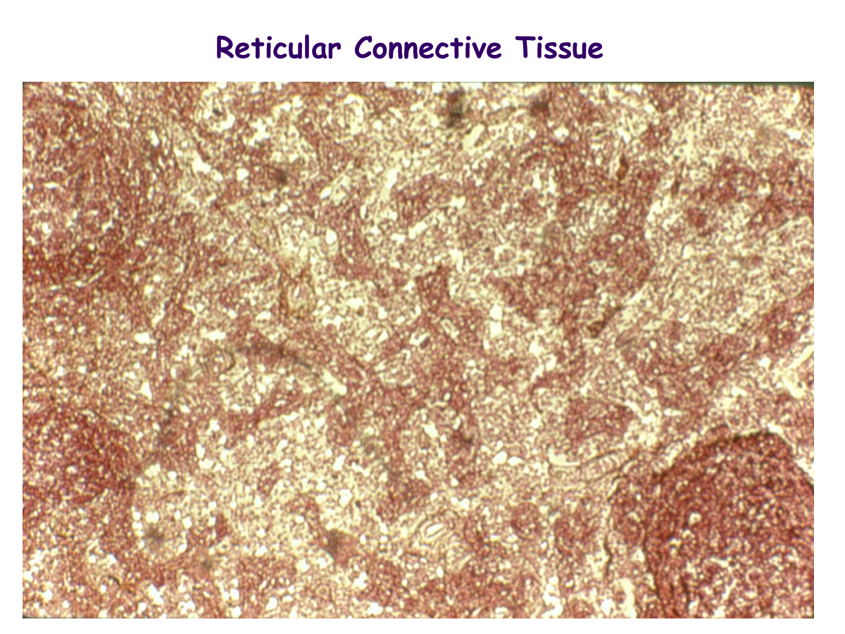
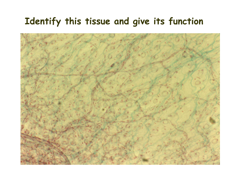
Reticular connective tissue is found in the lymphatic organs such as the liver, spleen and lymph nodes.
Pigmented tissue is reticular tissue with melanocytes and is found in the eye.
In the supportive connective tissue group, the primary bone cell is the osteocyte.
Others include osteoclasts and osteoblasts. The cell for cartilage is the chondrocyte.
The cell for dentin is the odontoblast and is found in teeth.
The fiber for bone, cartilage, and teeth is collagen. Elastin may also be found in elastic cartilage. There is just a small amount of fluid or ground substance, but dissolved substances are important to joint health. Some substances found in supportive connective tissues are chondroitin sulfate, hydroxyapetate, hyaluronic acid, and glucosamine.
You may recognize some of these substances in the joint health medications that are available in health food and other stores.
In blood, all cells come from a precursor called the hematocytoblast or pluripotent hematopoietic stem cell. This cell resides in the red bone marrow and makes the formed elements (cells) of blood. Formed elements make up approximately 45% of blood. These are red blood cells (erythrocytes), white blood cells (leukocytes), and platelets (thrombocytes). The fiber for blood is called fibrin and is important in the clotting mechanisms. The ground substance for blood makes up 55% of blood and is called plasma. Plasma has dissolved substances such as proteins, wastes, ions, gases, hormones, and enzymes all found in the solvent, water.
Histology of CT
Drawings of CT
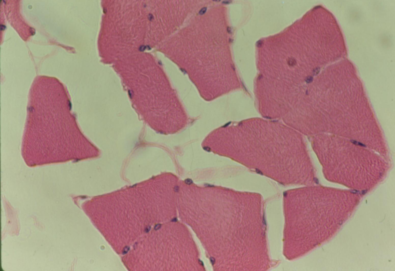
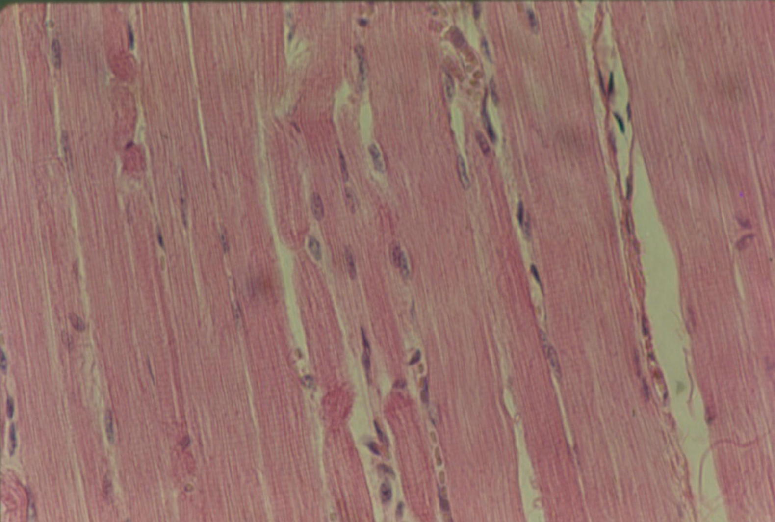
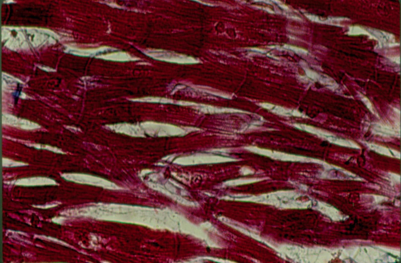
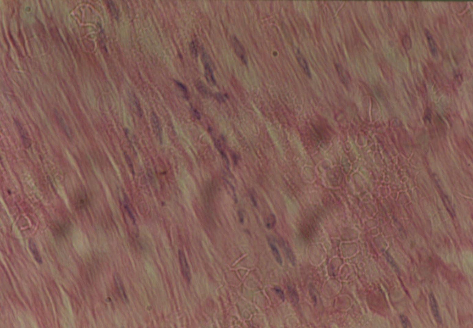
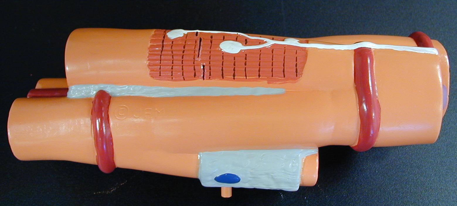
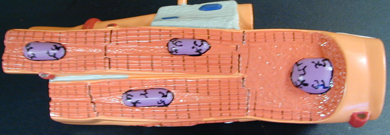
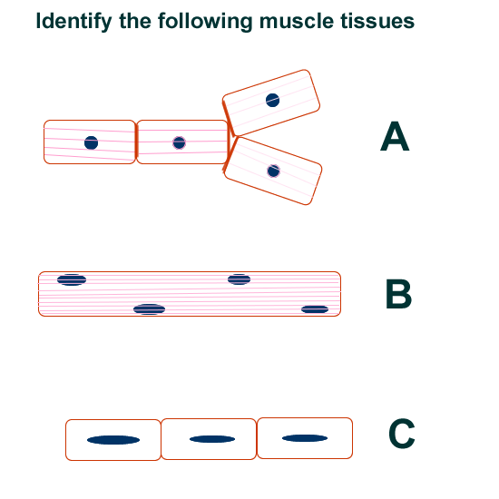
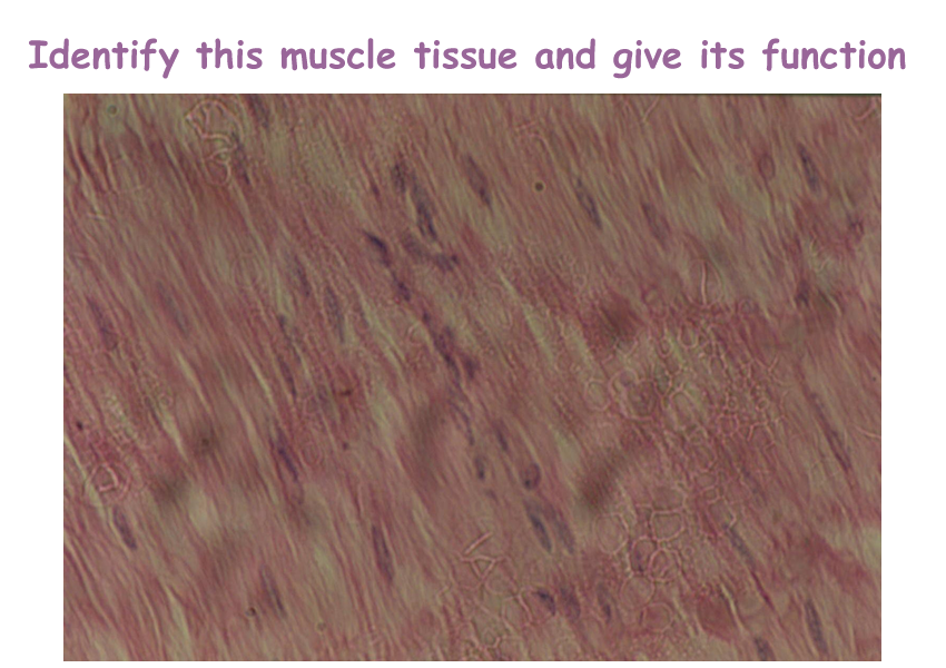
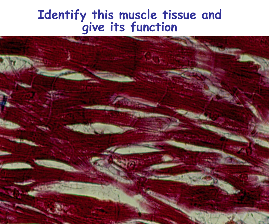
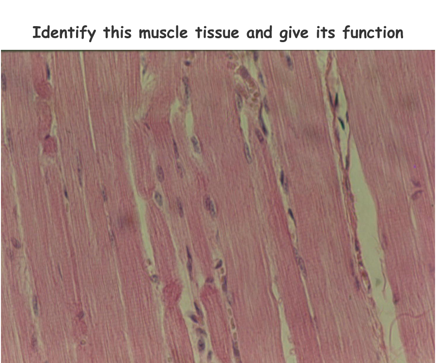
Muscle Tissue
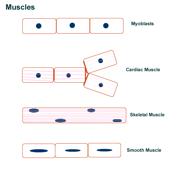
All muscle tissue comes from the embryonic precursor called the myoblast and then further develops into the three types of muscle: skeletal, cardiac, and smooth. All muscle shortens to produce a contraction, usually for movement. Depending on the location, the muscle will move related underlying structures such as bone or soft tissues.
Location and histology help differentiate the muscle groups.
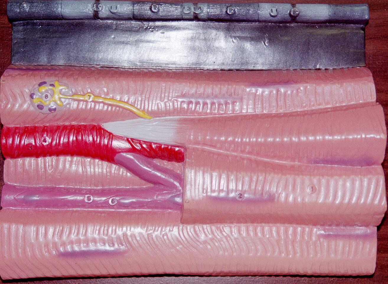
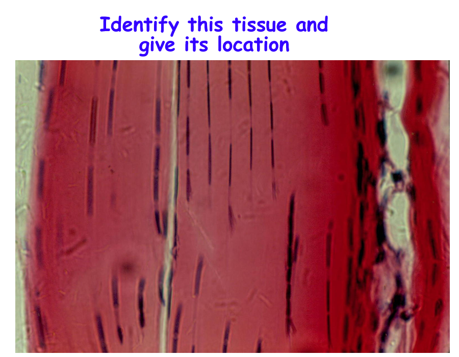
Skeletal located on bones multinucleated, striated
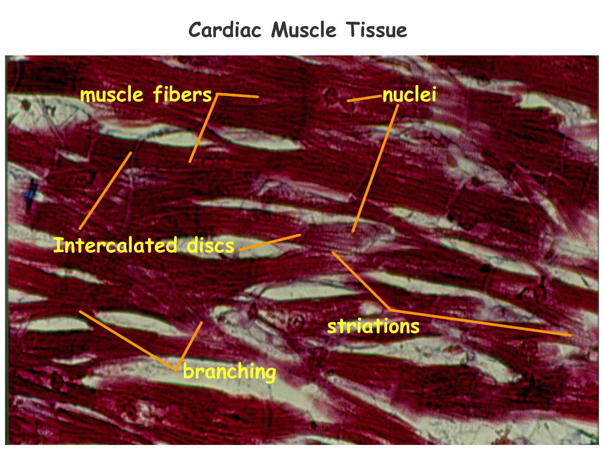
Cardiac located in the heart single nucleus, striated
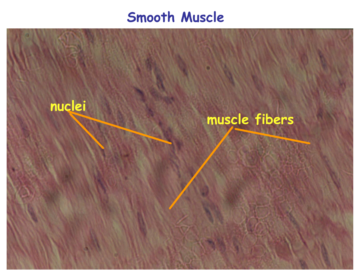
Smooth located in tubular organs single nucleus, non-striated
Muscles are also controlled and regulated by the nervous system. Skeletal muscle cannot contract without the nervous system signals. Cardiac and smooth muscle have internal specialized cells that signal their contraction. However, the nervous system still can regulate these particular specialized cells in order to provide feedback for homeostasis.
Cardiac muscles have another specialized structure called the intercalated disc. These are cell to cell membrane junctions that provide windows or holes between the cardiac muscle cells that allow the individual heart muscle cells to act as one unit.
Muscle can also be classified based on the nervous system controls. Since skeletal muscles must be controlled by the nervous system, it is called voluntary muscle.
Since cardiac and smooth muscle have internal controls that are modified by the nervous system they are referred to as involuntary muscle.
Voluntary muscle has conscious control and involuntary muscle has subconscious control, although through biofeedback, we can learn to control these “involuntary” muscles.
More discussion on individual muscle types will be available in specific chapters and exercises.
Functions of muscles
Skeletal move bones at joints, provide support for joints,
heat production
Cardiac pump (move) blood
Smooth move fluids for the cardiovascular, digestive, urinary, reproduction and change diameter of tubular organs for those systems listed as well as respiratory and the special senses. In order to be able to do this, smooth muscle is usually in two layers that run perpendicular to each other.
Histology of muscle tissue : Skeletal, Cardiac, Smooth
Drawings of Muscle tissue (Myoblast to muscle groups)
Muscle models of histology : Skeletal and Smooth
Enlarged muscle histology models
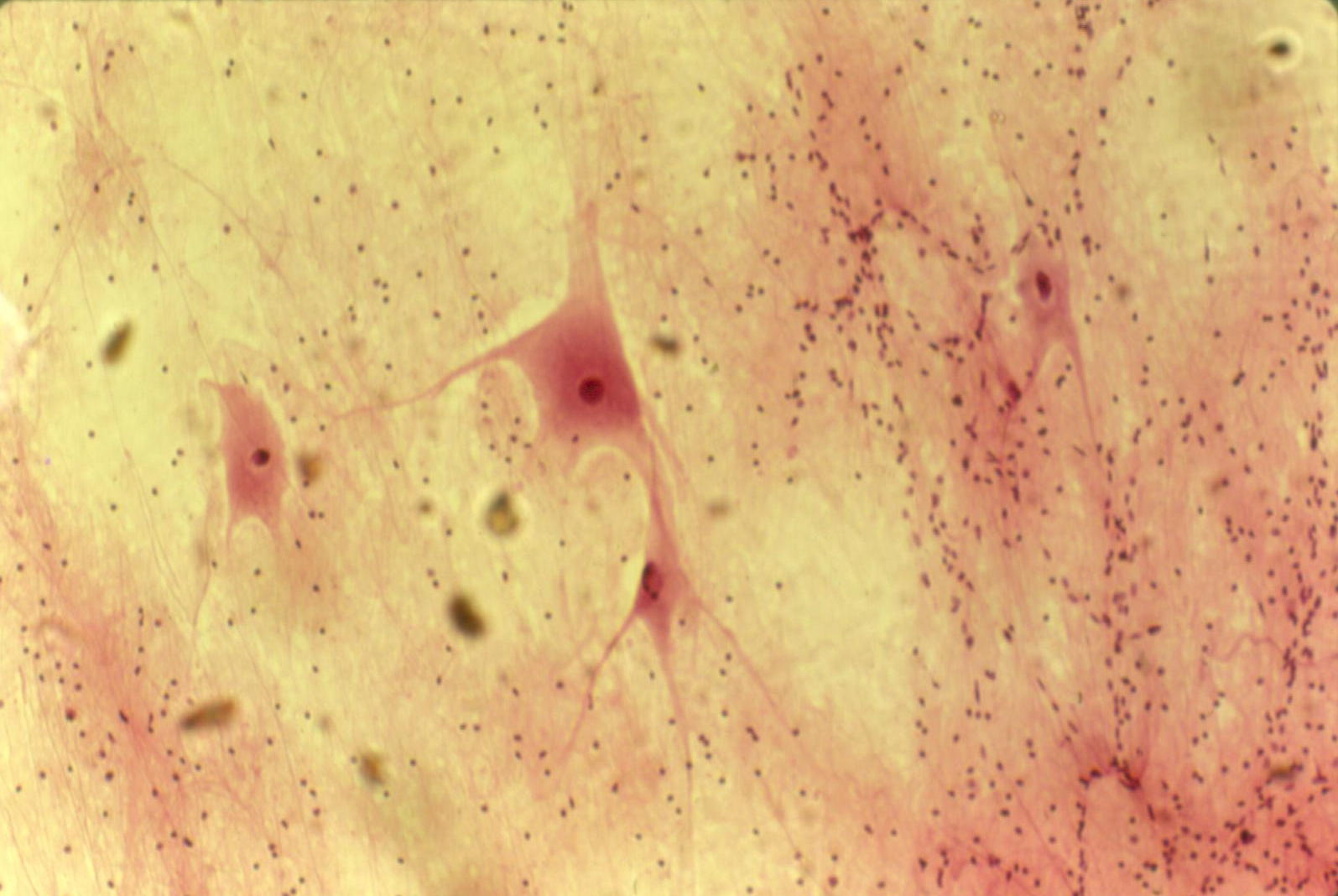
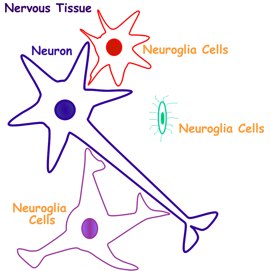
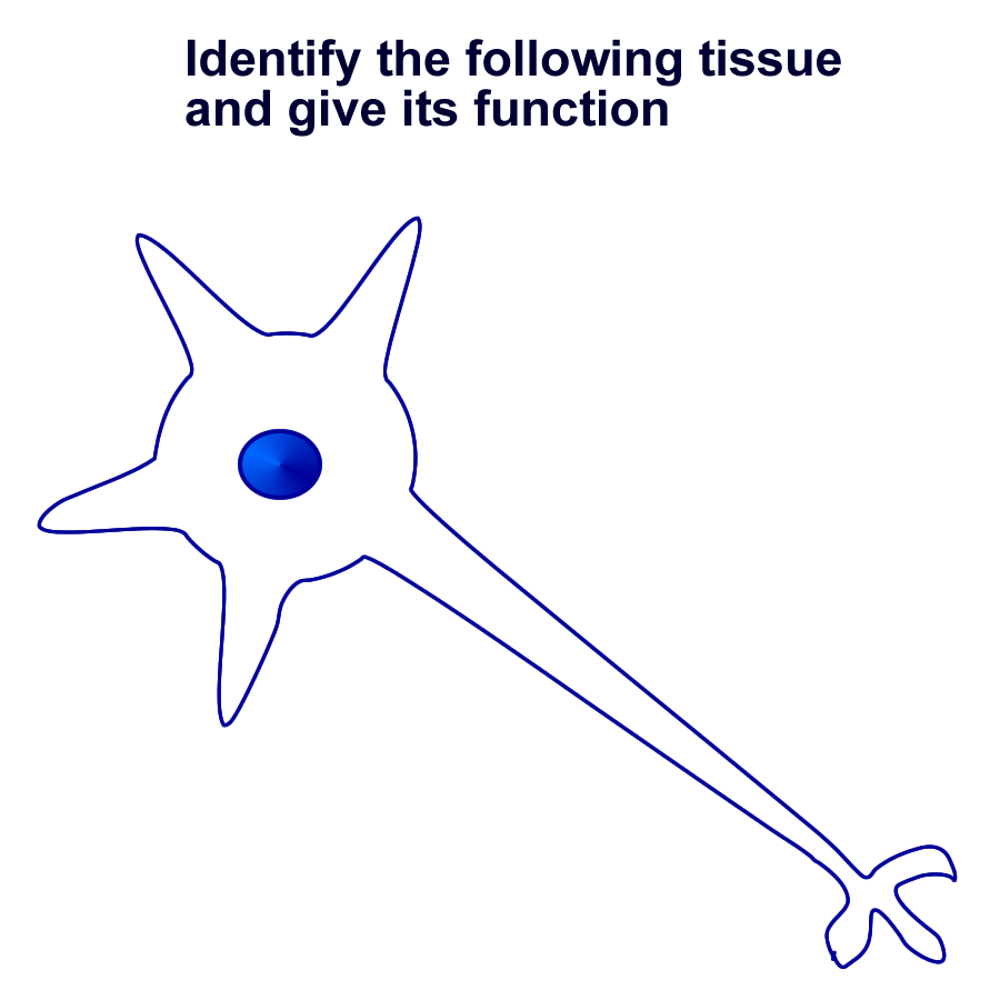
Nervous Tissue
Nervous tissue come from the embryonic tissue called neuroectoderm. It then develops into one of two major groups: neurons or neuroglial cells.
Neurons are specialized nervous tissue for communication. They are classified based on anatomical shape and function. Neuroglial cells are supportive cells and function primarily to insulate and protect neurons.
More discussion will be available in specific nervous system chapters and exercises.
Histology of neuron and neuroglial cells
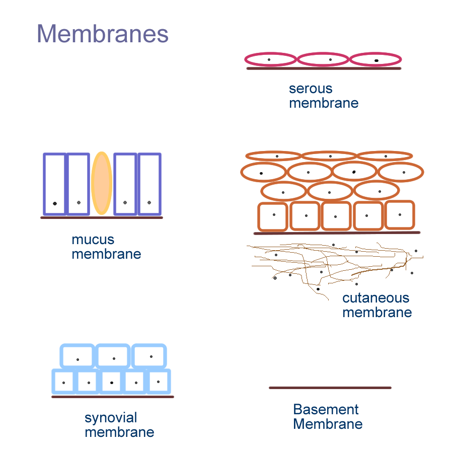
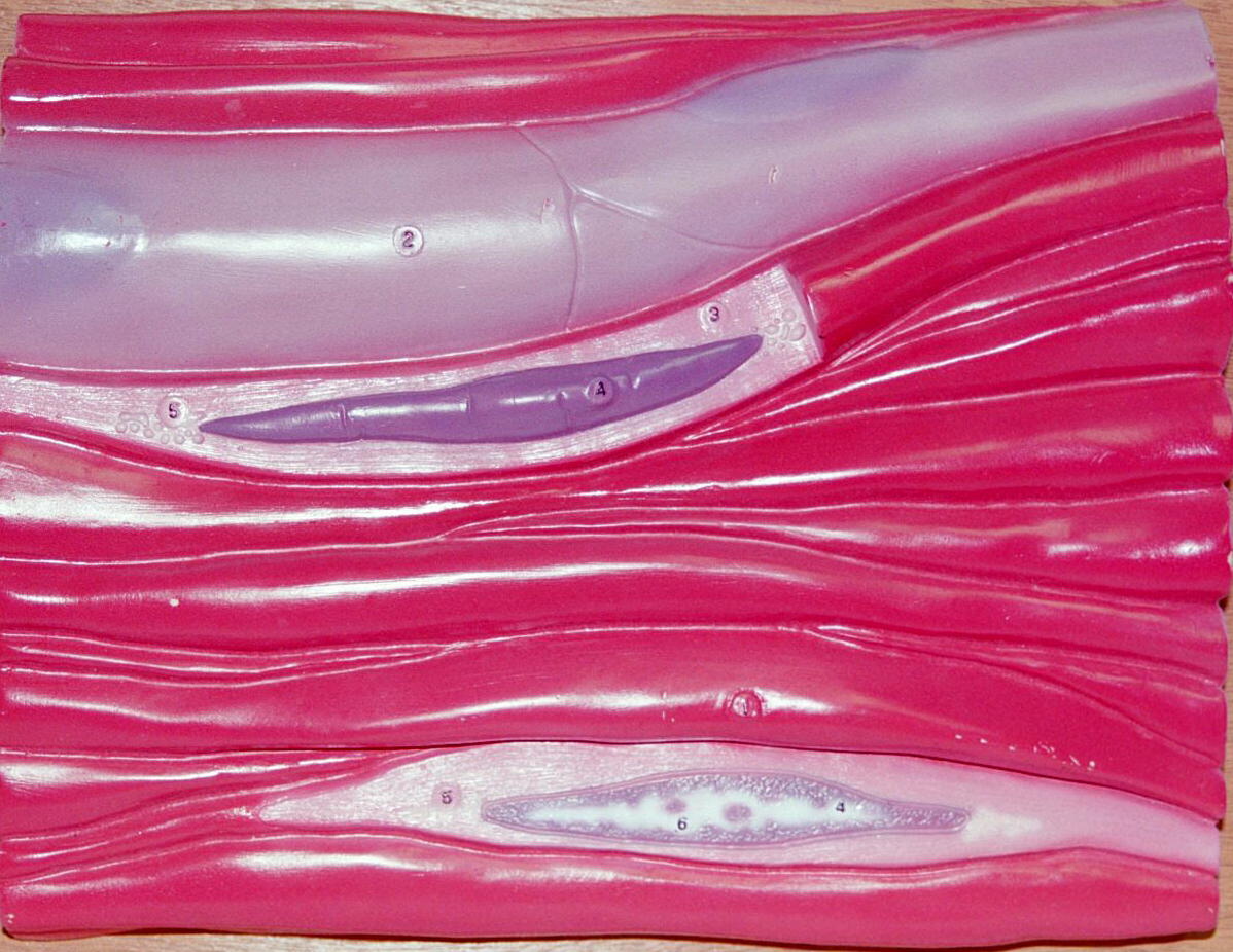
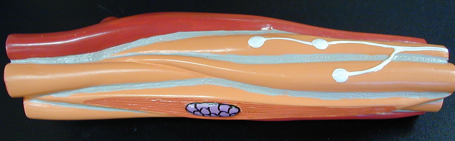
Membranes
Membranes are epithelial tissues with an underlying connective tissue support that
provides a lining that covers surfaces or can separate organs from other parts.
The five types of membranes are:
All membranes provide some type of secretion that gives them their function.
Basement membrane secretions provide adhesive forces to allows for diffusion of nutrients and wastes
Cutaneous membrane is also known as the skin. Secretions come fro Exocrine glands located in the dermis and helps to provide immune or defensive properties.
Mucus membranes secrete mucous and line open body cavities as the digestive, respiratory, urinary, reproductive systems. Mucous provides protection.
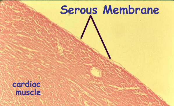
Serous membranes secrete serous fluid and line closed body cavities such as the dorsal and ventral body cavities.Serous fluid decreases friction between organs.
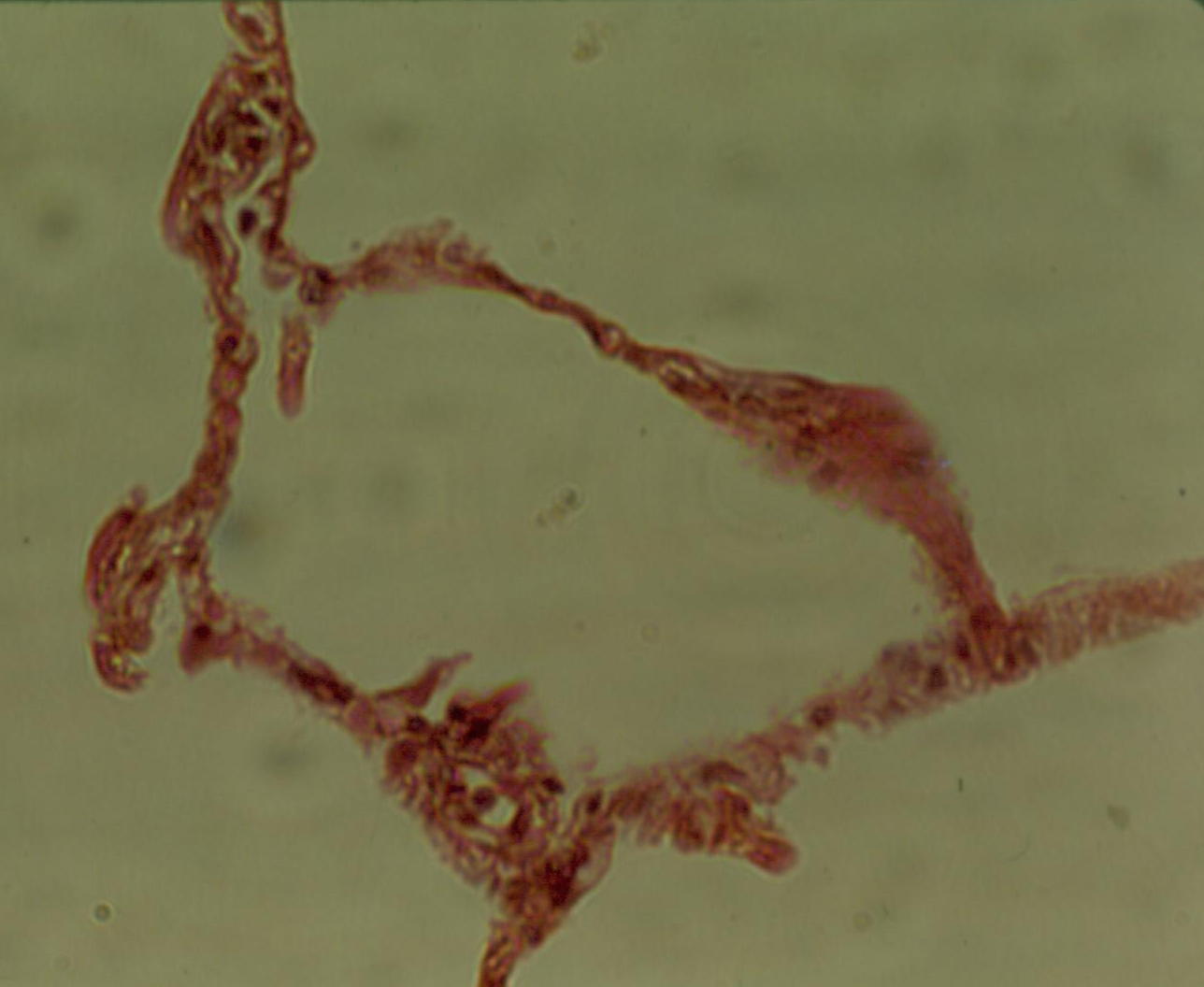
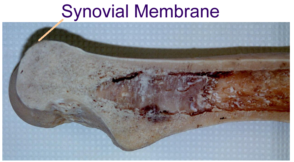
Synovial membranes secrete Synovial fluid in a joint capsule that lines moveable joints to provide nutrients to cartilage.
Histology
Synovial Membranes (joint specimen)
Hist/o- tissue adip/o- , lip/o- fat
Duct- lead blast- precursor
Aden/o- gland cardi/o- heart
Epi- on chondr/o- cartilage
Hidr- sweat -clast broken
-crine to separate odont/o-, dens tooth
lact/o- milk endo- inside
galact/o- milk hemat/o- blood
sial/o- saliva hyal/o- glass
myo- muscle neur/o- nerve
osteo- bone thromb/o- clot
ID drawings: Epithelial, Connective, Muscle, Nervous
ID histology:
Epithelial: Image One, Image Two, Image Three, Image Four
Connective Tissue:
CT Proper: Image One, Image Two, Image Three, Image Four, Image Five
Supportive and Other: Image One, Image Two, Image Three, Image Four, Image Five
Muscle: Image One, Image Two, Image Three
Compare and Contrast Table: Make a table comparing all 4 major tissue types with regards to cell type, function, location, repair (regenerate or scar tissue), and give 3 examples of each major tissue type.
Concept
Map: Make a concept map of the major tissue groups and their subtypes,
locations, and functions.
Include this map in your LAR lab report (if done) as an insert into the word
document or a PDF scan as an additional email attachment.
Construct a "new" organ using household items for each of the four tissues. Describe what you used and its function in your new organ. Send this description (photo optional) for extra credit (+5 points) on tissue lab quiz.
Biopsy punch
Needle biopsy
SX biopsy
Tissue culture
Specimen collection
Regeneration vs Scar tissue
Adhesions
Stem Cell research
Pathologist
Surgeon
Surgery Tech
Internal Medicine
Medical Examiner
http://www.udel.edu/Biology/Wags/histopage/colorpage/colorpage.htm
http://www.udel.edu/Biology/Wags/histopage/empage/empage.htm
http://www.udel.edu/Biology/Wags/histopage/illuspage/illuspage.htm
http://www.med.uiuc.edu/histo/medium/atlas/slides.htm
http://www-medlib.med.utah.edu/WebPath/HISTHTML/EM/EM.html
http://www.medinfo.ufl.edu/year1/histo/
http://www-medlib.med.utah.edu/WebPath/ORGAN.html#1
http://www.science.ubc.ca/~biomania/tutorial/skin/outline.htm
http://www.science.ubc.ca/~biomania/tutorial/exogland/outline.htm
http://www.kcmetro.cc.mo.us/maplewoods/Biology/Bio110/Labs.htm
1. Name the embryonic or germ tissues that form adult tissues.
2. Name the four major tissues and give their general function.
3. Define tissue and give two diagnostic procedures that use tissues.
4. Define membrane and give the five major groups.
5. Give three examples of epithelial tissue and give the location, and function.
6. Give three examples of connective tissue and give the location, and function.
7. Give two examples of muscle tissue and give the location, and function.
8. Give two examples of nervous tissue and give the location, and function.
9. Name the two types of epithelial glands and give their location and function.
10. Define the following terms :
a) adhesion
b) biopsy
c) scar tissue
d) regeneration
e) histology
f) embryology