Biology 2404 A&P Basics Lab Exercise 9 Skeletal System Dr. Weis
| Objectives | Background | Medical Terms | Activities | Applications | Careers | WWW | Review Questions |
Students should be able to
* List the functions of the skeletal system
* Describe the histology of bone
* Name and identify the bones of the axial skeleton
* Name and identify the bones of the appendicular skeleton
* Name and describe the types of joints in the body
* List the components of a Synovial joint and their function
* Define related word parts and medical terms
Read related information in textbook
The skeletal system is formed from connective tissue mesoderm and functions to
Provide a lever system for movement
Protection for internal body organs
Store minerals
Support the body’s internal framework
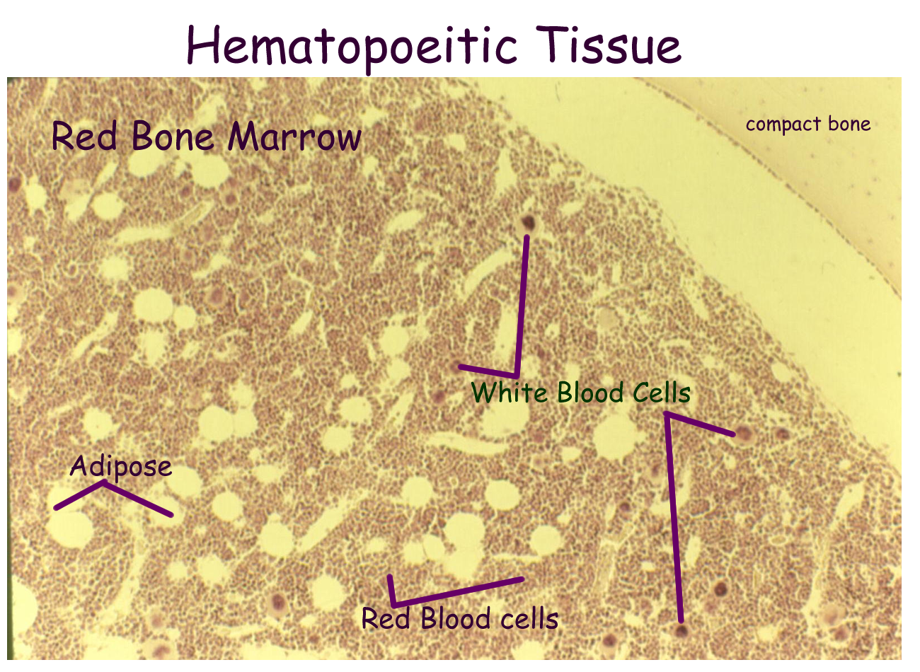
Provide a site for blood cell formation
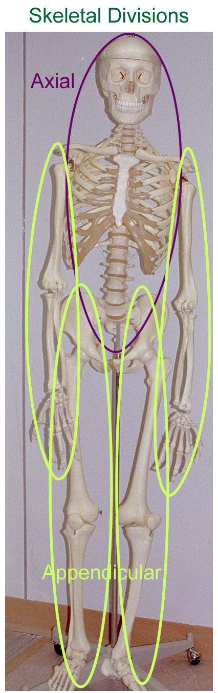
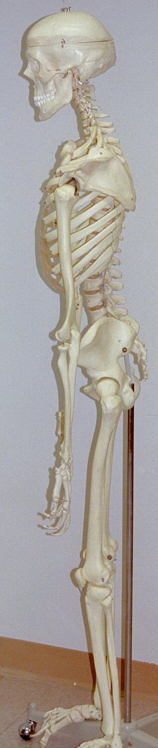
The skeletal system is divided into two parts: axial skeleton and appendicular skeleton.
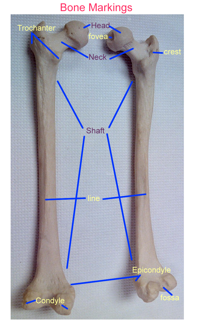
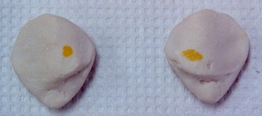
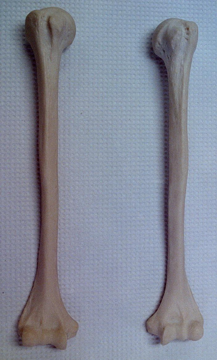
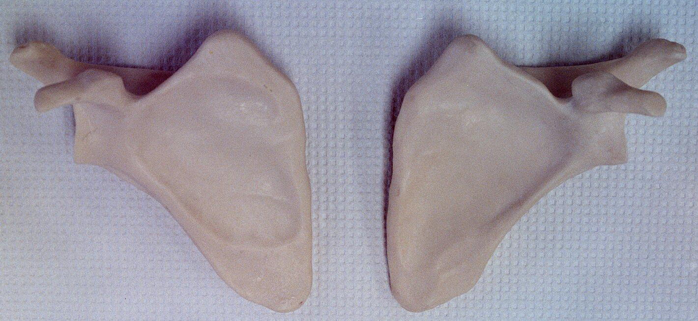
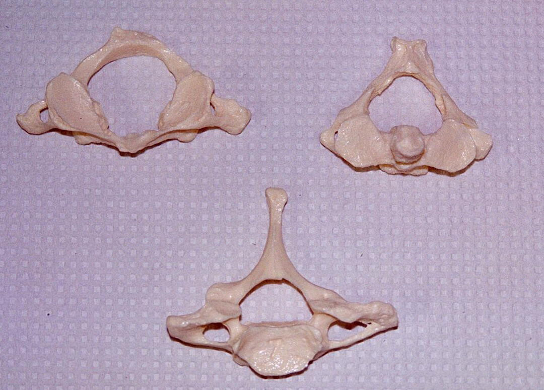
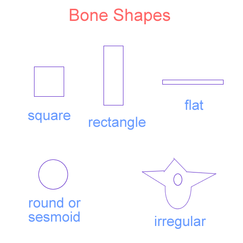
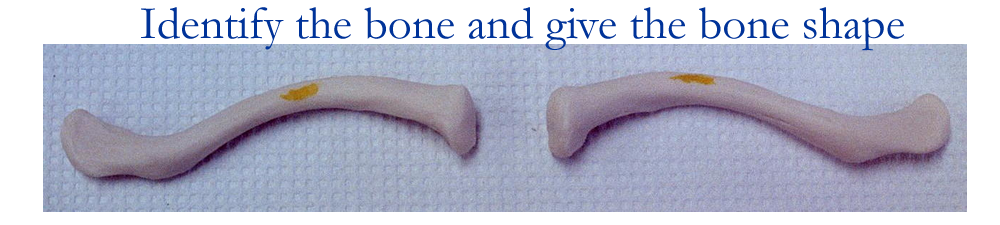
The bones that make up the skeleton can be one of five distinct shapes: square, long, round, flat, and irregular. Bones also have distinct markings that help identify them.
Examples of these markings are:
Head rounded articular process at the proximal end of a bone
Condyle rounded articular process at the distal end of a bone
Epicondyle a small raised area above a condyle for joint capsule attachment
Foramen a short passageway through bone for vessels and nerves
Meatus a long canal like passageway
Fossa a depression in bone
Sinus a cavity in bone lined by a mucous membrane
Tuberosity a large rounded projection for muscle attachment
Tubercle a small rounded projection
Fissure a slit like opening through bone
Associated structures that are seen with bone are cartilage, tendons, and ligaments.
Cartilage is the basis for most bone formation and remains at the ends of many bones to help form joints. Tendons are epimysial connective tissue extensions of muscle that attach to bones. Ligaments are periosteal connective tissue extensions of bone that help join bone to bone.
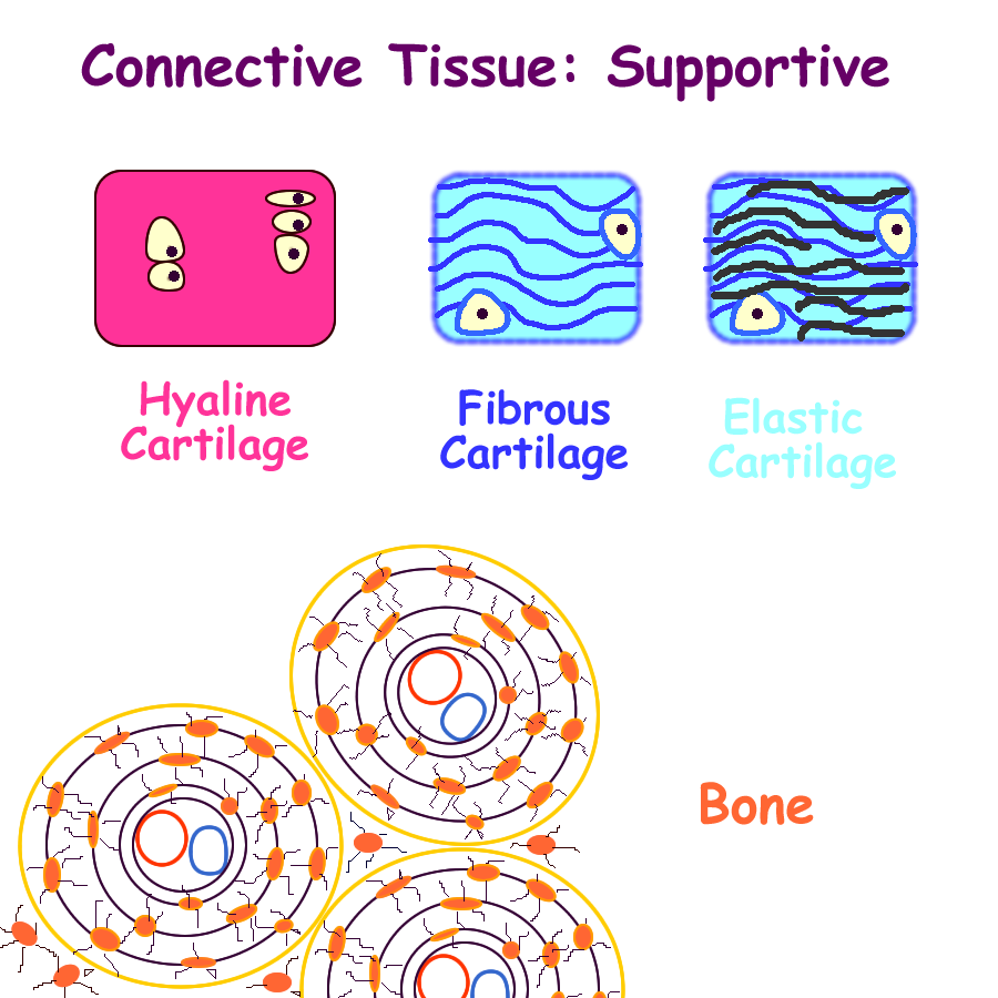
Drawing of Supportive Connective Tissues
Classification of Cartilage
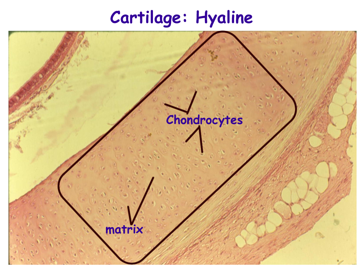
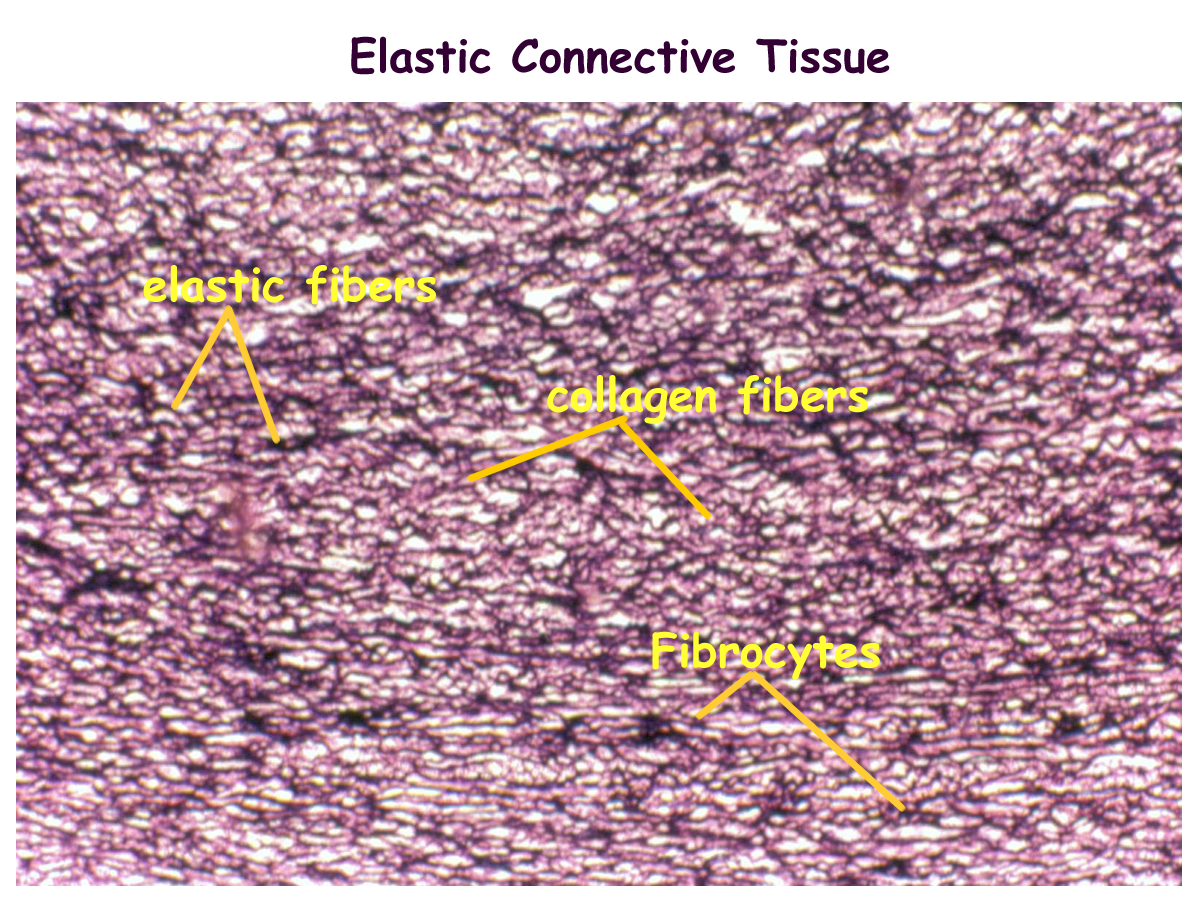
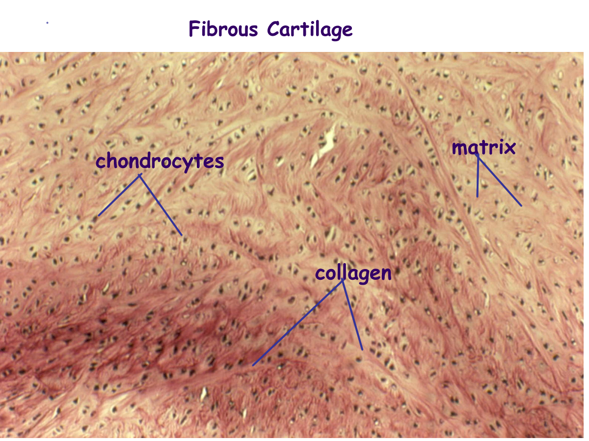
Cartilage cells or chondrocytes produce collagen fibers and proteoglycans such as chondroitin sulfate to form the cartilage matrix that create the three types of cartilage.
Hyaline cartilage is the most abundant type of cartilage. It forms most of early fetal skeleton and will remain over articular surfaces of joints, in the respiratory system airways, and at the ends of ribs. Elastic cartilage contains elastic fibers within the collagen and is found in the ears and epiglottis of the larynx. Fibrous cartilage has thick layers of collagen and is found in areas of great weight bearing pressure such as the intervertebral discs and the menisci of the knee.
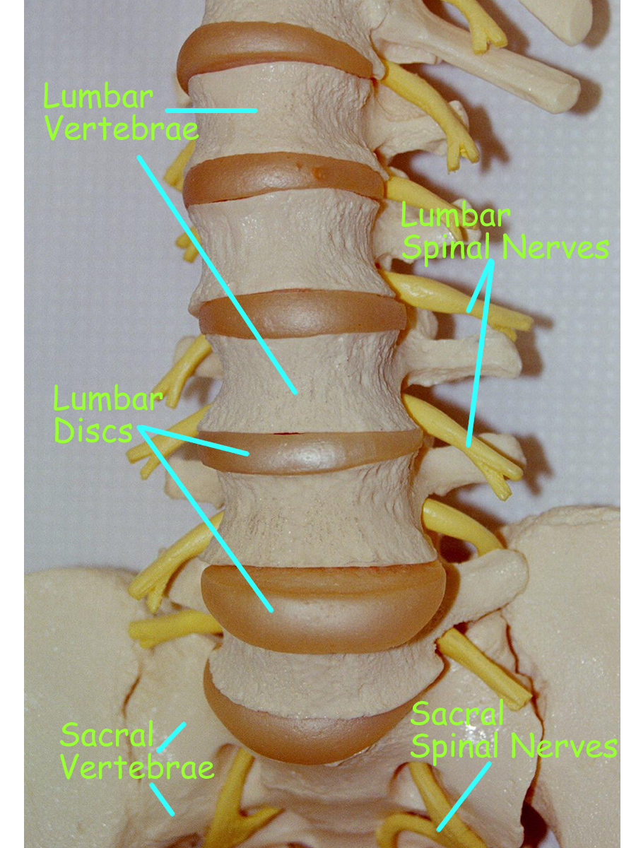
Intervertebral Disc Model : Lumbar
Classification of Bones
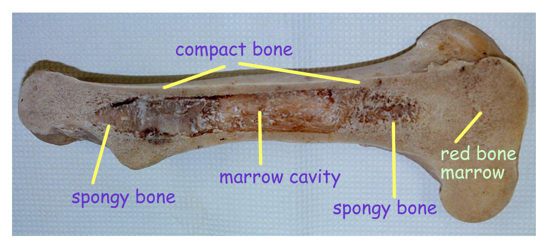
Bone shapes determine bone classification. For most bone shapes, there is a spongy arrangement internally and a compact bony arrangement externally. This arrangement is seen in short (square), round, flat, and irregular bones.
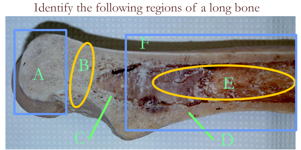
For long bones, the matrix arrangement is modified. Long bones do have spongy centers, but most of the center is hallowed out to create a space or marrow cavity. This cavity along the length or shaft of bone is called the yellow marrow cavity because it is filled with fat or adipose tissue.
The ends of long bones or the epiphyseal regions look just like the other bones with spongy filling the center and compact bone on the outside.
Bone Shapes
Formation of Bone
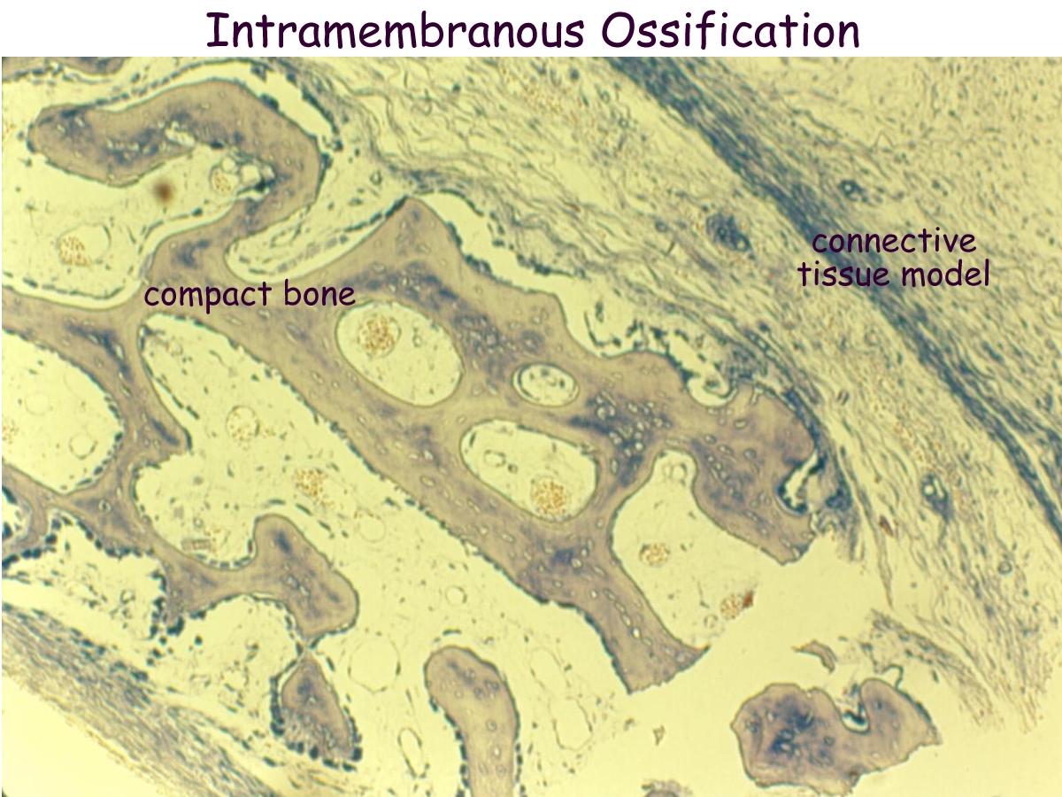
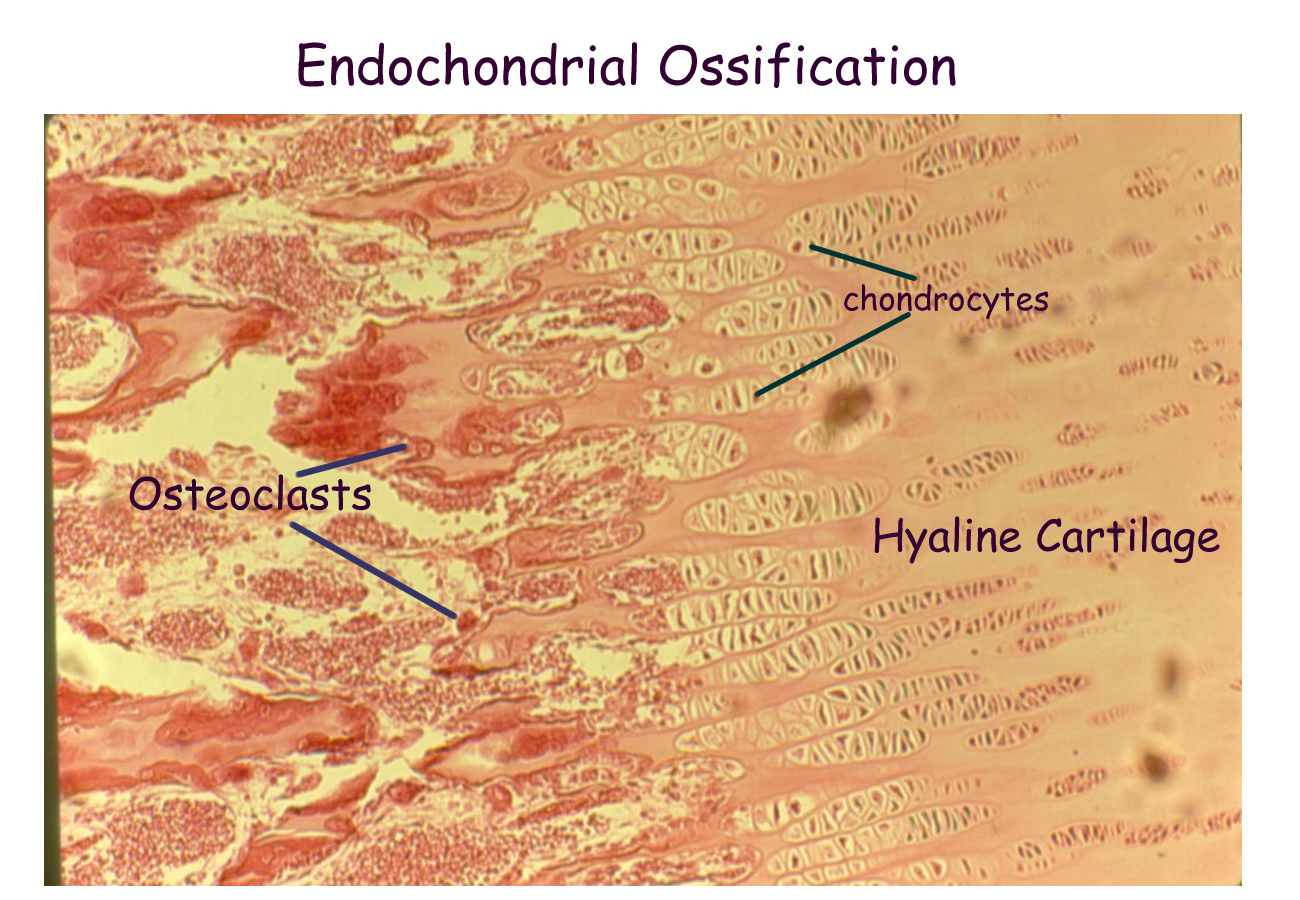
Bone formation is based on a connective tissue plate or model. Ossification is the process where one tissue is turned into bone. Precursor bone cells are deposited in the connective tissue matrix and begin secreting bone. This process is called intramembranous ossification. If cartilage cells are deposited on the connective tissue matrix and then later changed to bone, the process is called endochondral ossification.
Both occur in the fetus in order to develop the skeleton. Ossification steps will also occur in bone fracture repair.
The bone cells responsible for ossification are called osteoblasts. Remember that bone is a connective tissue and the cells secrete their fiber and ground substance fluid to form a matrix. The fiber for bone is collagen and the ground substance contains hydroxyapetate salts such as Calcium Carbonate and Calcium Phosphate in addition to other substances.
As osteoblasts lay down their foundation, the matrix can take one of two arrangements: compact or spongy. External forces such as gravity work on the fetus during development to help determine the matrix arrangement as well as shape and projections of individual bones.
All bones will have both an outer compact arrangement and an internal spongy arrangement.
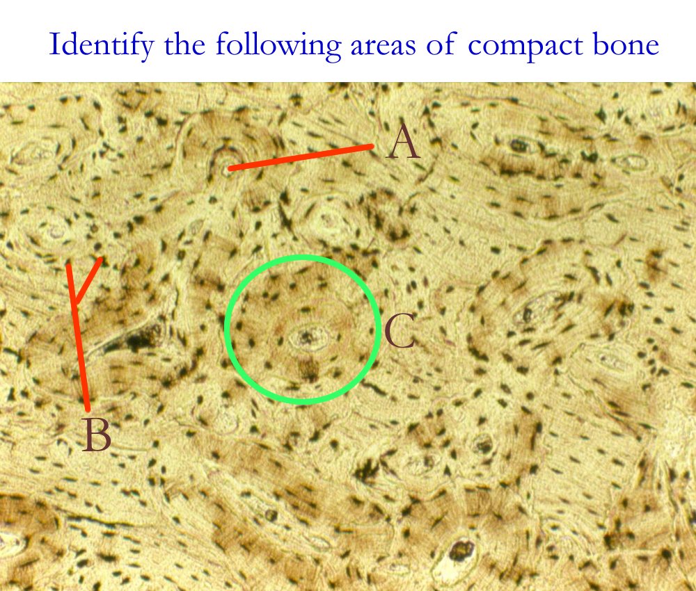
Compact bone is regularly arranged into functional units called osteons that makes it dense. Spongy bone has a lattice like arrangement called trabecular or cancellous. Blood supply is important to bone, since it is a living tissue and replaces itself yearly. Blood vessels will follow the structure of the bone, such as the Haversian canals seen in compact bone.
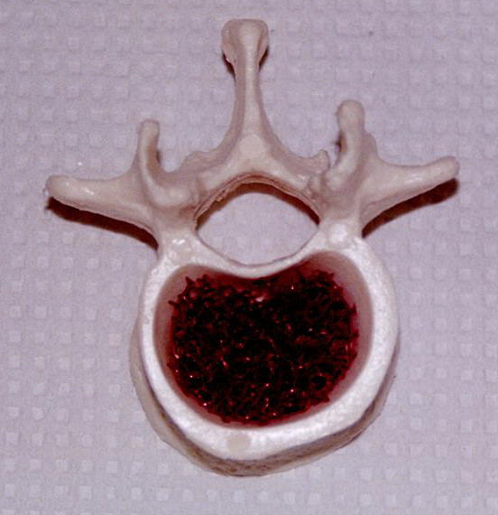
Red marrow containing blood precursor cells can be found in areas of some spongy bone.
Once the osteoblasts secrete its matrix, the bone cell is trapped. These cells then mature and are called osteocytes whose function is to maintain, repair, and replace the bony matrix.
As ossification occurs, there is still left over connective tissue and cartilage. The left over connective tissue is found both inside and outside the forming bone. It will become the endosteum and periosteum of bone. These connective tissue layers will house bone cells such as the osteoblasts and a modified white blood cell called the osteoclasts. The periosteum will also form the ligaments for bone. Osteoblasts and osteoclasts in these regions are under hormonal control by thyroid hormone, growth hormone, Parathormone, Calcitonin, and the reproductive hormones. Osteoblasts build bone while osteoclasts tear down and destroy bone. It is a dynamic process that is usually balanced, especially when dealing with calcium levels in the body.
The cartilage that is left at the ends of the bone helps in joint formation to provide a smooth surface, especially in moveable, Synovial joints.
Chemical analysis of bone reveals approximately 33% organic compounds, mainly collagen and approximately 67% inorganic compounds, mainly salts. The minerals found in the inorganic matrix of bone are as follows: 99% Calcium, 88% Phosphate, 80% Carbonate, 50% Magnesium, 35% Sodium, and 4% Potassium.
Bone Growth
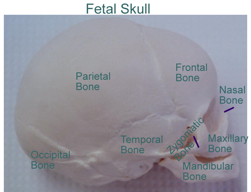
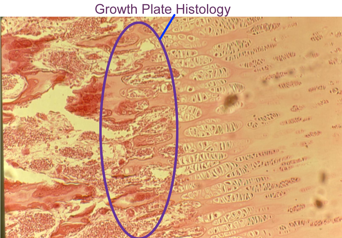
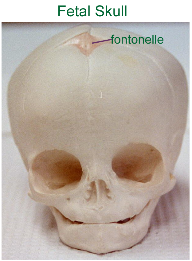
Bone growth can occur in two planes: length and width. Lengthening of bone occurs during the original ossification processes at growth plates or metaphyseal regions in bones that use endochondral ossification processes and at fontanels for bones that use intramembranous ossification processes. Both growth plates and fontanels have the original tissue model that is still being replaced. At growth plates for short, round, long, and irregular bones, cartilage is being replaced by bony tissue. At fontanel regions for flat bones, connective tissue is being replaced by bony tissue. Growth plates and fontanels will close based on hormonal signals, thus ending longitudinal growth.
Width or appositional bone growth can happen at any time in life. Factors that influence this type of growth are gravitational forces, stress burdens, injury, and weight changes.
Bone is deposited or removed at the periosteal and endosteal layers based on the new needs of the body in order to overcome the changing requirements for support and movement.
Bone Plate to repair broken Bone
Factors that affect bone
Although we will discuss these hormones again in the endocrine system, it is good to be aware of the hormones that affect osteoblasts and osteoclasts. These hormones are:
Growth Hormone signals osteoblasts at metaphyseal growth plates
Signals Chondrocytes at growth plates
Thyroid Hormone works with Growth hormone
Signals energy production and protein synthesis
Reproductive Hormones signal the closing of the growth plates
Helps retain calcium in bone to maintain strength
Calcitonin signals osteoblasts to build bone
use calcium from blood, lowers blood Ca++ level
Parathormone signals osteoclasts to tear down bone
release calcium from bone, raises blood Ca++ level
Heredity factors (genes) will control the potential for a person’s height although nutritional factors (environment) are also important during development.
Vitamin D is needed for calcium absorption
Vitamin A and C help in matrix formation
Weight bearing exercises are also important to keep bone healthy and maintain calcium.
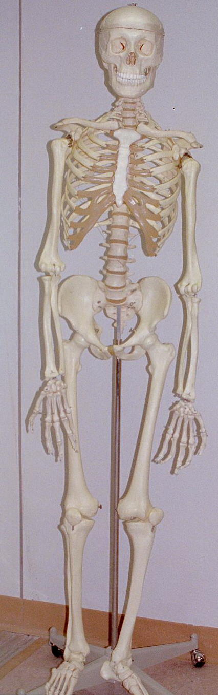
The Skeleton
Axial Skeleton
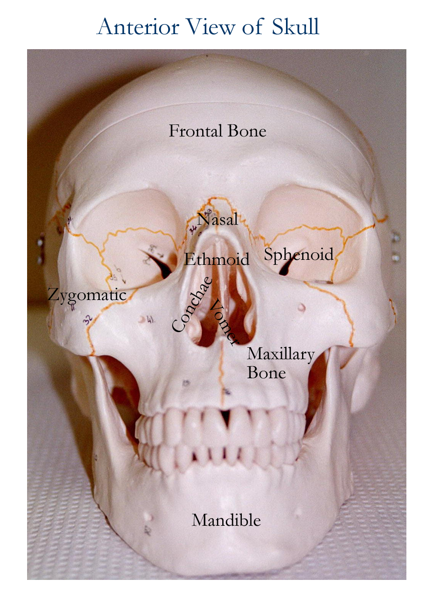
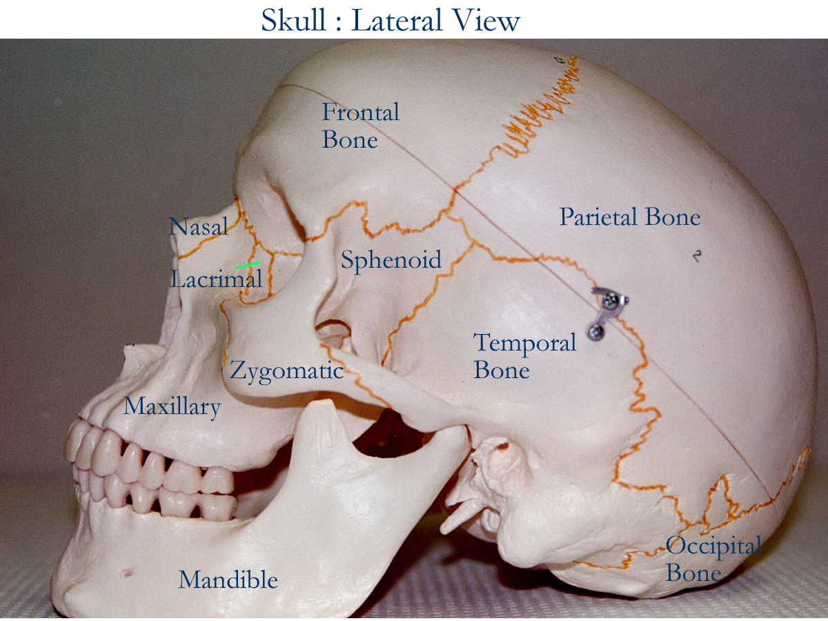
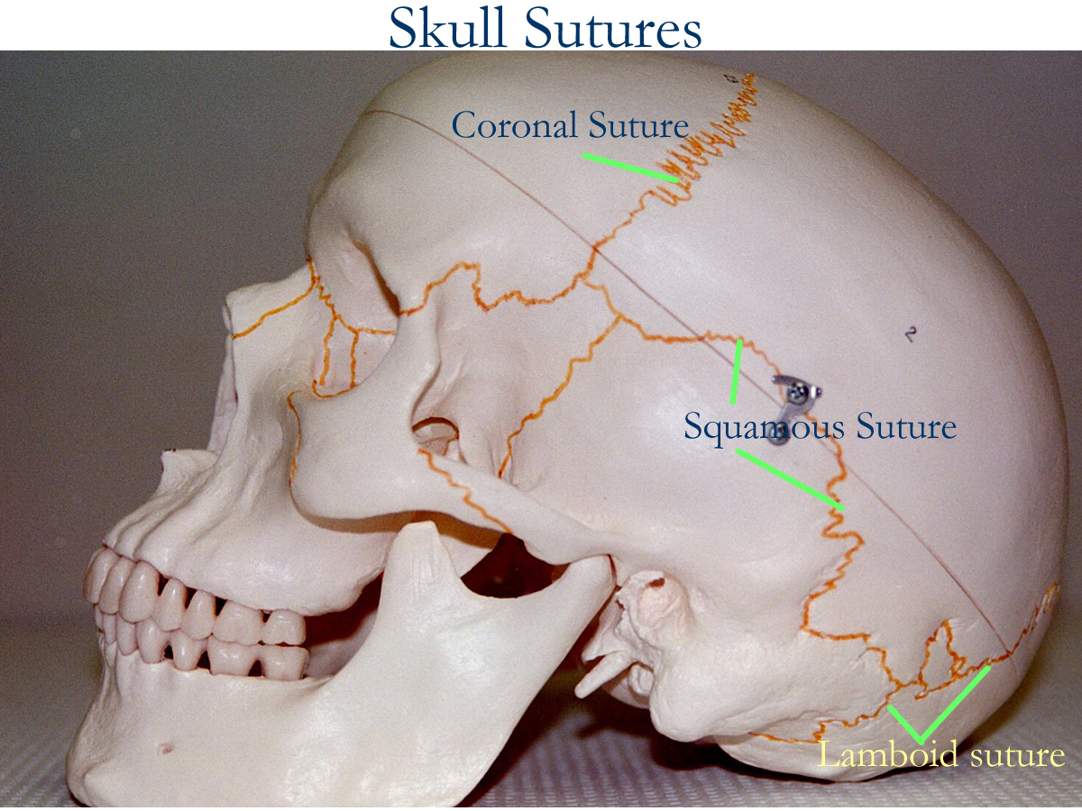
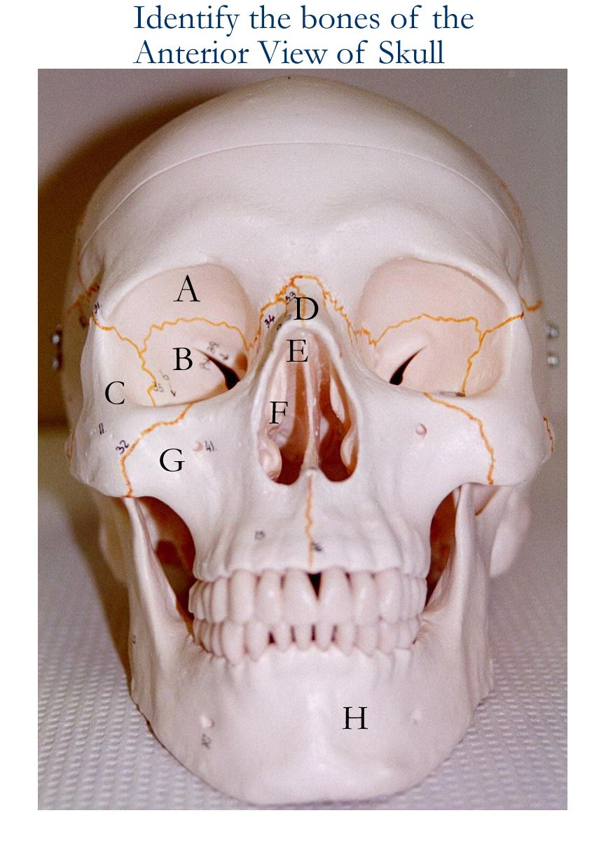
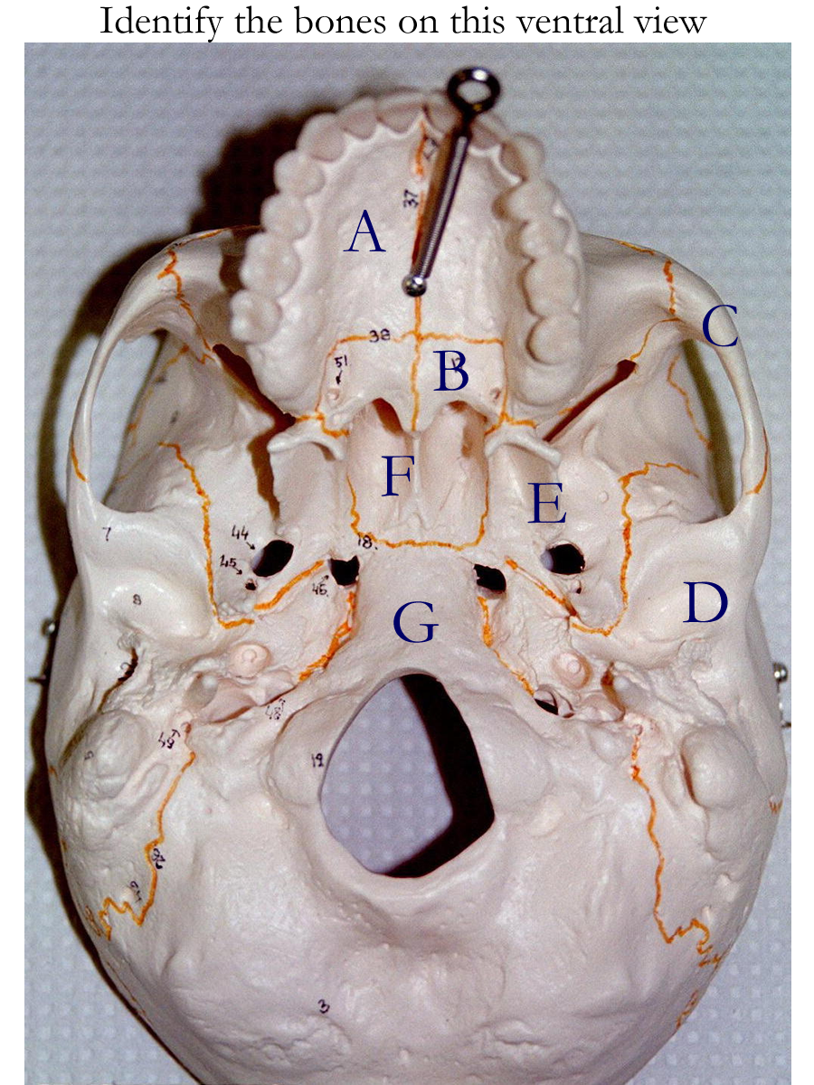
Skull: Cranial and Facial Bones
Anterior, Lateral, Exploded Anterior, Exploded Lateral, Ventral, Interior, Sagittal, Medial
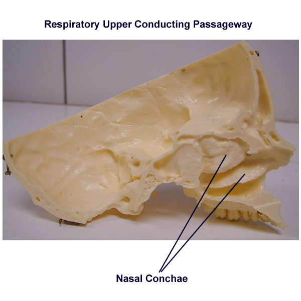
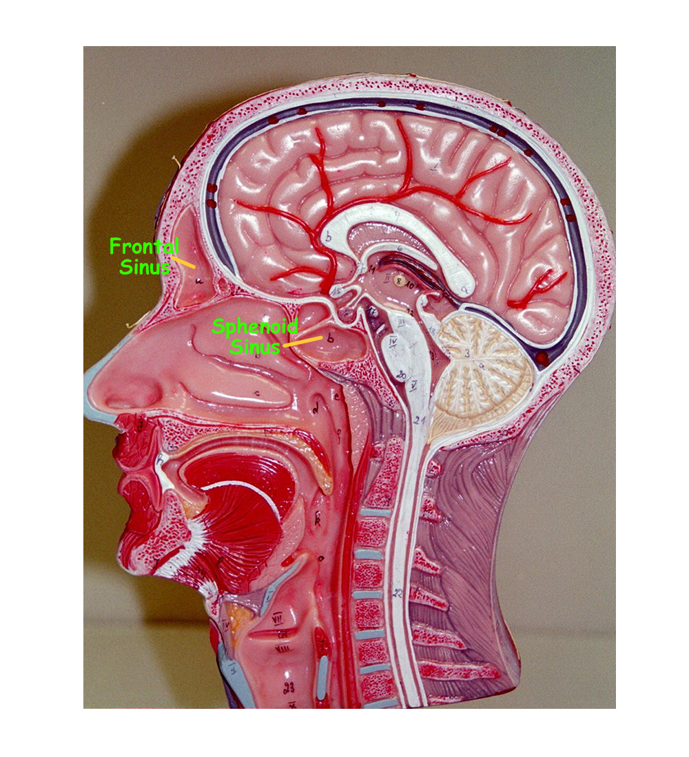
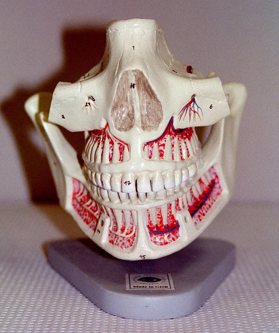
Paranasal Sinuses: Medial View, Transverse View
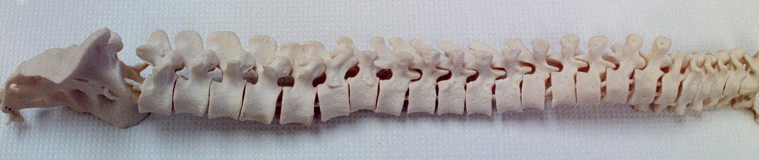
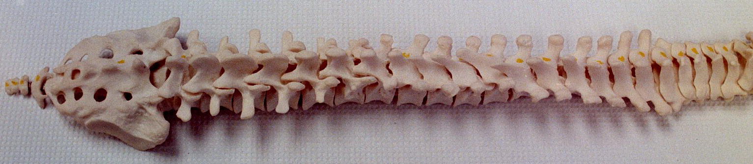
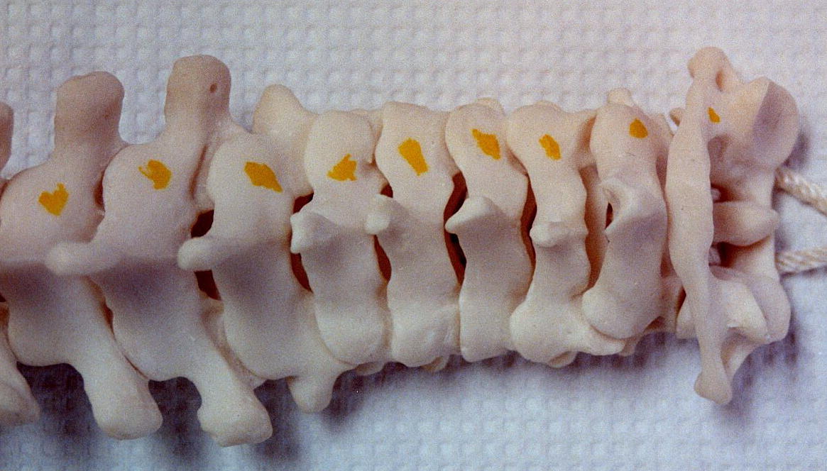
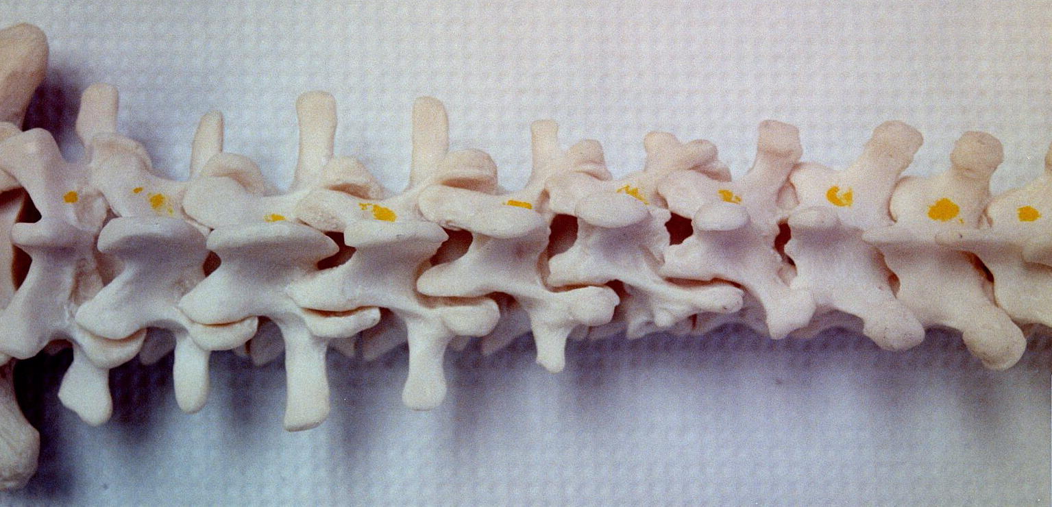
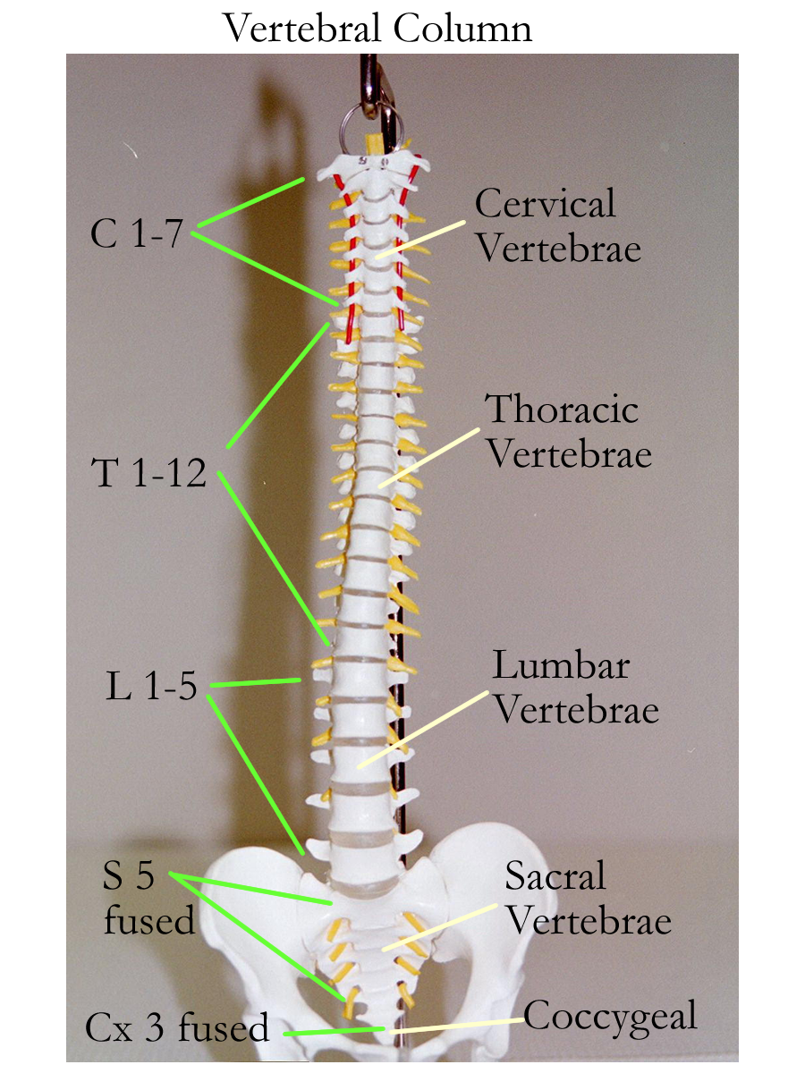
Vertebral regions= Cervical, thoracic, lumbar, sacral, coccygeal
Thoracic cage:
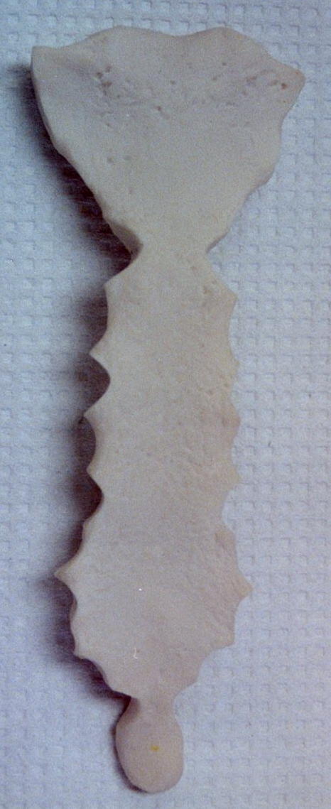
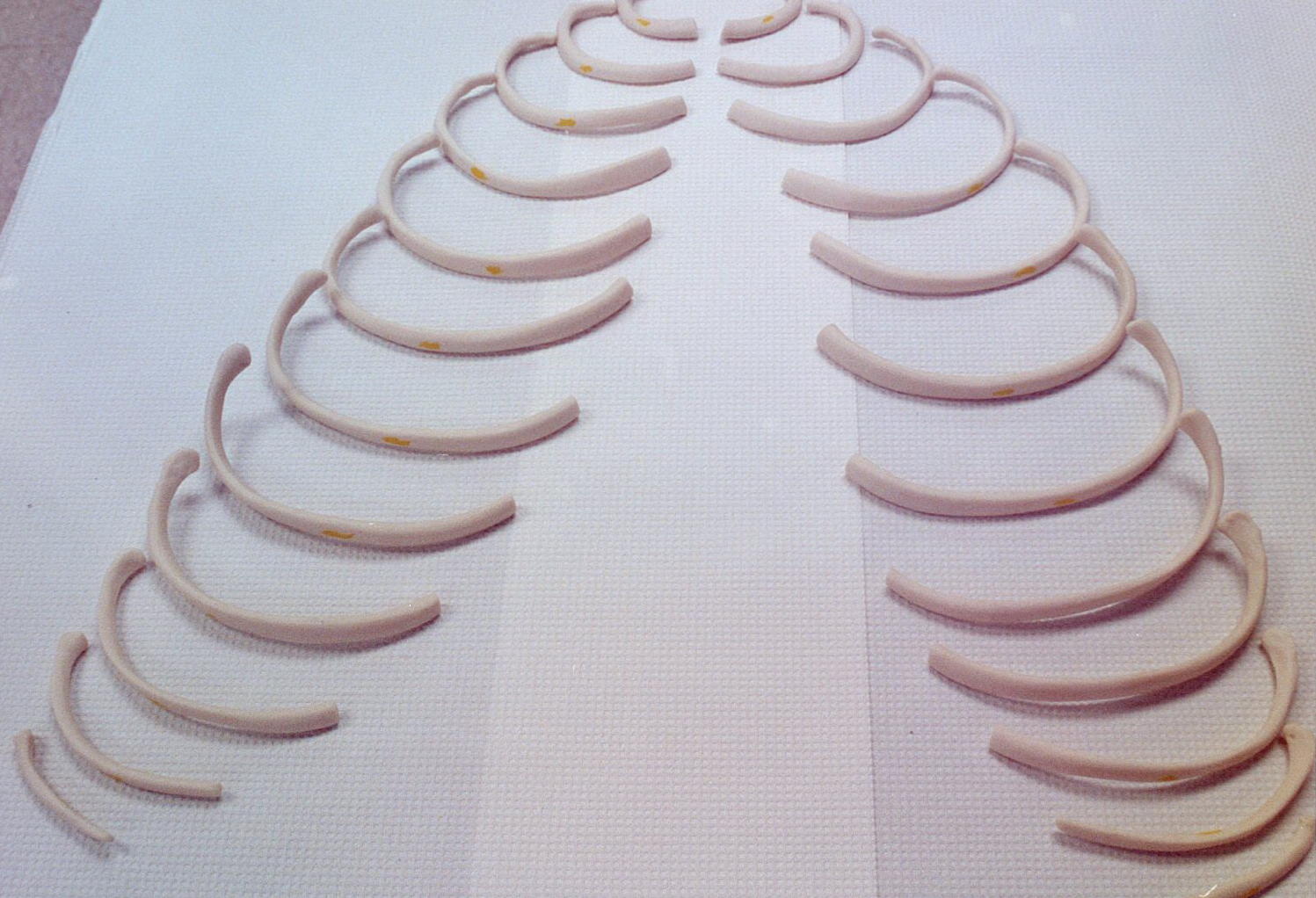
Sternum, Ribs, Thoracic vertebra, Hyoid bone
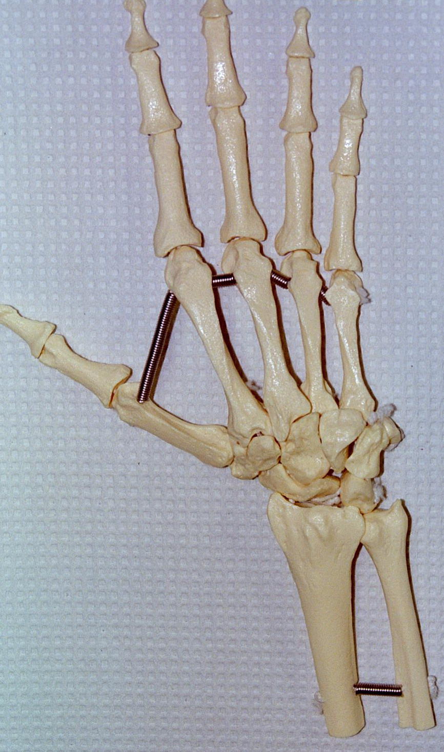
Appendicular Skeleton
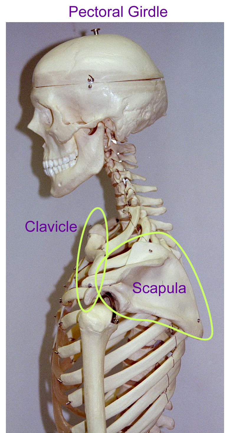
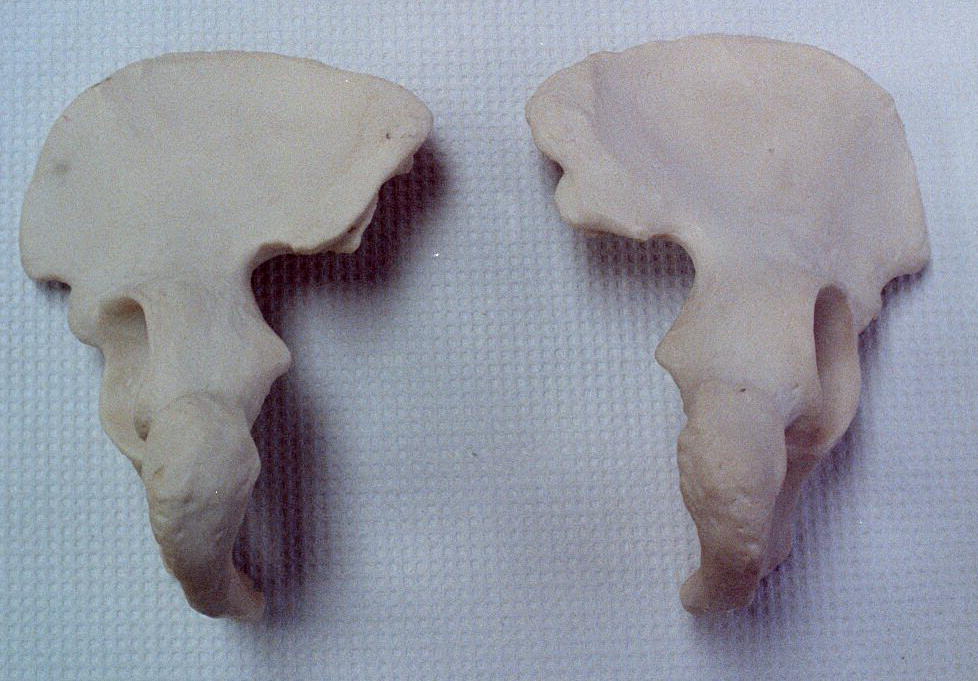
Girdles:
Pectoral Girdle: Scapula, clavicle
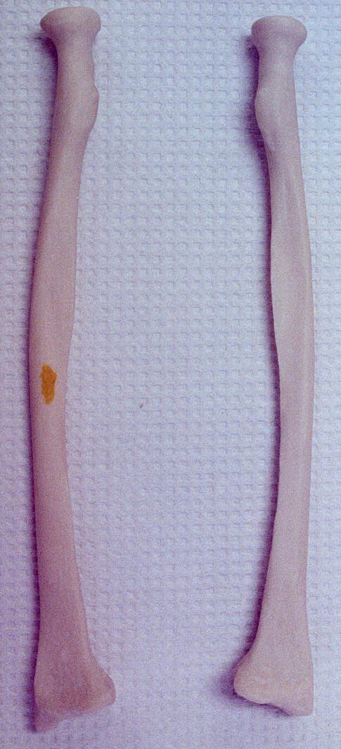
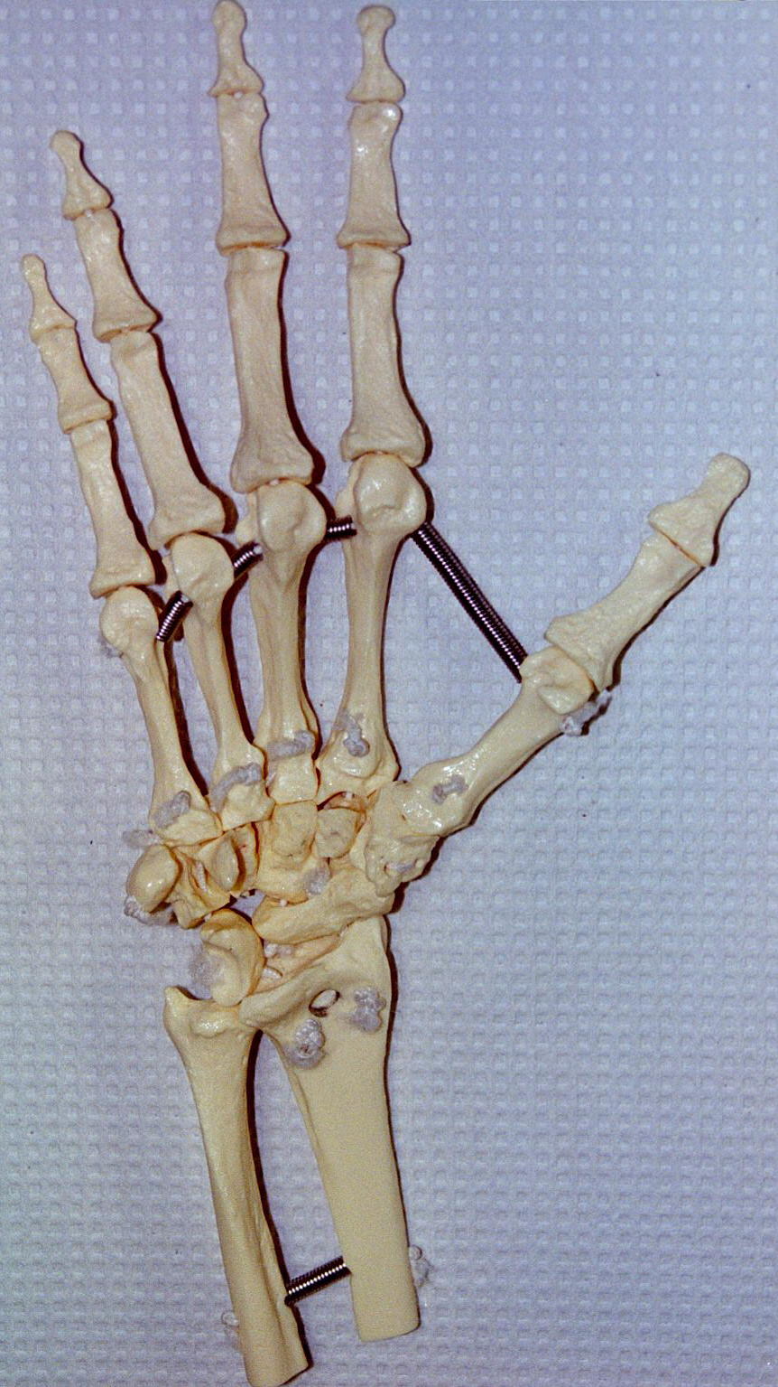
Arm:
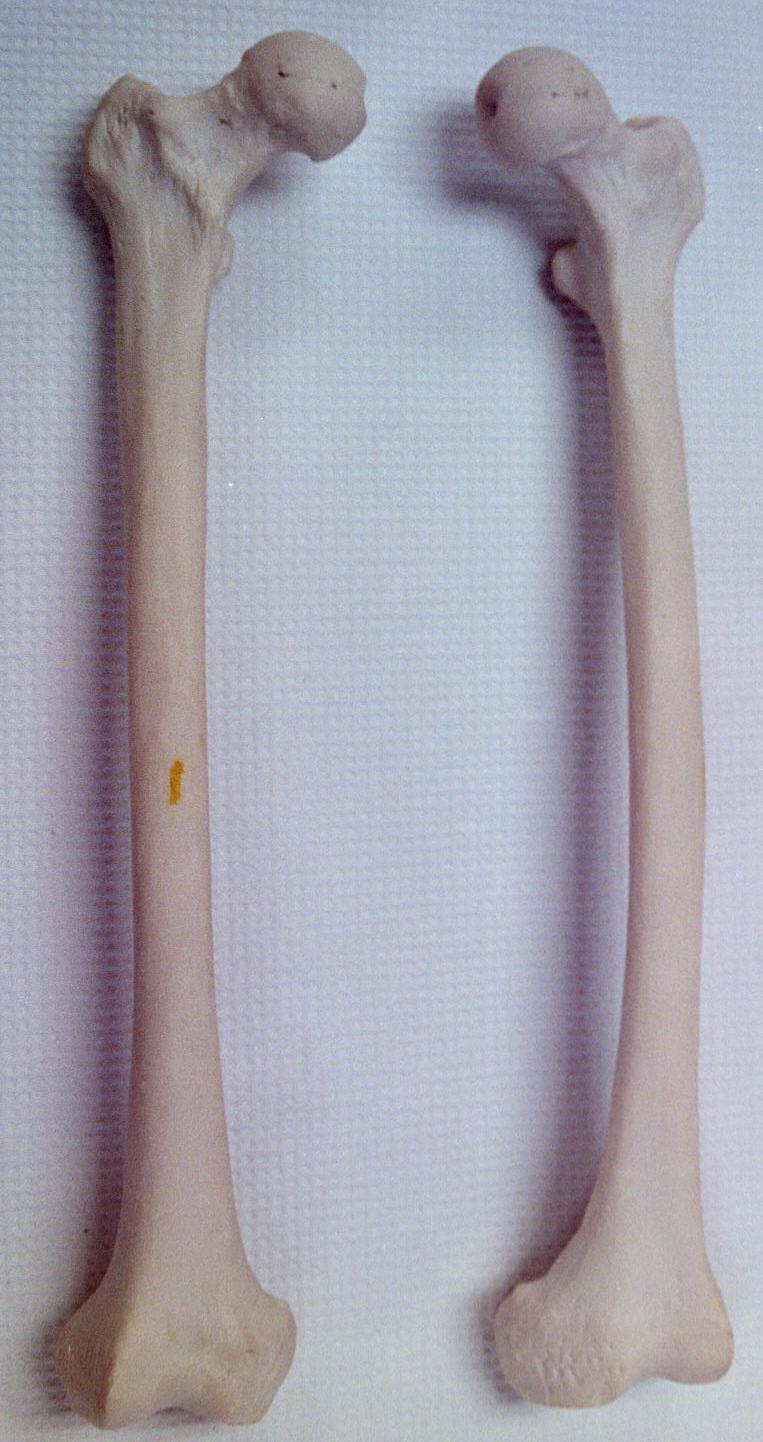
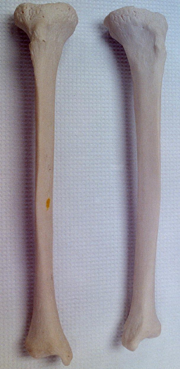
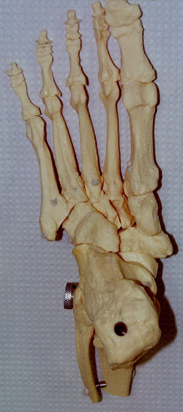
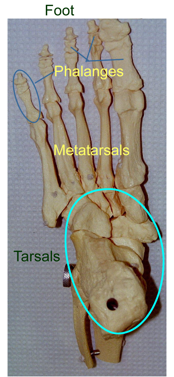
Leg:
Skeletal System Web Site click on the skeletal system for review and a quiz
A sinus is a mucus membrane lined air space in bone. In the skull, sinuses help lighten the skull and help in the tonal quality of the voice.
There are five skull bones that contain sinuses around the nasal cavity and together are called the paranasal sinuses. The paranasal sinuses are named for the bones involved which are the frontal, maxillary, sphenoid, ethmoid. The other bone that contains a sinus is the mastoid process of the temporal bone. The mastoid process can be felt behind the ear.
Joints
Joints are defined as a bone or components of a bone meet. They provide support for the skeleton and can provide movement for the body.
Joints can be classified based on anatomical structure and physiological movement.
Anatomical classifications are: Fibrous, Cartilaginous, and Synovial.
Because the structure dictates the function, this also determines the joint classifications.
Fibrous joints have fibrous connective tissue between the bones. They hold the bones together and do not usually allow movement. These joints are also known as synarthroses.
Cartilaginous joints have cartilage tissue between the bones. They can allow for limited movement if necessary. These joints are also known as amphiarthroses.
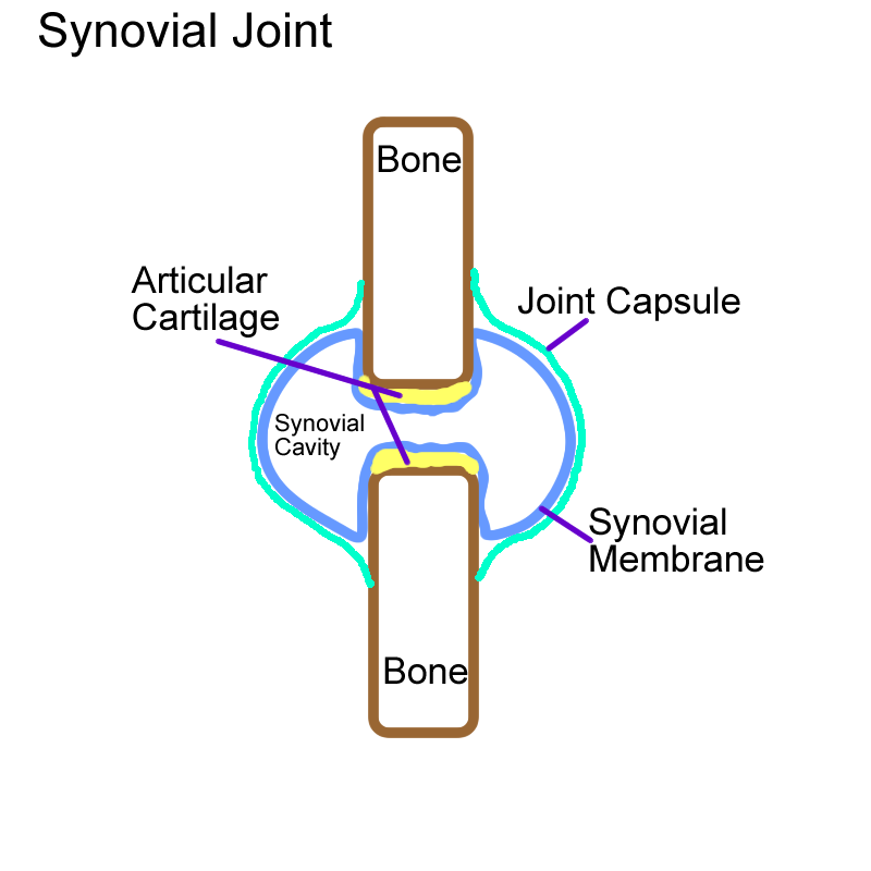
Synovial joints have a unique structure that allows a space between the bone. They have a joint capsule, joint fluid, and other supporting structures such as ligaments that allow for free movement. Synovial joints are also known as diarthroses.
Examples of each joint type are as follows:
Fibrous Synarthrotic Joints
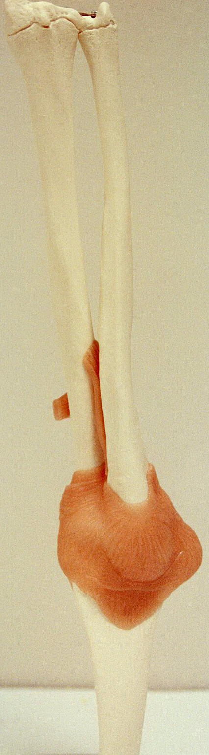
Syndesmosis interosseous membrane between radius/ulna & tibia/fibula
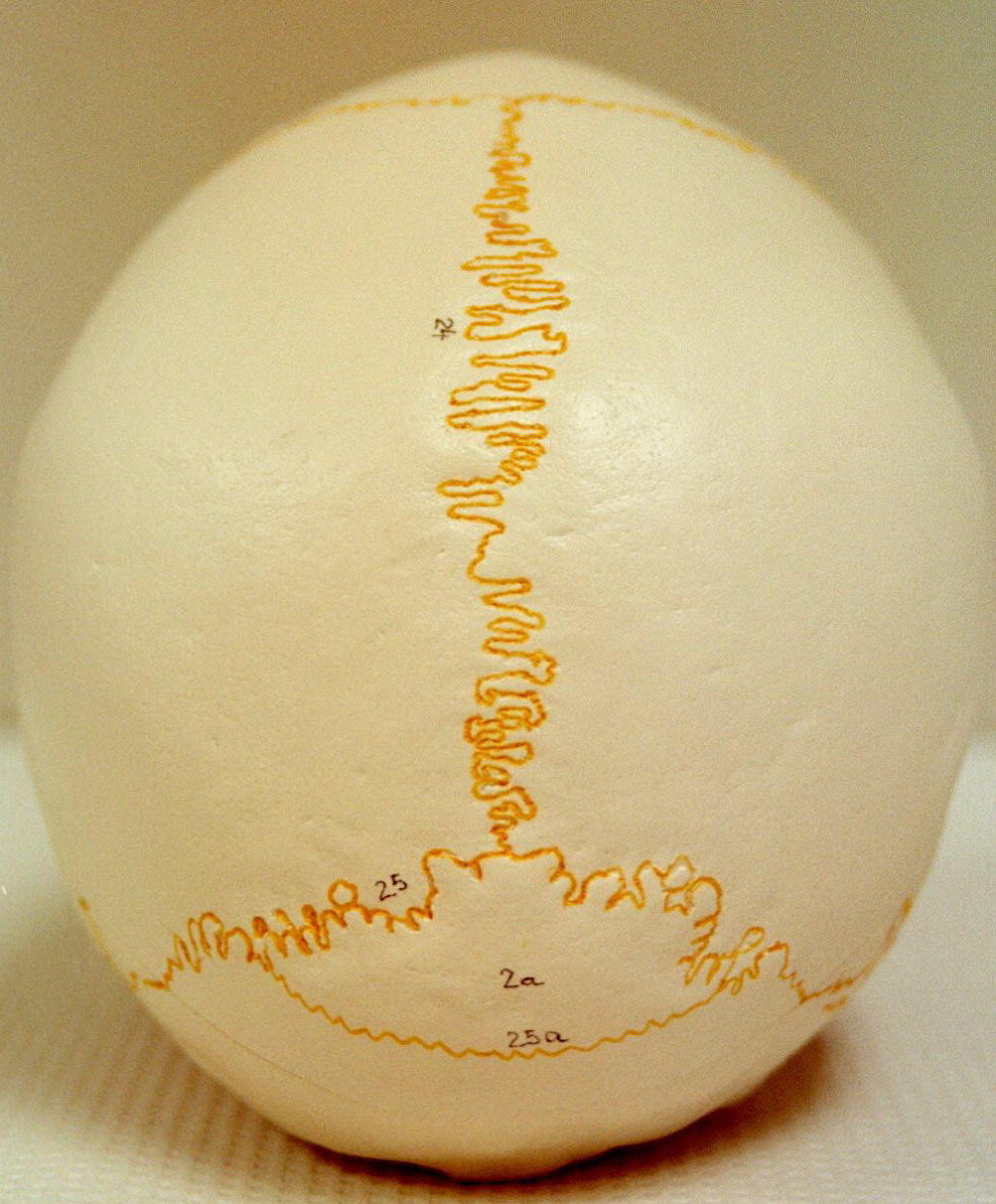
Synostosis sutures between bones of the skull
Gomphosis periodontal ligament between skull and teeth
Cartilaginous Amphiarthrotic Joints
Symphysis at intervertebral discs and between pubic bones of pelvis
Synchondrosis metaphyseal or growth plate region, a temporary joint
Synovial Joints all freely moveable joints that have movement in a body plane and within a certain range.
Synovial Joint Structure:
* articular hyaline cartilage
* joint cavity or space
* articular capsule
* Synovial membrane that lines the capsule
* Synovial fluid produced by the membrane
* additional structures for Synovial joints (some may or may not be present)
- ligaments: reinforce, strengthen and help direct bone movement
- fat pads : cushion
- fibrocartilage discs (menisci) : increase stability
- bursa : flat Synovial sacs that help to decrease friction
- tendon sheath: elongated bursa sacs wrapped around tendons to decrease friction
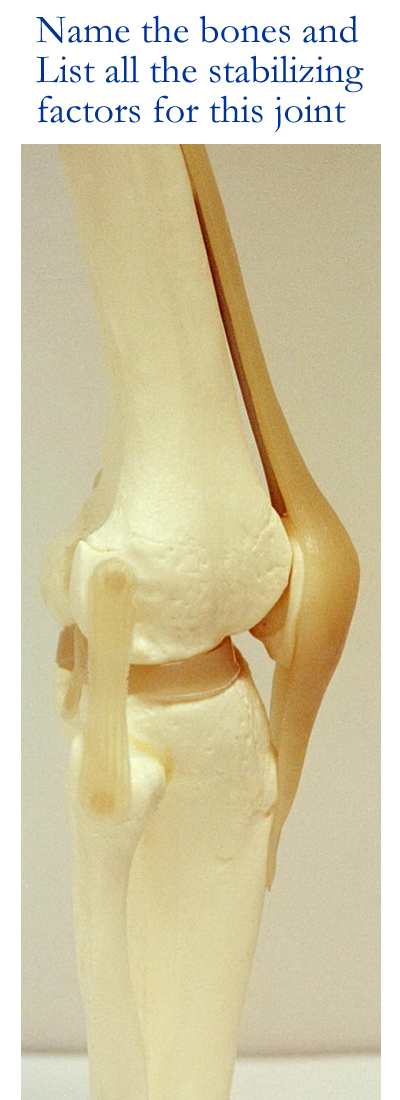
Synovial Joint stability depends on
1) Shape of articular surfaces (how well the bones fit together)
2) Ligaments
3) Muscle tone via tendons of origin and insertion
4) Joint capsule thickness
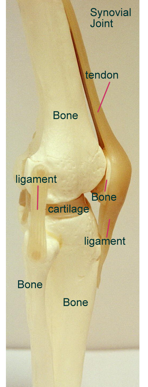
Synovial Joint models
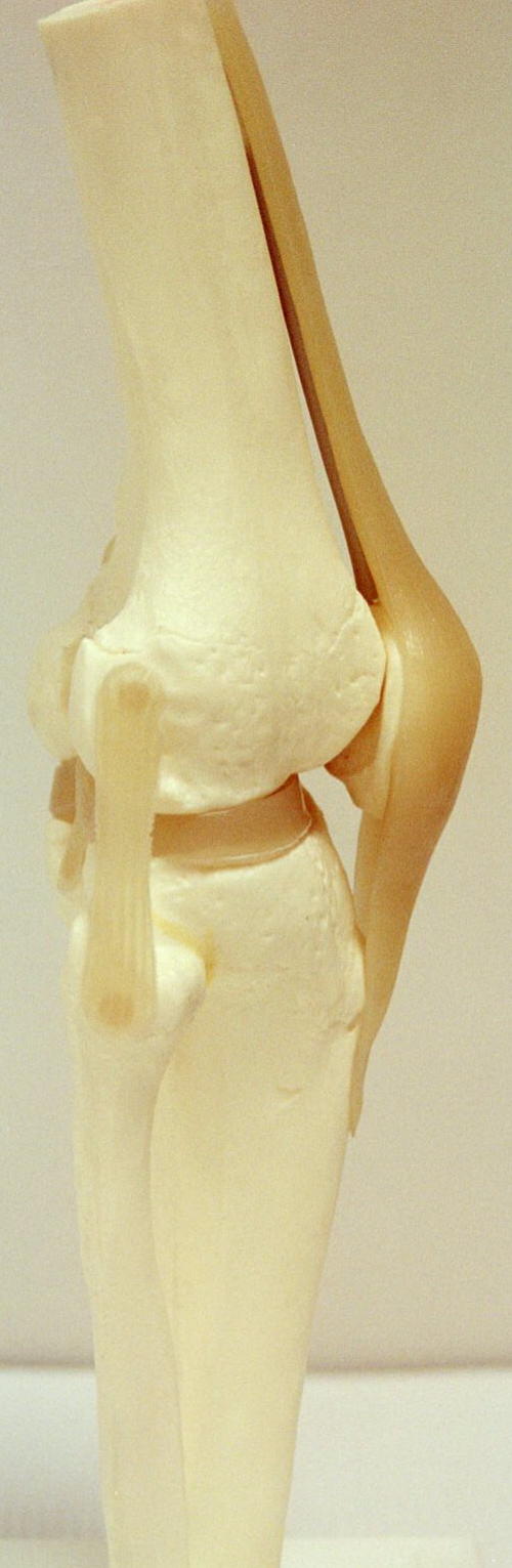
Synovial Joint Radiograph: the Knee
Movement by Synovial Joints
Movement achieved by Synovial joints is based on muscle shortening for an isotonic contraction that produces motion. Muscles are anchored by their tendons of origin in order to pull (never push) their tendons of insertions which are attached to bones across a moveable joint. Movement is created when a more moveable bone is pulled toward a less moveable or stationary bone. Movement can be summarized as the plane of motion + the range of motion.
Planes of motion are along the axis formed by the three major body planes which are transverse, frontal, and sagittal.
Range of motion [ROM] can be nonaxial, uniaxial (one plane), biaxial (two planes), and tri or multiaxial (all three planes).
With the planes and range of motion, three general types of movement can be identified: gliding, angular, and rotational.
Gliding :: one flat surface slips over another
Angular :: changing the angles that form a circle
Flex, extend, abduction, adduction, circumduction
Rotation :: turning of a bone along its long axis
Pivot, rotate
Other specialized movements can be seen at certain joints ::
Supination / pronation radius/ulna
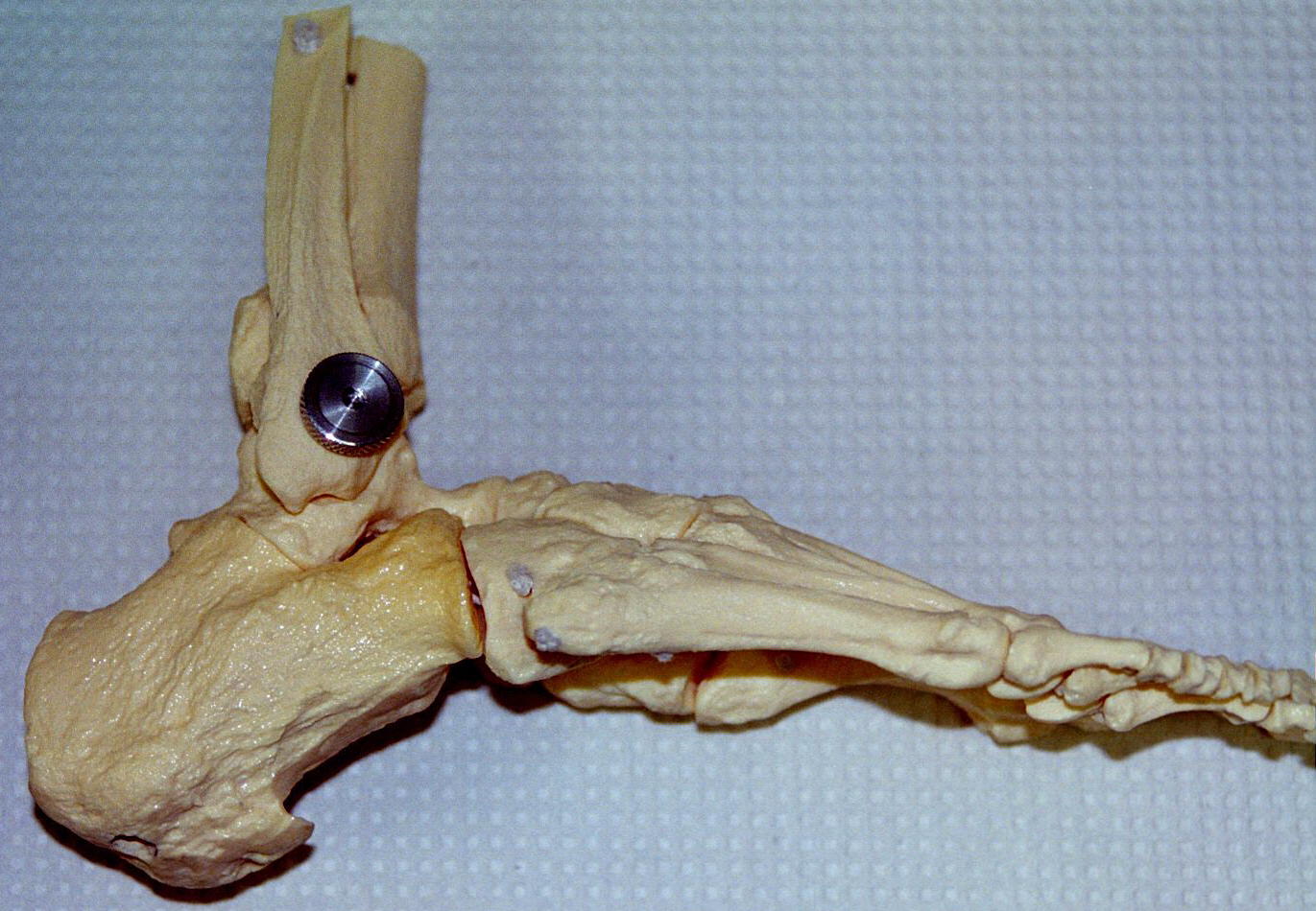
Inversion / eversion foot
Protraction / retraction mandible
Elevation / depression mandible
Opposition thumb
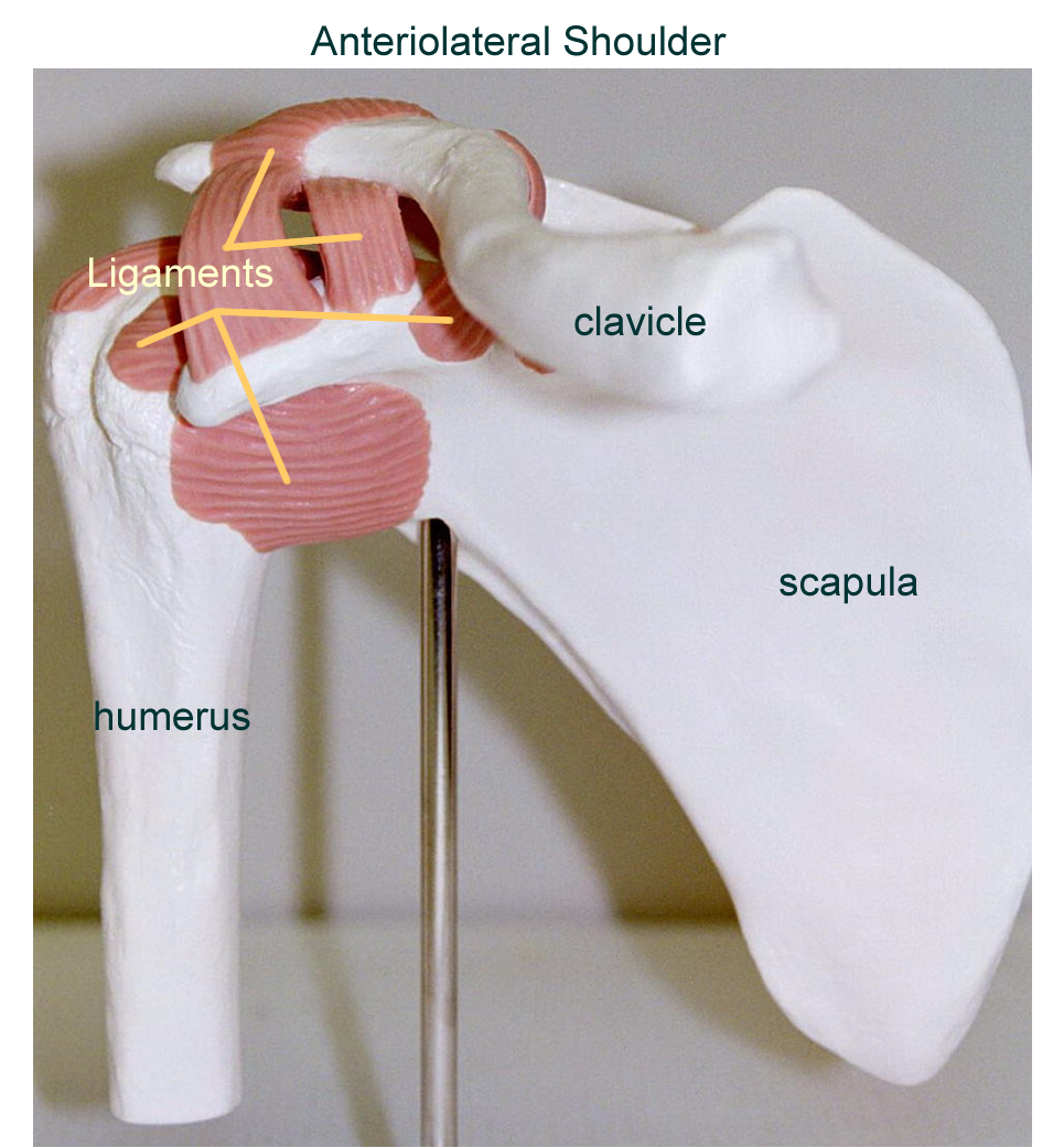
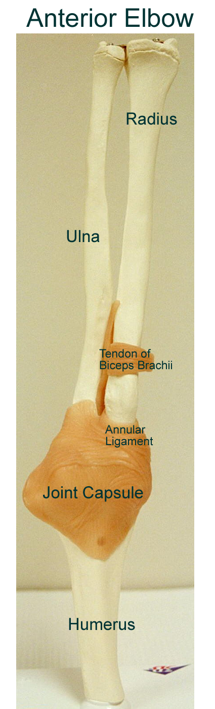
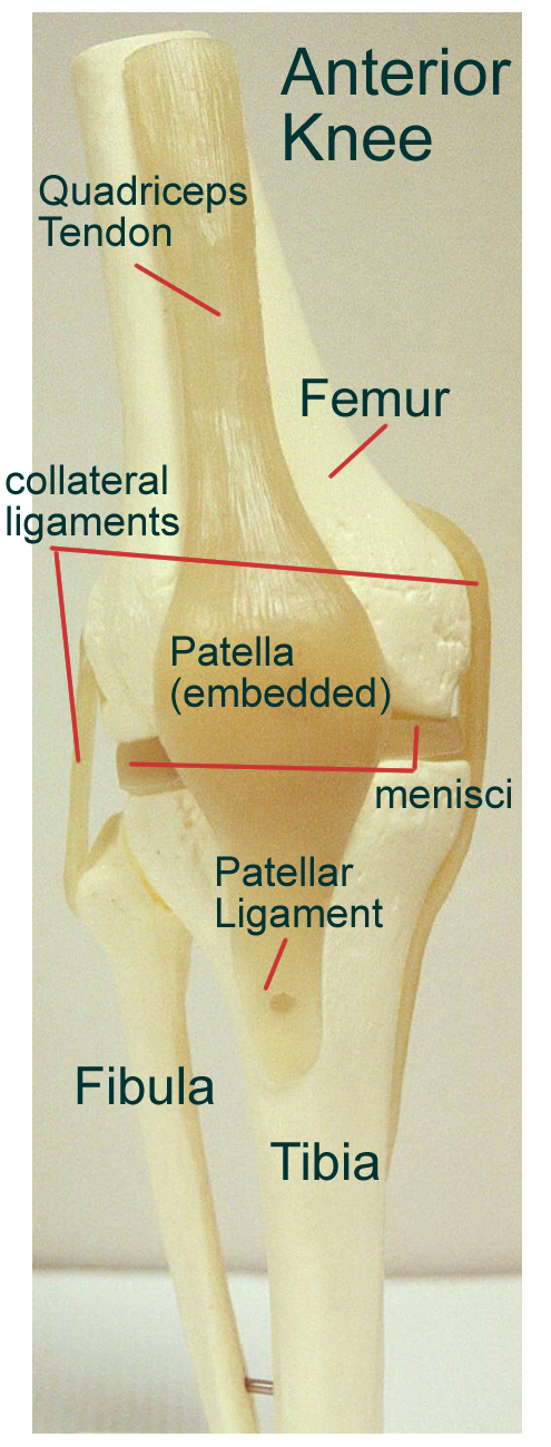
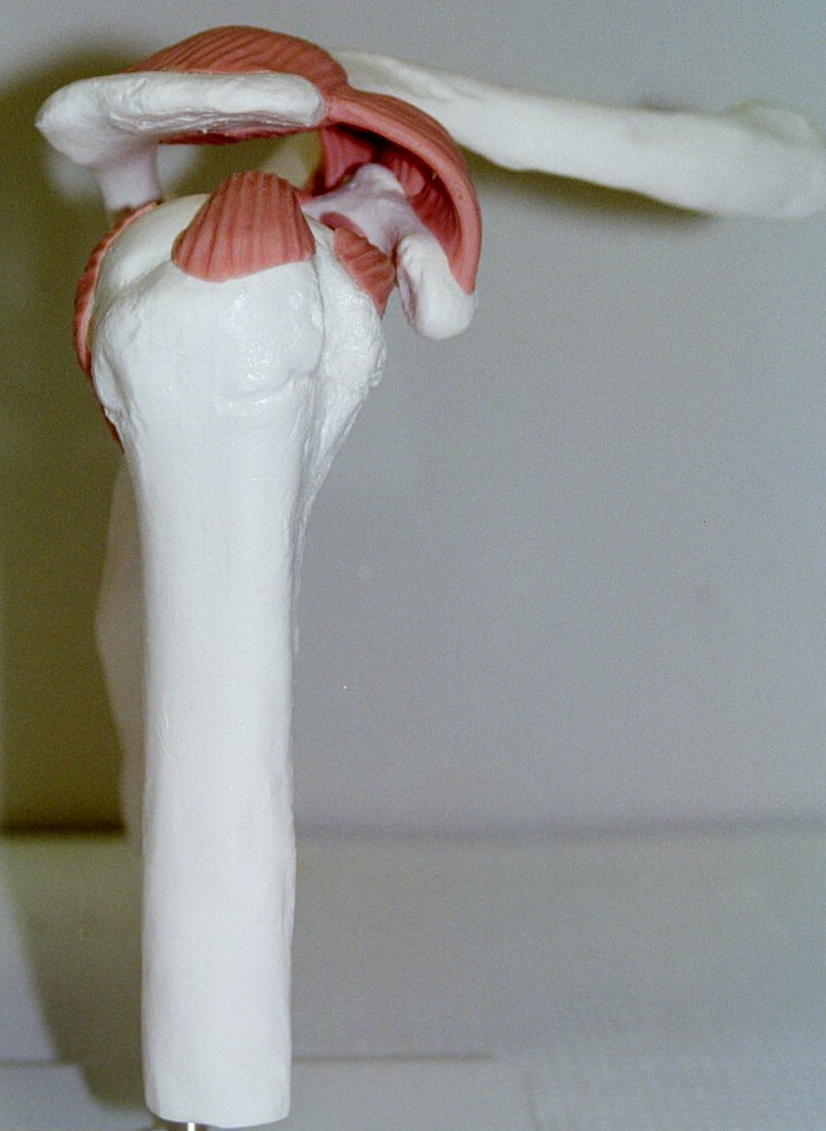
The major joints identified in the body are the shoulder, elbow, hip, and knee.
Each has alternate surgical names and can be identified either by bone fit or physiological movement. Joints are usually named for bones involved from proximal to distal.
Shoulder Glenohumeral Joint Ball and socket Multiaxial
Elbow Cubital Joint Hinge uniaxial
Hip Coxal Joint Ball and socket multiaxial
Knee Stifle Joint hinge biaxial
Due to bone fit and other stabilizing factors, each joint has unique movement as well as problems.
Ad- to, toward amphi- both
Ab- away, from ankyl/o- bent, crooked
Arthro- joint articul/o- joint
Brachi- arm cost/o- rib
Circum- around fract- break
Crani/o- skull gnath- jaw
Genicula- knee like structure man- hand
Ili/o- ilium nas/o- nose
Myel/o- marrow os, or/o- mouth
Os, osteo bone orth/o- straighten
Pterygo- wing rach/i- spine
Sacr/o- sacrum synov/i- synovial
Spin/o- spine, vertebral column lumb/o- lumbar
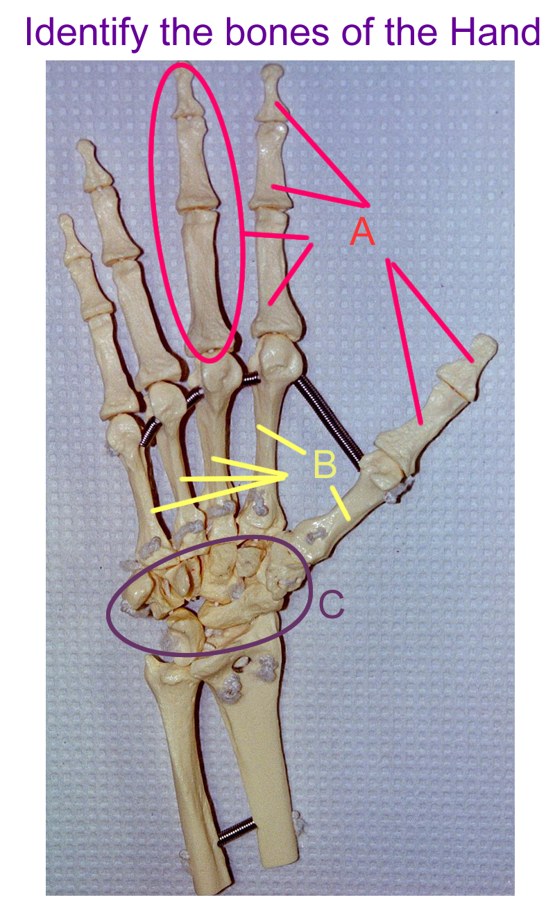
Scapul/o- scapula phalang/o- phalanges
Mandibul/o- mandible rad/i- radius
Brachy- short hyo- u-shaped
Styl/o- stake, pole ante- before
Crur- shin, leg circum- around
Identify Bones of axial and appendicular skeleton
Example One, Example Two, Example Three, Example Four, Example Five
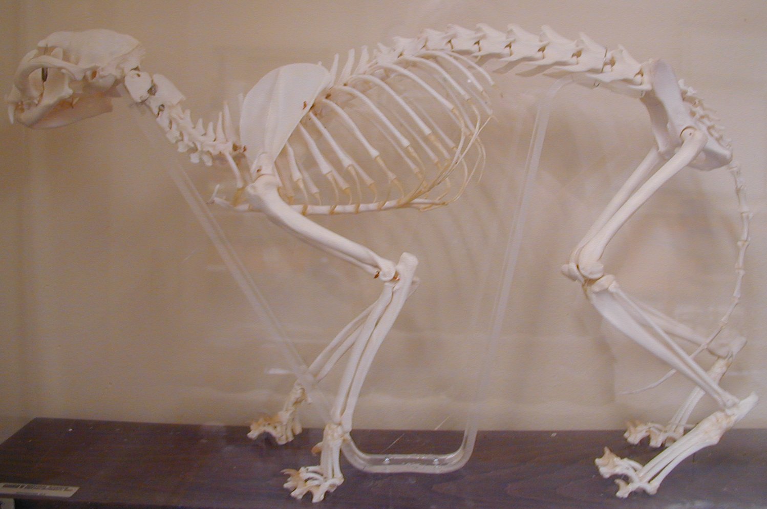
Compare and contrast the human and cat articulated skeleton using 6 similarities and 6 differences.
Ox (beef) tail: Purchase an ox tail from the grocery store.
Describe the vertebra
and intervertebral disc parts seen and give their function. Use gloves to handle
specimen!
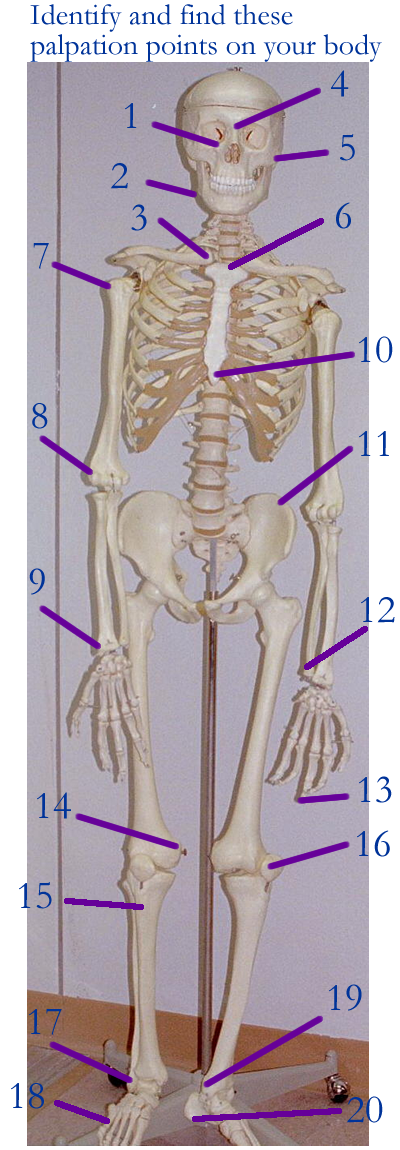
Palpation points: Identify these palpation points. Pick 6 of them and explain how they are used clinically.
ROM : give the range of motion for all major types of joints
synovial : shoulder, elbow, foot, vertebral column
ID bones and stabilizing factors for a major joint
Ingredients in a Joint health label : Go to the store (GNC, Whole Foods, Pharmacy section of various stores) and read the label for a Joint Health product. Write down two ingredients that you do not recognize and research their use in this product.
Skeletal review and quizzes : click on appropriate system to begin
Concept Map: Make a concept map of the skeletal system (histo and gross) anatomy using the bone tissue and bones involved. Include joint types and movements as well. This concept map is part of your LAR lab report (if selected) included as a document insert or as an additinal PDF scan of the map.
Sprains
DJD
Dislocations
Aging changes
Arthritis: Osteoarthritis, Rheumatoid
Bursitis / Tendonitis
Cartilage tears
Sports injury
Bone fractures
Deformities of the Spine: Lordosis, Kyphosis, Scoliosis
Herniated Intervertebral Disc
Osteoporosis
Osteomalacia
Osteomyelitis
Paget’s Disease (Osteitis Deformans)
Gout
Carpal Tunnel Syndrome
Physical Therapist
Chiropractor
Prosthetics/Orthotics
Biomedical Engineer
Podiatry
http://www-medlib.med.utah.edu/WebPath/HISTHTML/ANATOMY/ANATOMY.html
http://www9.biostr.washington.edu/da.html
http://www.ohsu.edu/cliniweb/A2/A2.html
http://rpiwww.mdacc.tmc.edu/ap/frontAnatomy.html
http://www.medtropolis.com/VBody.asp
http://www.arthroscopy.com/sp12001.htm
http://www.kcmetro.cc.mo.us/maplewoods/Biology/Bio110/Labs.htm
http://www.track0.com/canteach/links/linkbodysystems.html
http://www.innerbody.com/htm/body.html
http://medstat.med.utah.edu/kw/sol/sss/
http://www.aaos.org/wordhtml/pat_educ/kneeanat.htm
http://www.medem.com/MedLB/article_detaillb.cfm?article_ID=ZZZKBBGBGJC&sub_cat=258 skeleton
http://www.medem.com/MedLB/article_detaillb.cfm?article_ID=ZZZCIHWBGJC&sub_cat=523 skull
http://www.medem.com/MedLB/article_detaillb.cfm?article_ID=ZZZDLCB46JC&sub_cat=181 carpal tunnel
1. Name four minerals or ions seen in bone matrix.
2. Name the bones based on their shape classification.
3. Name the two methods for embryonic bone ossification.
4. Identify the bones of the axial skeleton
5. Identify the bones of the appendicular skeleton
6. Be able to identify bones and their markings as part of surface palpation points.
7. Define joint and name the three major types.
8. Give the five components of every Synovial joint
9. Compare compact and spongy bone
10. Give three functions of the skeletal system.
11. Name 2 hormones and 2 vitamins that are important to bone and explain each of their functions.
12. Define related word parts and terms of the skeletal system.
13. Be able to list several bone diseases or conditions
14. Explain how the skeletal system interacts with other body systems.
15. Be able to define bone fracture and name or classify two types.
(open and closed bone fractures are not breaks in the bone)