Biology 2404 A&P Basics Lab Exercise 12 Blood Dr. Weis
| Objectives | Background | Medical Terms | Activities | Applications | Careers | WWW | Review Questions |
Students should be able to:
* give the functions of blood
* name the formed elements of blood and their specific function
* give the components and function of plasma
* name the phases of hemostasis
* give the major steps of coagulation
* explain the differences between agglutination and coagulation
Anatomy and Physiology Background
Read related information in textbook
Blood is a connective tissue consisting of a fluid ground substance, fiber, and cells formed in the red bone marrow by hematopoiesis.
The cells of blood are called formed elements, the fiber is the clotting protein called fibrin, and the ground substance is a fluid called plasma. Blood is transported in vessels that create vascular circuits in order to distribute substances to the proper organs. Pressure gradients allow for blood flow and are initiated by the heart muscle contractions. Together, the blood, vessels, and heart form the cardiovascular system.
The general functions of blood are transportation, regulation, and protection.
Transportation of nutrients, waste products, gases, and hormones
Regulation of ion balance, acid-base (pH) balance, and temperature
Protection by blood clotting to prevent hemorrhage
Protection by white blood cells for immune defense
General Characteristics:
Volume of blood in humans is ~ 5 liters
of which 45% is composed of solutes (solids) and 55% solvent (liquid)
pH of blood ranges from 7.35-7.45 (for venous and arterial blood, respectively)
viscosity or thickness of blood is 3-5 times that of water
Blood Components
A. Plasma
Makes up 55% of the blood volume
90% is water
10% are composed of dissolved substances
~7% are proteins such as hormones, globulins (alpha, beta gamma), and albumin
along with clotting factors such as prothrombin and fibrinogen
The remaining 3% of plasma is conprised of
respiratory gases such as O2 and CO2
ions such as Na+, K+, Ca++, Cl-, HCO3-
nitrogen wastes such as urea and uric acid
nutrients such as glucose and amino acids
Functions in transport and regulation
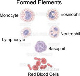
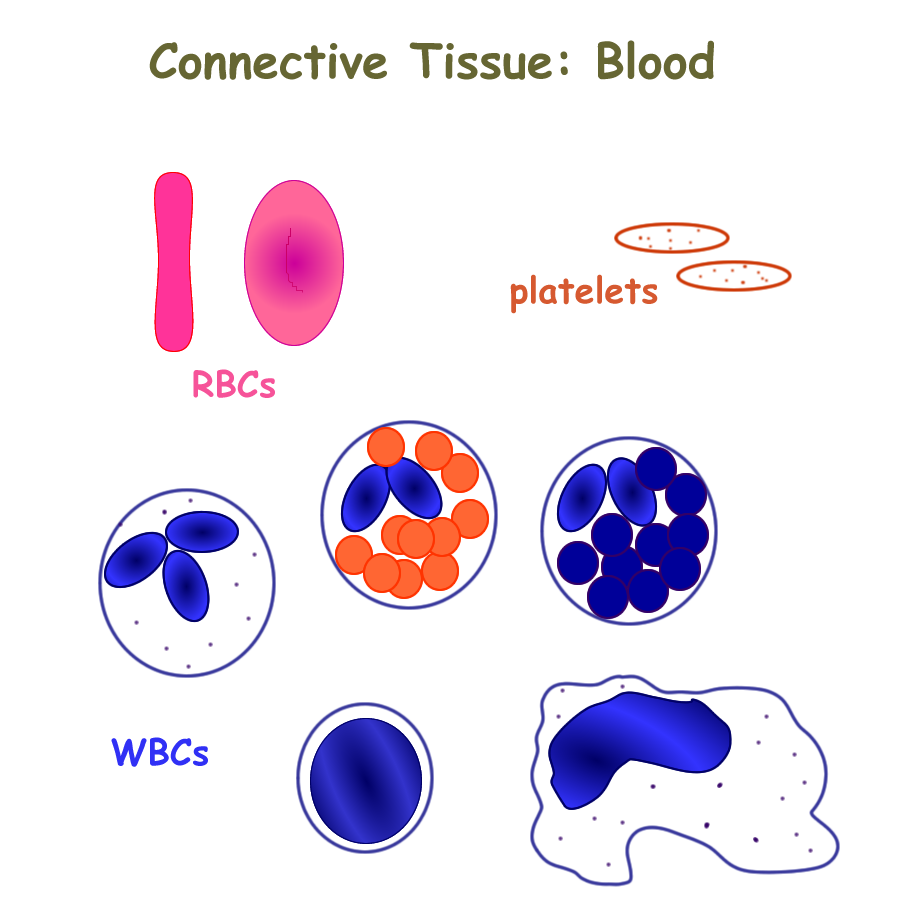
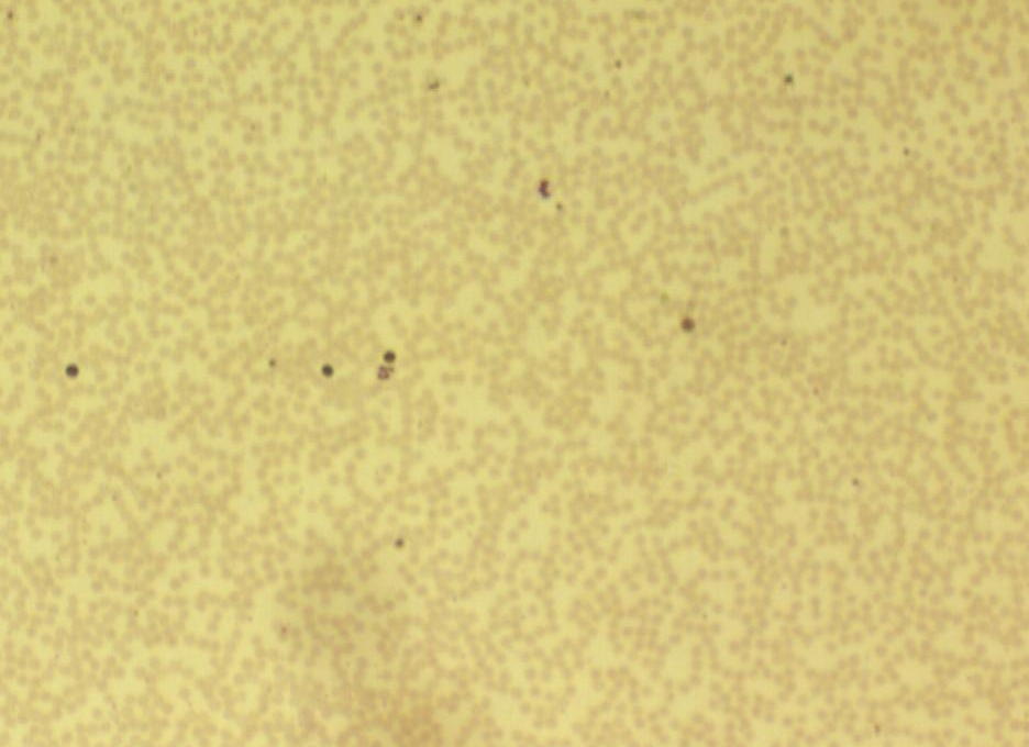
B. Formed Elements : Red Blood Cells
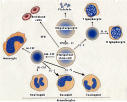
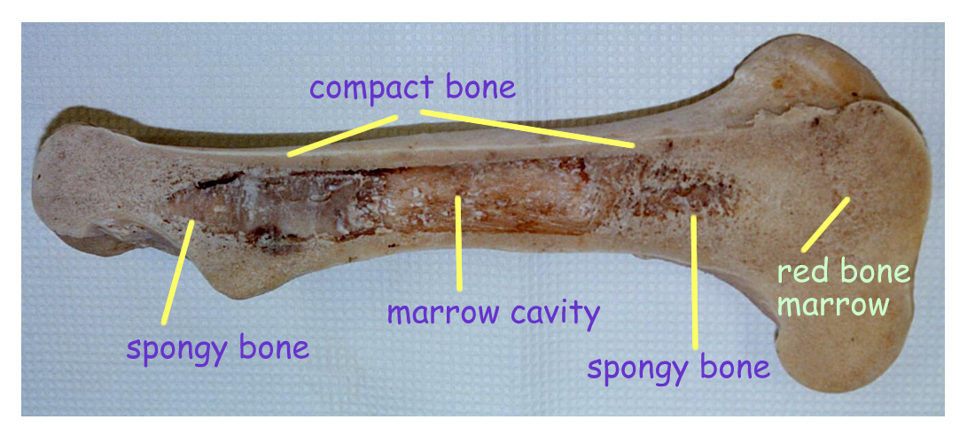
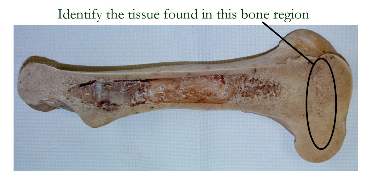
All blood cells (formed elements) come from the hemopoietic tissue located in the spongy region that forms the red bone marrow and lymphatic tissue found in the lymph nodes, spleen, and thymus. Stem cells in the red bone marrow create most of the blood cells and the formation process is under hormonal control. Some of these blood cells will migrate to the lymphatic tissues and under immune signals, can create additional products or defense cells. The normal adult blood cells are red blood cells, white blood cells, and the formed elements: platelets.
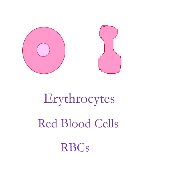
Red Blood Cells (erythrocytes, RBCs)
RBCs come from the stem cell’s myeloid cell line and production regulation is under the influence of the hormone Erythropoeitin (EPO). EPO is made in the kidneys in response to low O2 levels. Maturation of erythrocytes takes approximately one week and is dependent on several B vitamins such as B12 and folic acid. Immature RBCs such as reticulocytes or nucleated RBCs can sometimes be seen in a blood smear and are important indicators of bone marrow response in blood loss conditions.
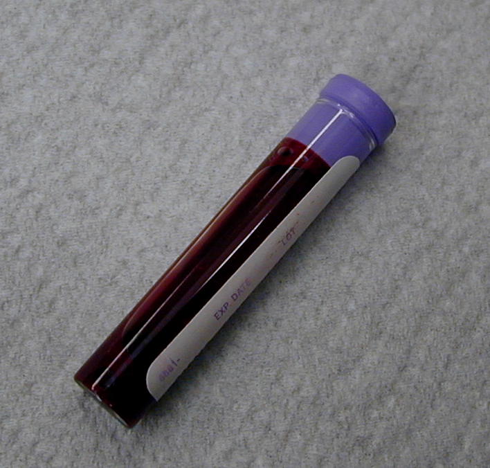
Mature human red blood cells are biconcave discs about 8 µm in size in order to fit single file through a capillary bed. They appear clear in color when seen on a wet mount, but red in color when observed during collection or on a stained smear. A mature RBC has lost all its organelles including the nucleus and ribosomes, and contains only hemoglobin protein molecules and some enzymes such as carbonic anydrase. Most RBC enzymes are located on the plasma membrane and function in transport of nutrients into the cell or to maintain plasma membrane integrity. Because they can not repair their cell membranes, the lifespan of a RBC is only about 120 days. Old, fragile red blood cells are recycled by the tissue macrophage (reticuloendothelial) system in the liver and spleen. Most of the hemoglobin is reused, but the pigment portion contains nitrogen and is eventually converted to a waste product called bilirubin.
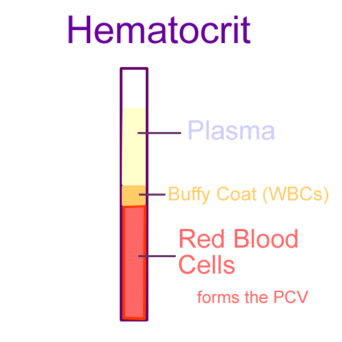
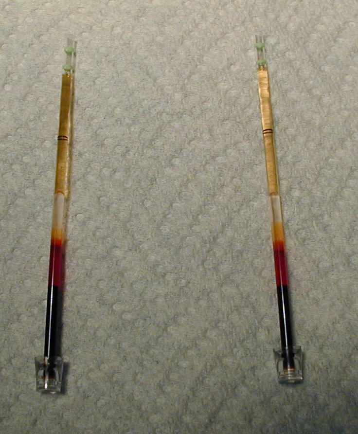
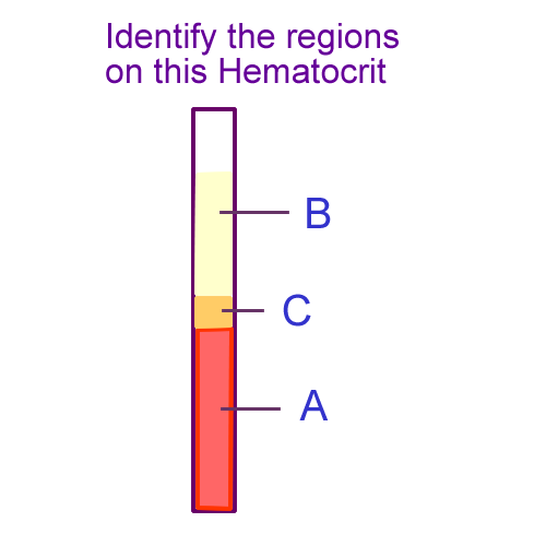
Normal RBC count is between 4.5 – 6.0 cubic millimeters and makes up almost 45% of the blood volume. These cells function to carry respiratory gases and help control blood pH by acting as a buffer that ties up hydrogen ion. A blood test used to determine the percentage of red blood cells is the hematocrit or HCT. This the percentage is calculated based on other information such as RBC number and MCV (mean corpuscular volume). If the % RBC is dtermined by spinning down a blood capillary tube and read using a scale, it is called the packed cell volume or PCV.
Another test performed using RBCs is blood
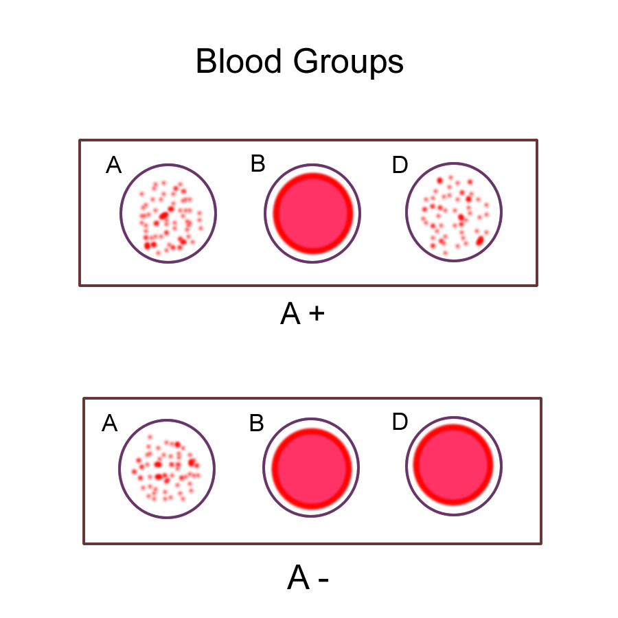
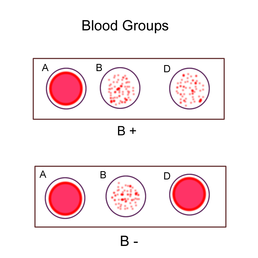
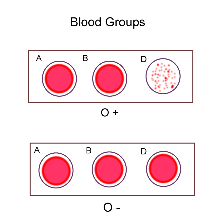
typing. Blood types are genetically determined proteins that are on the cell
membrane as an immune marker. The most important proteins to identify belong
to the ABOD grouping. If these proteins interact, they can cause severe, even
life threatening problems. The ABO group contains proteins identified as Type
A, Type B,
Blood typing also identifies the D antigen known as the Rh factor. If the RBC has the dominant “D” antigen, the blood is identified as Rh+. If the antigen is the recessive “d”, the blood type is identified as Rh-. There are other Rh antigens, but they do not cause severe transfusion reactions. The blood tests performed to identify blood types are called typing and cross matching. Blood typing is determined by an agglutination reaction on the slide between the donor’s red blood cells and the recipient’s plasma antibodies. Cross matching looks for any agglutination between the recipients red blood cells and the donor’s plasma antibodies.
Red Blood cells in different types of solutions: No solution, Isotonic, Hypotonic, Hypertonic
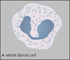
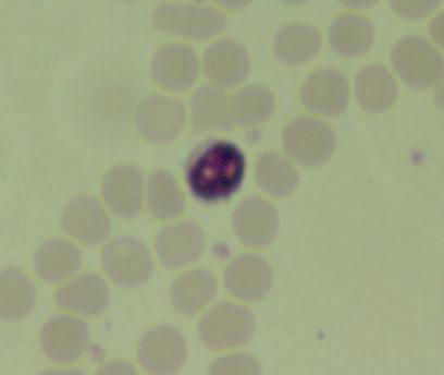
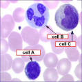
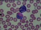
C. Formed Elements: White Blood Cells (leukocytes, WBCs)
White blood cells come from either the myeloid cell line or the lymphocytic cell line. Production regulation is under the influence of various hormones called colony stimulating factors known as colony stimulating factors, or CS-F.
Leukocytes average about 5-9 thousand per cubic millimeter of blood and the 5 different WBC types are divided into two major classes that help to identify them in when a differential white cell count is performed. The two groups of WBCs are granulocytes and agranulocytes.
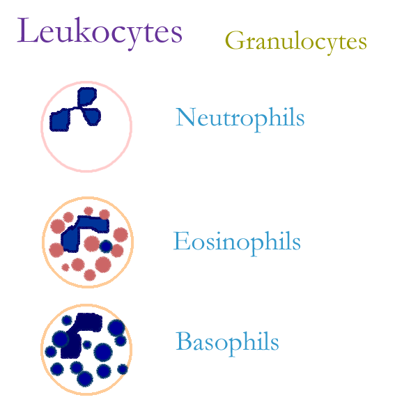
Granulocytes are approximately the same size as a RBC. Their nucleus is divided into at least two segments or parts and their cytoplasm contains granules that give them their function. The granulocytes consist of:
Neutrophils 3-5 lobed nucleus, neutral staining granules
Function in nonspecific, general defenses, acute phagocytosis
70% of the WBC count, Life span 12-24 hours
Eosinophils 2 lobed nucleus, pink staining granules
Functions in specific defenses for allergens antigens
3% of WBC count, life span varies
Basophils 2 lobed nucleus, blue staining granules
Functions in specific defenses for allergens (Type I hypersensitivities) and antigens on parasites
0.5-1% of WBC count, life
span varies
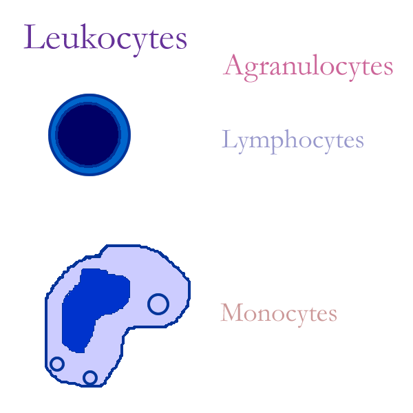
Agranulocytes do not contain granules in their cytoplasm and the size and nucleus varies. The agranulocytes are:
Lymphocytes round nucleus, small amount of cytoplasm, 20 % of the WBC count
Three sizes= small, medium, large. Medium is the same size as a RBC.
Some large lymphocytes function in nonspecific defenses as Natural Killer cells
Most function in specific defenses as T cells or B cells
Life span varies, sometimes life long
Macrophage bean shaped nucleus, large amount of cytoplasm,
8-10% of WBC count (monocytes in the blood move to the tissues to mature into macrophages)
2-3 x the size of a RBC.
Nonspecific defense that ties to specific defenses, Antigen Presenting Cell (APC)
Life span varies, sometimes life long
Fixed or Free (wandering) macrophages
Drawings:
Neutrophils
Monocytes
Eosinophil
with Monocyte
Basophil
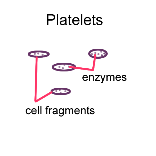
D. Formed Elements: Platelets
Platelets or thrombocytes are packages of enzymes that come from fragments of megakaryocyte cells located in the red bone marrow. Production regulation is under the influence of the hormone thrombopoietin (TPO). Life span of platelets is about 10 days.
The normal platelet count is 150-400 thousand per cubic milliliter of blood.
Enzymes contained in the membrane sac function to aid in the clotting mechanisms
Blood Tests
CBC : Equipment
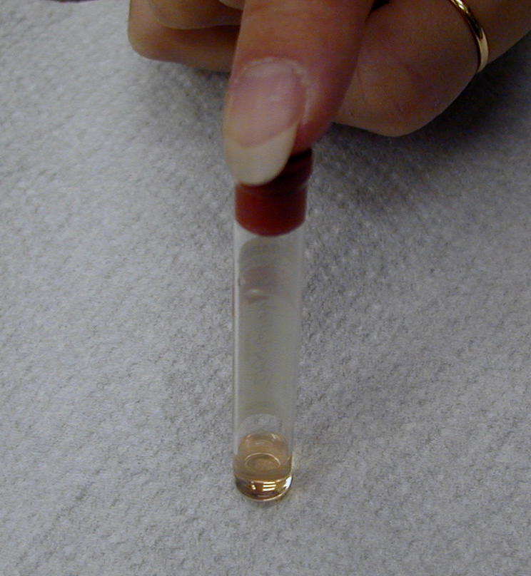
PCV: Equipment
E. Hemostasis
Hemostasis is the control and stoppage of blood loss. It is under positive feedback control and once started should go to completion and end the cycle. The four phases of hemostasis are:
1. Vascular spasm: When a vessel is injured, the smooth muscle in the wall constricts.
Collagen is exposed due to the injury and vascular chemotaxis chemicals are released.
2. Platelet Plug: Platelets are attracted to the injured vessel by chemicals and the rough surface of the
collagen causes the platelets to enlarge and become sticky. The platelets form a temporary
barrier to close off the opening in the vessel and then release factors that attract other
platelets to the area.
3. Coagulation: Clotting factors in the plasma are activated by the roughened vessel surface and the platelet plug. Two pathways of coagulation are then activated.
One phase called the extrinsic pathway takes about a minute and the second called the intrinsic pathway takes longer, at approximately 3 minutes.
The end result of both coagulation phases is the formation of fibrin from the clotting factor,
fibrinogen. Fibrin strands are stabilized and form a mesh or net at the injury site.
This fibrin mesh traps RBCs to help form the blood clot.
4. Clot Retraction and Resolution: Once the injury site is closed off, the clot retracts to pull the
vessel lining together. Once the site is repaired, the blood clot is no longer needed.
Chemicals are released to cause fibrinolysis and the clot is dissolved.
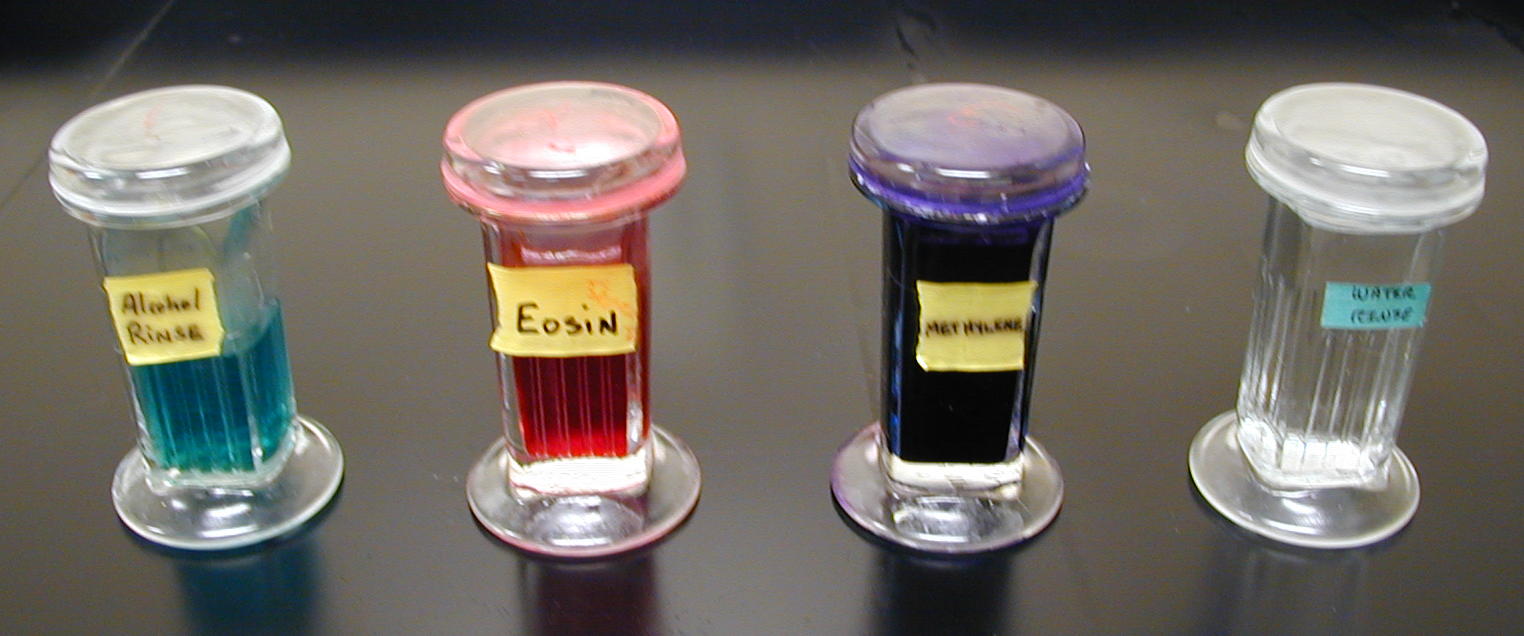
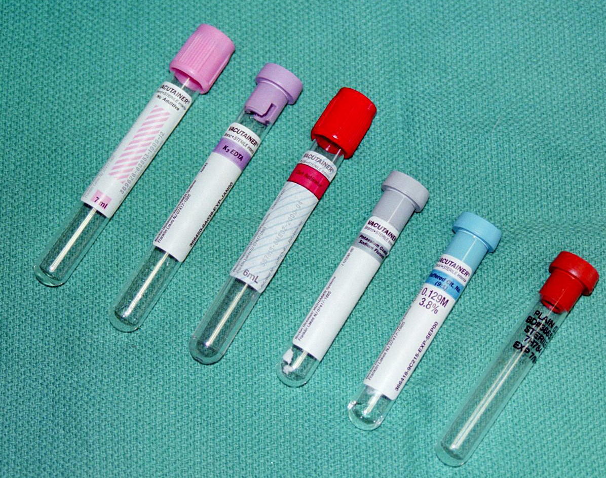
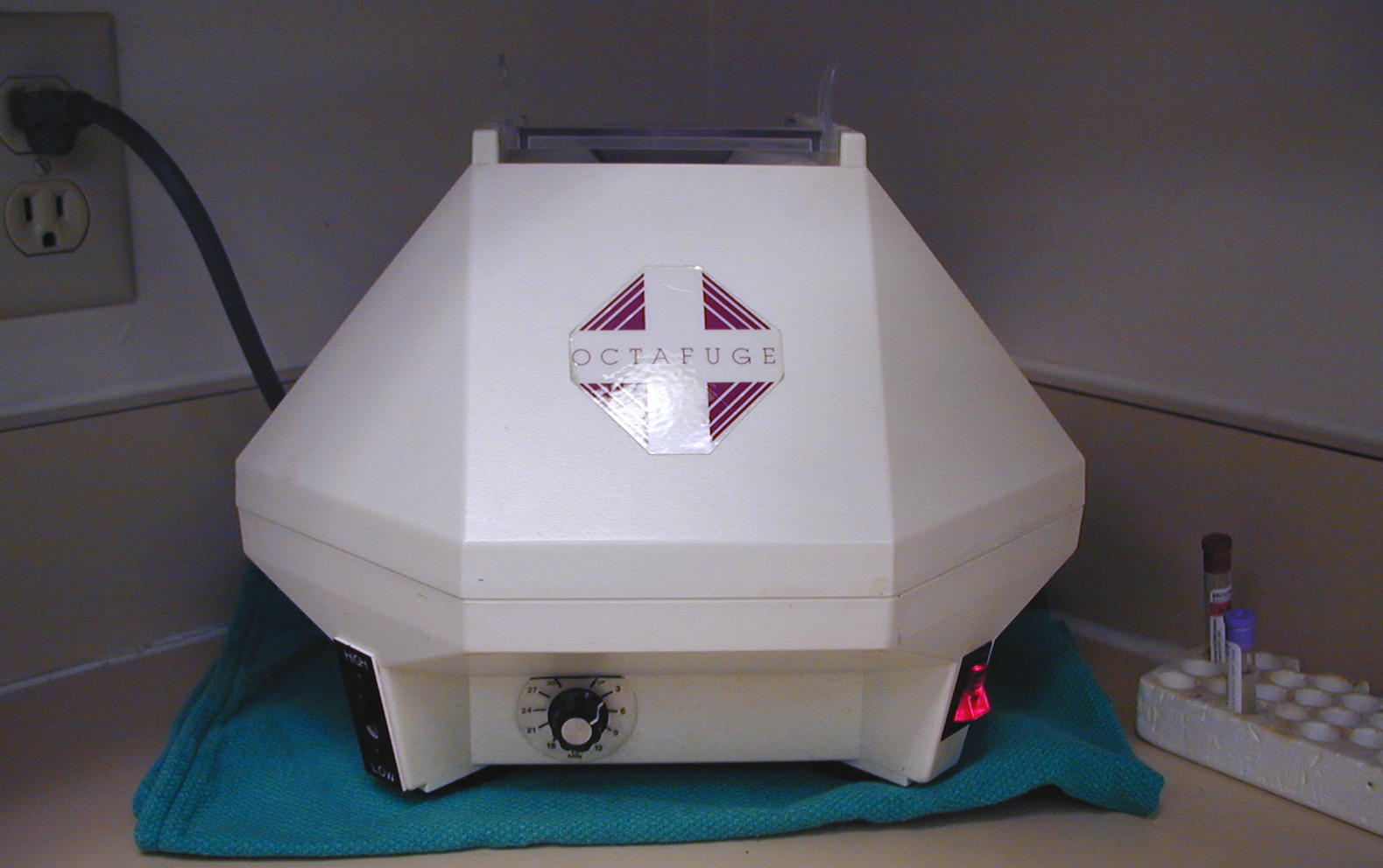
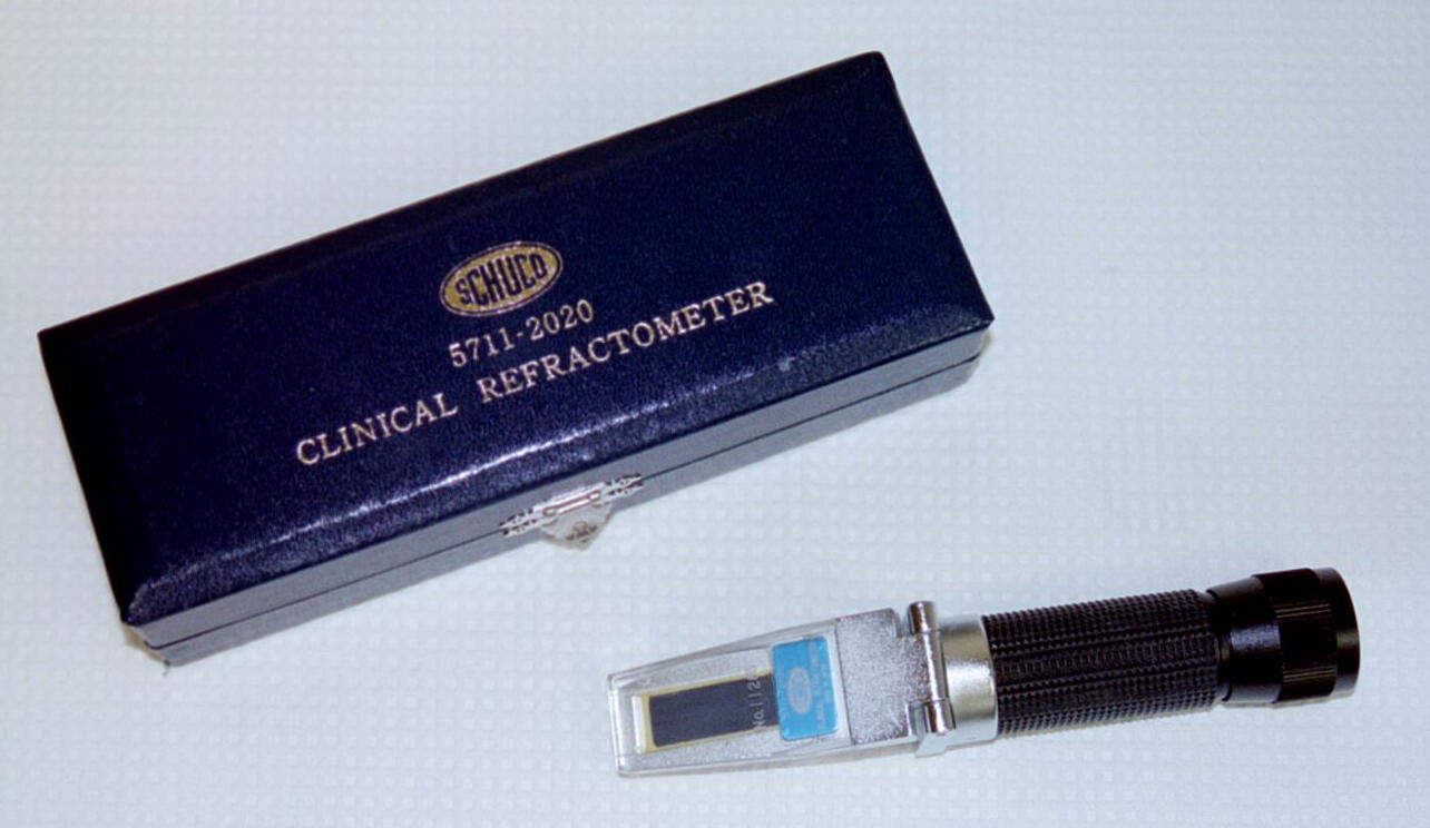
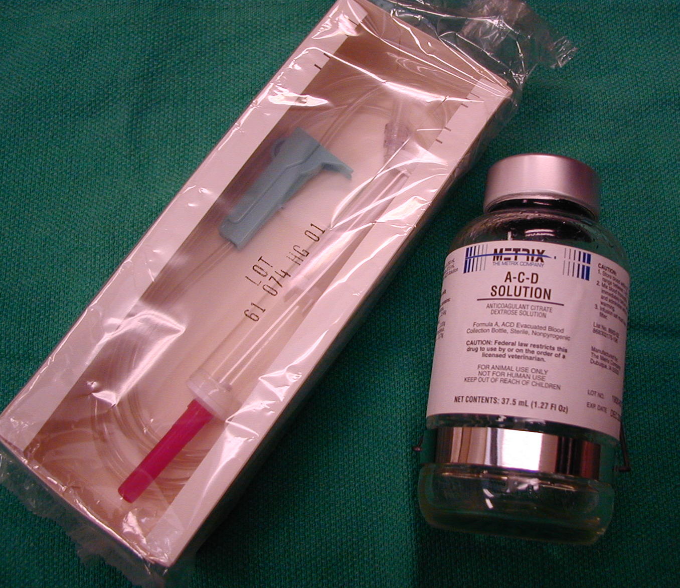
Equipment: stain, blood tubes, centrifuge, refractometer
Blood Web Site : click on specific formed elements of blood
Erythema- flushed pan- all
Erythr/o-. rubr/o- red -penia lack
Leuk/o- white -phil, -philia love
Macro- large -poiesis make
Mono- single -rrhage flow
-osis abnormal condition bol- throw
sanguin/o- blood thromb/o- clot
hemat/o- blood -crit separate
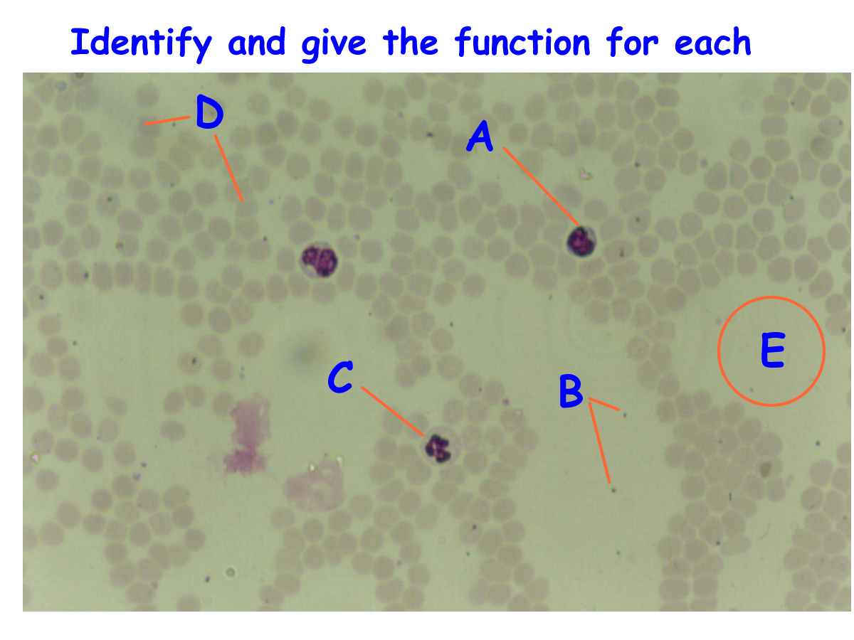
A) Drawings of Blood cells to ID
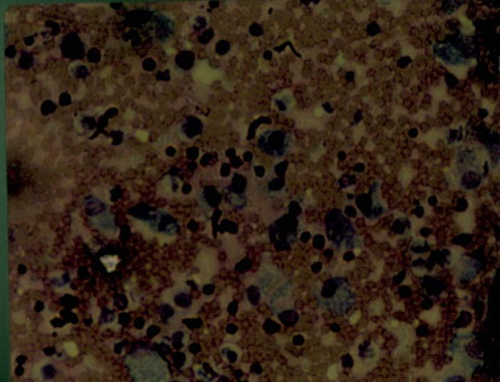
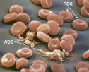
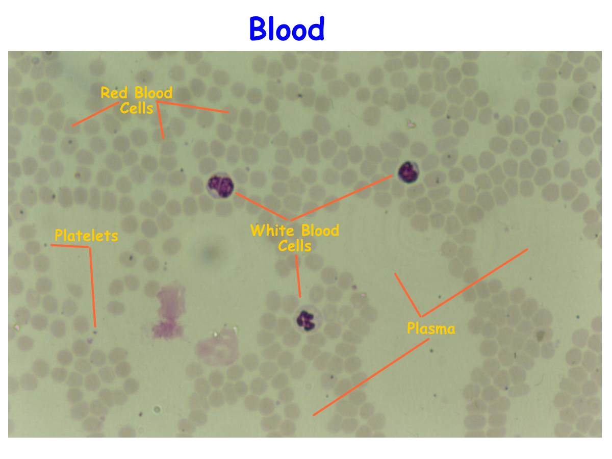
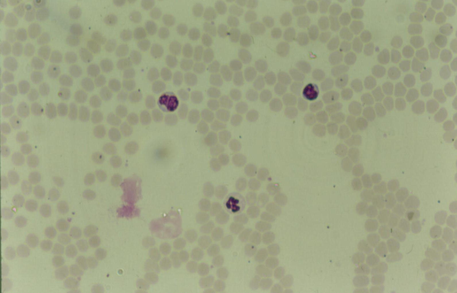
B) Histology ID of Blood cells
D) Blood type Scenario (go to this web site and treat your patient!)
E) Specimen : Identify region labeled
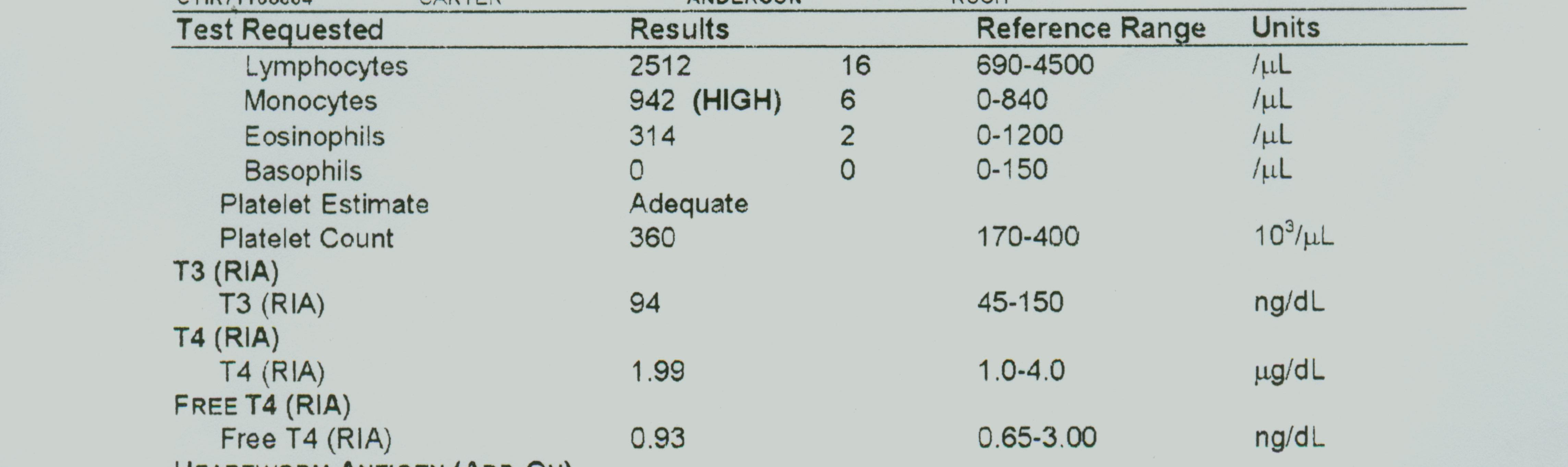
F) CBC reports to analyze: Report # 1, Report # 2 , Report #3
Blood lab (click here to quiz yourself on blood)
Concept Map: Make a concept map of the blood (source, cells, location, function). Include this map in your LAR lab report (if selected) as a document insert or as an addtional PDF document scan.
Anemia: Iron deficiency, Pernicious anemia, Sickle cell anemia, aplastic anemia,
hemolytic anemia, Folic Acid Deficiency anemia,
Polycythemia Vera
Jaundice: hepatic, prehepatic, hemolytic, posthepatic, obstructive
Hemolytic disease of the newborn (erythroblastosis fetalis)
Leukemia: acute lymphocytic leukemia (ALL), chronic myeloid leukemia, acute monoblastic,
Acute myeloblastic leukemia (AML), chronic lymphocytic leukemia (CLL)
Thrombus, Embolism
Hemophilia
Thalassemia
Malaria
Infectious Mononucleosis
Leukocytosis
Neutrophilia, Neutropenia
Eosinophilia, Eosinopenia
Basophilia
Macrocytosis
Lymphocytosis, Lymphopenia
Thrombocytosis, Thrombocytopenia
Bone Marrow Transplant
Phlebotomist
Blood Bank Technician
http://www.lumen.luc.edu/lumen/meded/histo/frames/histo_frames.html
http://www.mc.vanderbilt.edu/histo/blood/
http://www.nlm.nih.gov/medlineplus/bloodlymphaticsystem.html
1. Give four functions of blood
2. Give the composition and % of plasma
3. Give the description and specific functions of RBCs and two tests that can be used using RBCS.
4. Name the RBC antigens and corresponding plasma antibodies for the various blood types.
5. Give the description and specific function for platelets
6. Name each of the five WBCs and give their description and unique function
7. Define hemostasis and name the four stages
8. Define coagulation
9. Explain the difference between agglutination and coagulation
10. Name a hormone that affects blood.