Biology 2404 A&P Basics Lab Exercise 13 Heart Dr. Weis
| Objectives | Background | Medical Terms | Activities | Applications | Careers | WWW | Review Questions |
Students should be able to
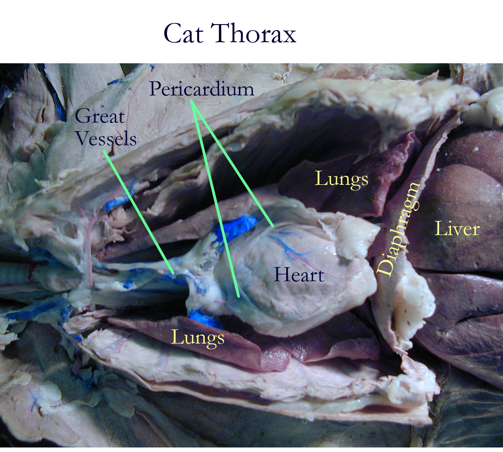
* describe the location of the heart and the pericardial membranes
* name the structures and associated functions of the heart wall
* name the chambers of the heart and their function
* name the 4 valves of the heart, their location and function
* describe blood flow through the heart
* name the components of the cardiac conduction system
* recognize a normal Lead II ECG tracing and describe the waveform meanings
* describe the cardiac cycle and its events
* Define cardiac output and give its formula
* List the factors that affect Stroke volume and Heart Rate
Read related material in textbook
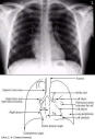
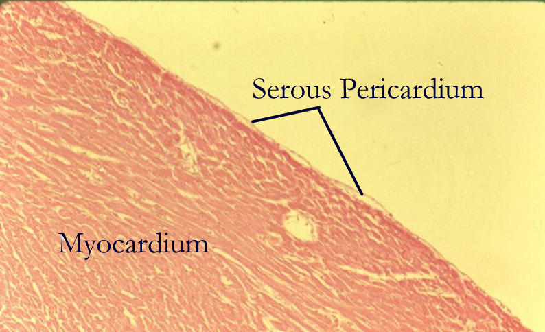
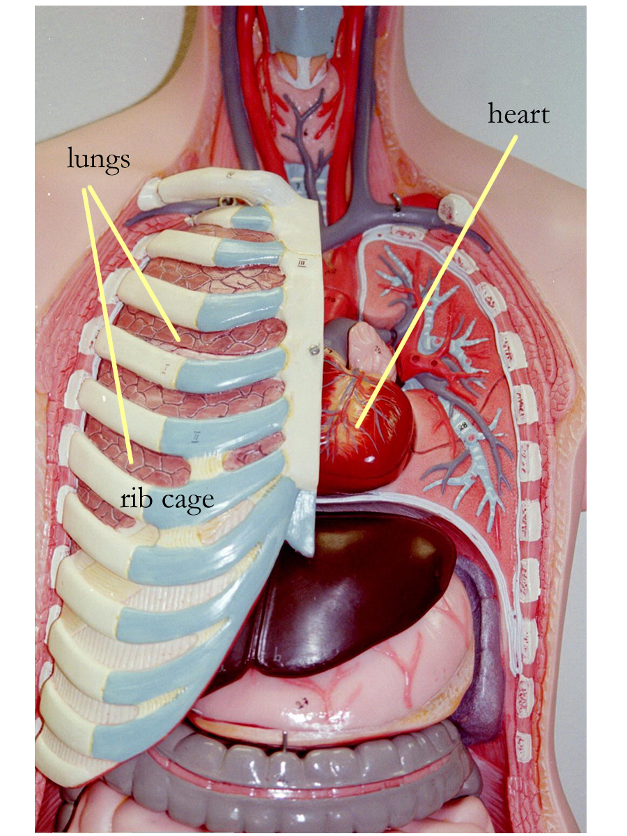
The embryonic development of the heart begins as two tubes that fuse and twist to form a functional pump by four weeks of age. The heart, located in the ventral chest, sits obliquely in a region called the mediastinum. The cone shaped, fist sized organ has a broad base where major vessels enter and leave and a pointed apex tip that lies mainly to the left of midline. Recall that most organs enclosed in a closed body cavity are covered by a serous membrane sac that secretes a watery, serous fluid. The sac that surrounds the heart is called the pericardial sac and is actually folded twice over the heart to create an outer fibrous pericardium, a parietal pericardium that is in contact with the throacic wall, and the visceral pericardium on the heart surface known as the epicardium. The serous fluid secreted by this membrane sac is called pericardial fluid which functions to decrease frictional forces of movement between the heart and lungs.
Normal Heart Size
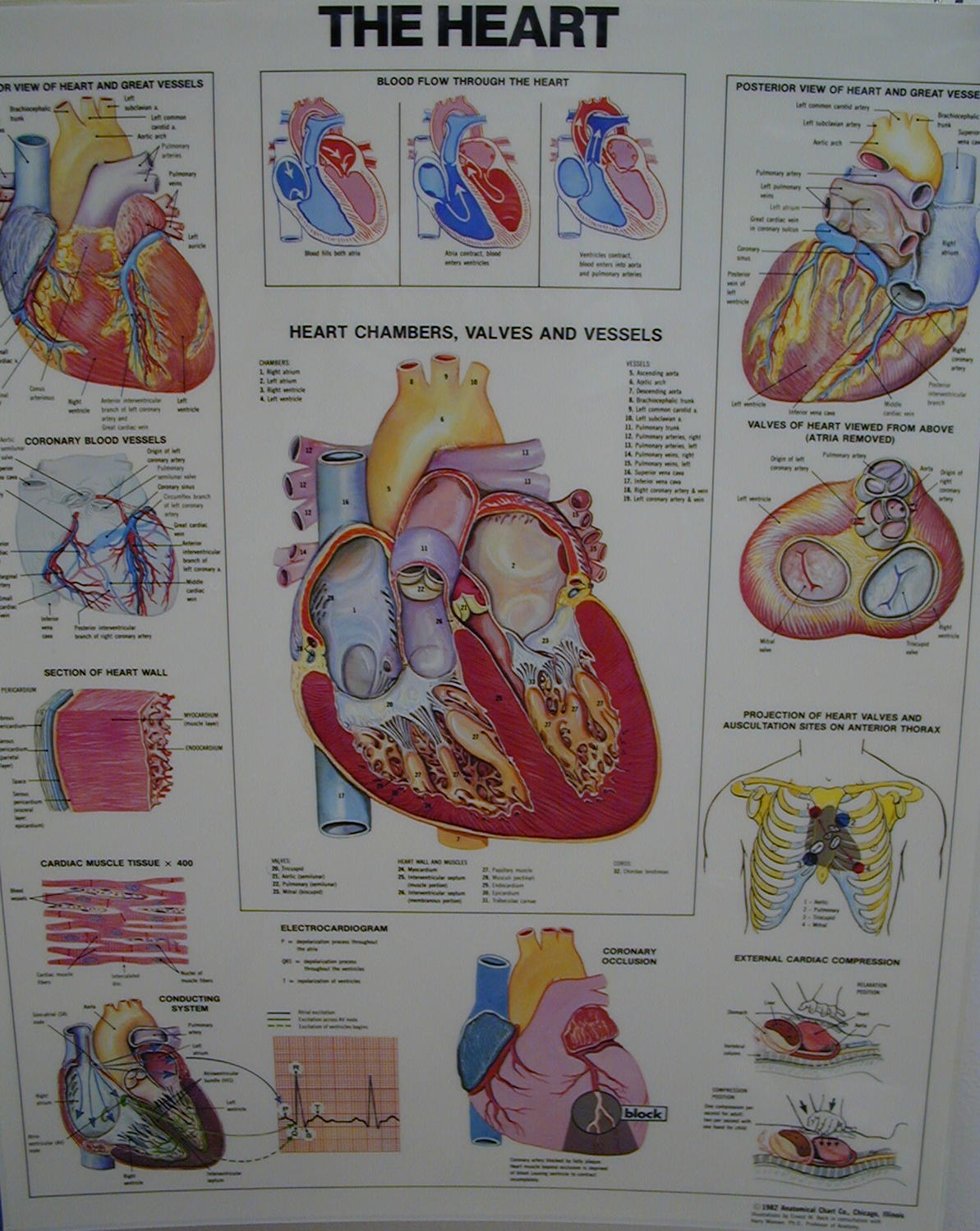
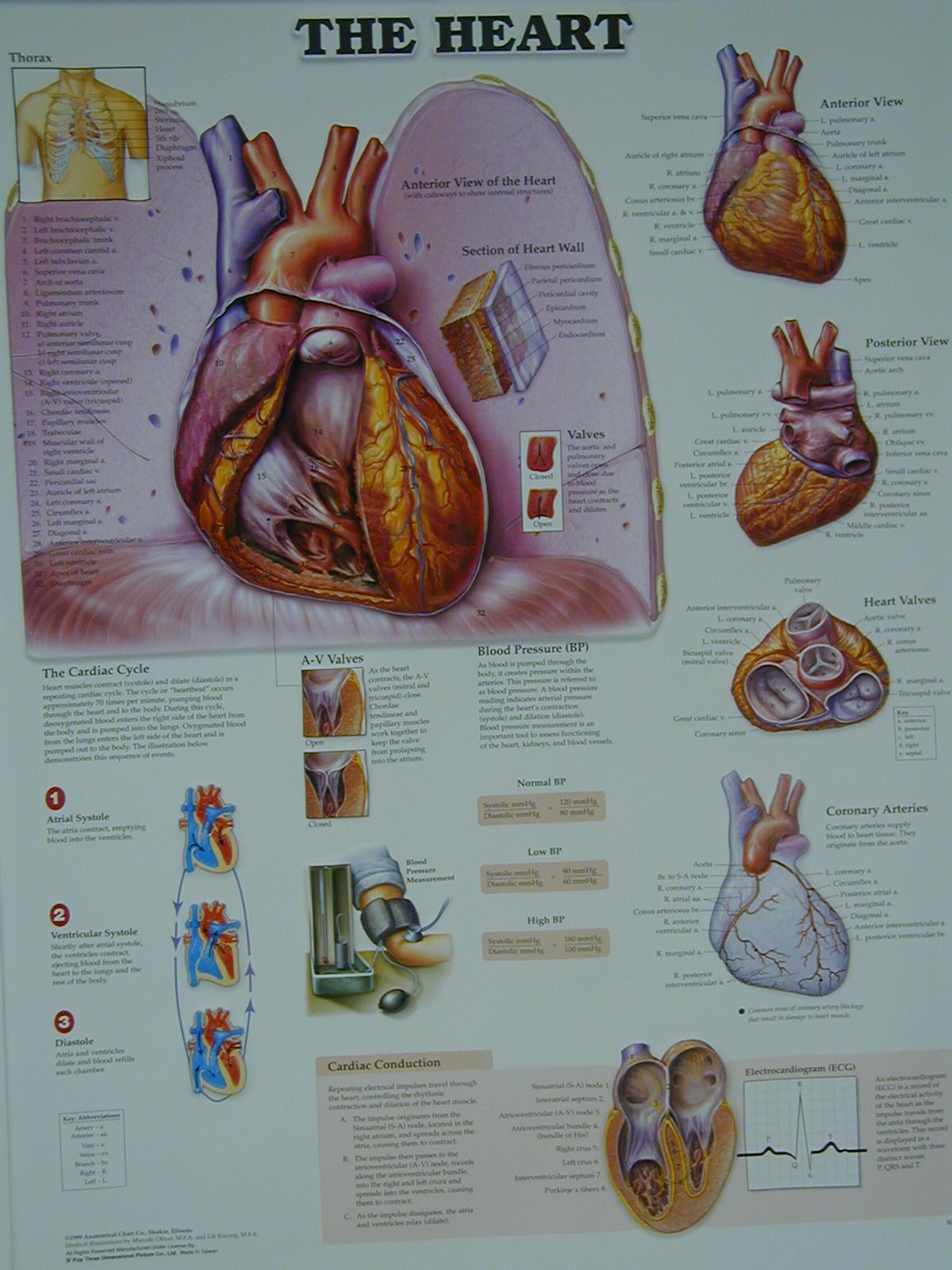
The heart wall is composed of three layers:
Epicardium simple squamous epithelium that forms the visceral pericardium
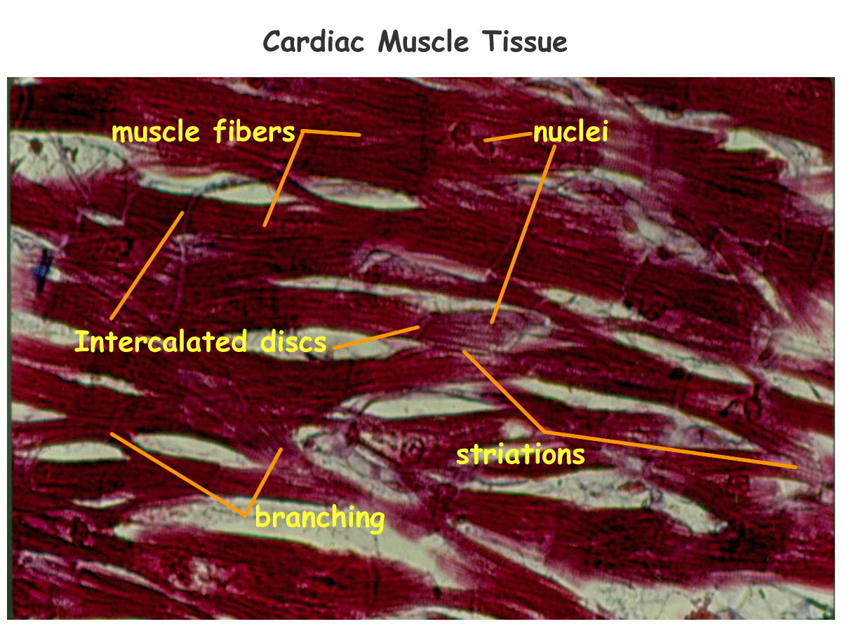
Myocardium cardiac muscle that forms the chamber pumps
Endocardium simple squamous epithelial lining of the cardiovascular system
As the heart develops, it forms walls or septums that create the 4 chambers. The upper chambers called atria are divided into right and left sides by the interatrial septum.
The lower chambers called ventricles are divided into right and left sides by the interventricular septum. The septum grows together from top and bottom portions that meet in the middle. Failure to close will create a septal defect between the chambers known as either an ASD (atrial septal defect) or VSD (ventricular septal defect)
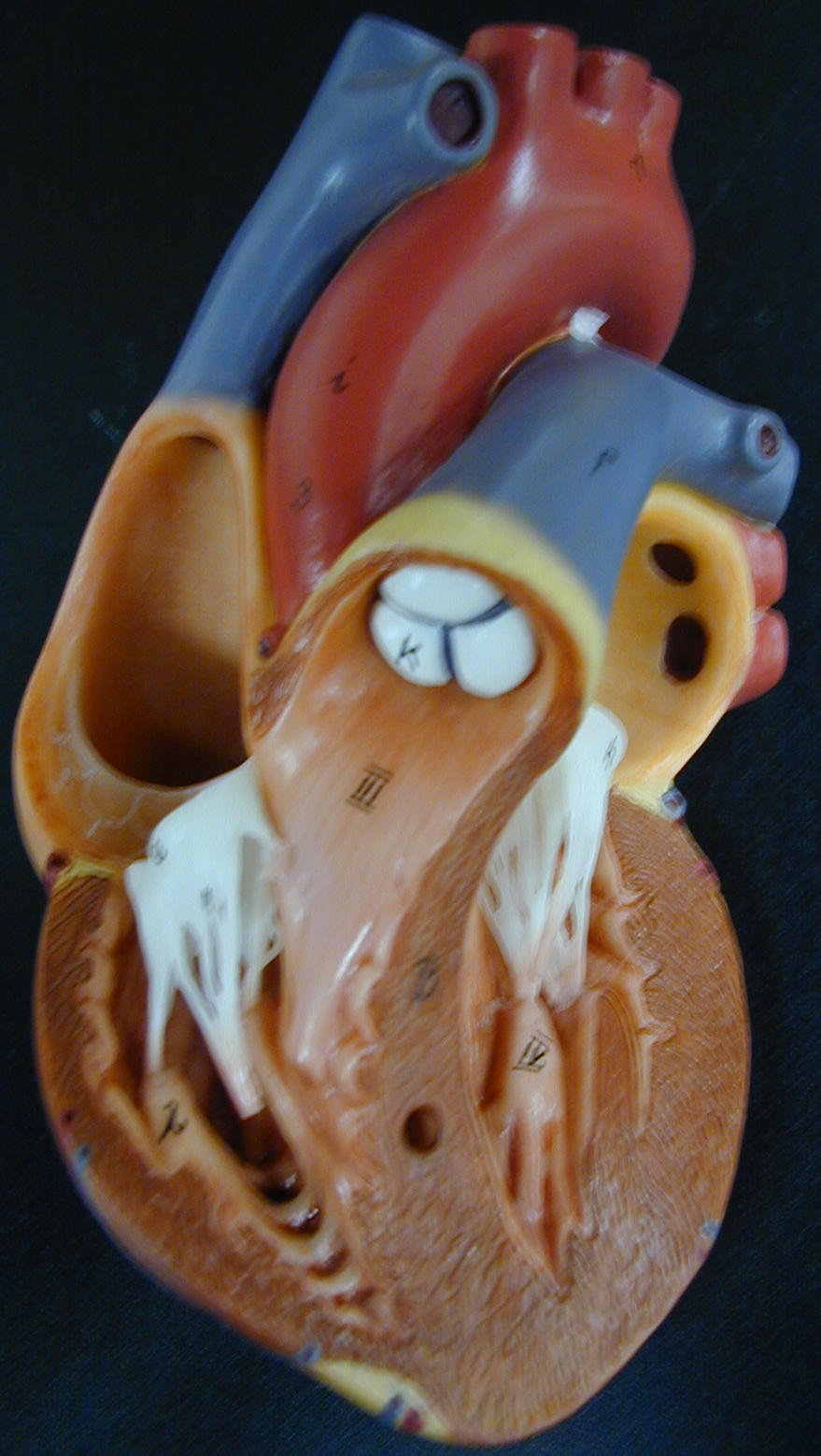
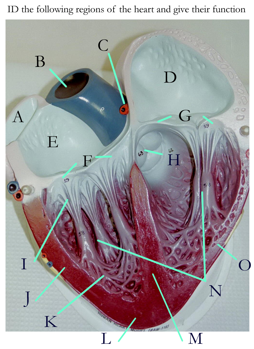
Normal Heart Interior Transverse Section
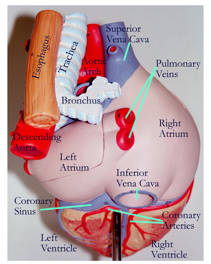
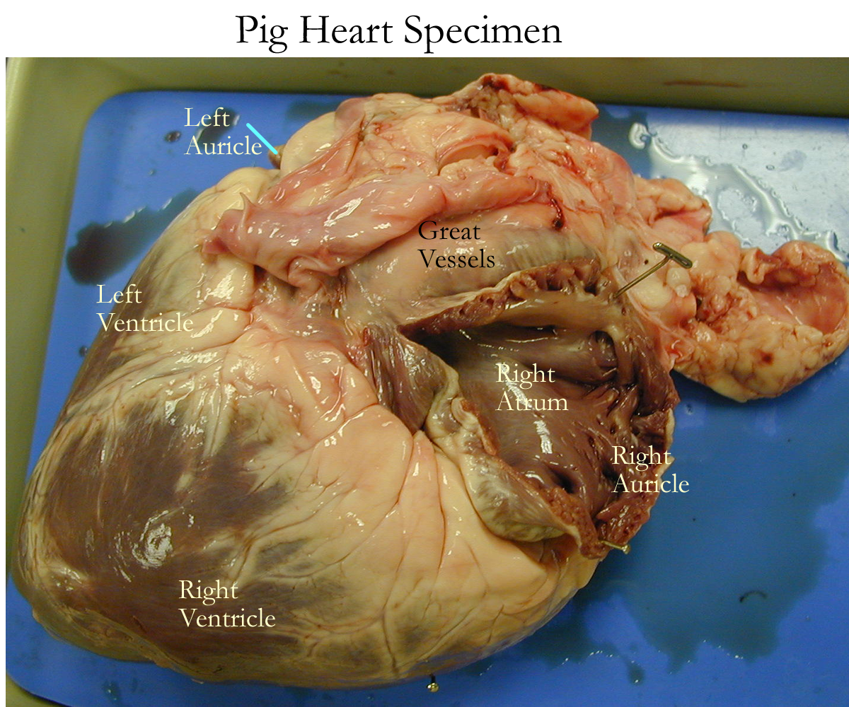
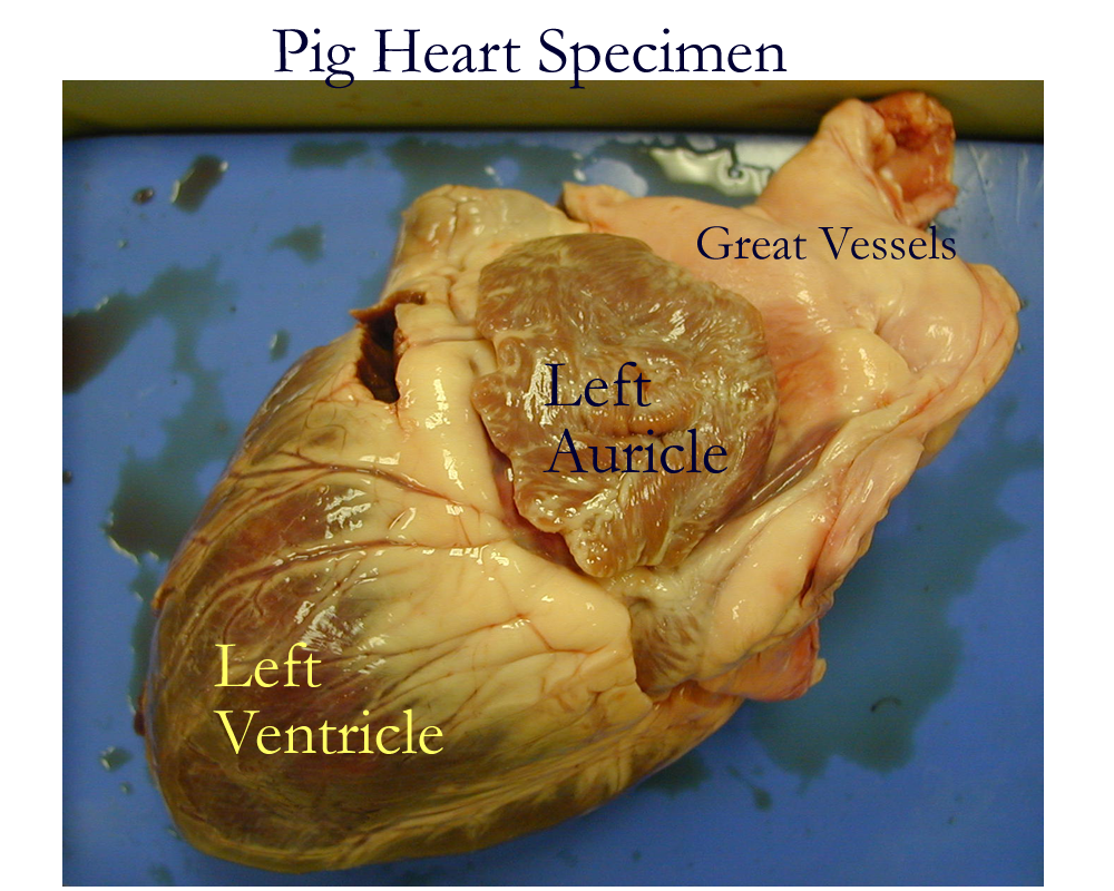
External extensions of the atria form ear flap appendages called auricles which function to help increase the volume of blood that can be received by the atria. The atrial lining is smooth except in the regions of the auriclar appendage where the muscle wall forms bundles called pectinate muscle. Vessel openings in the atria allow blood to be returned from body organs. The right atrium has three vessel openings: the coronary sinus, the superior vena cava, and the inferior vena cava that return blood from all the body organs. The left atrium has four vessel openings: two right and two left pulmonary veins that return oxygenated blood from the lungs back to the heart to be pumped to body organs.
The functions of the atria are:
a) to collect blood returning from the body
b) to pump remaining blood to the ventricles
c) to produce ANP (atrionatriuretic) peptide hormone (by the right atria) that helps regulate blood pressure
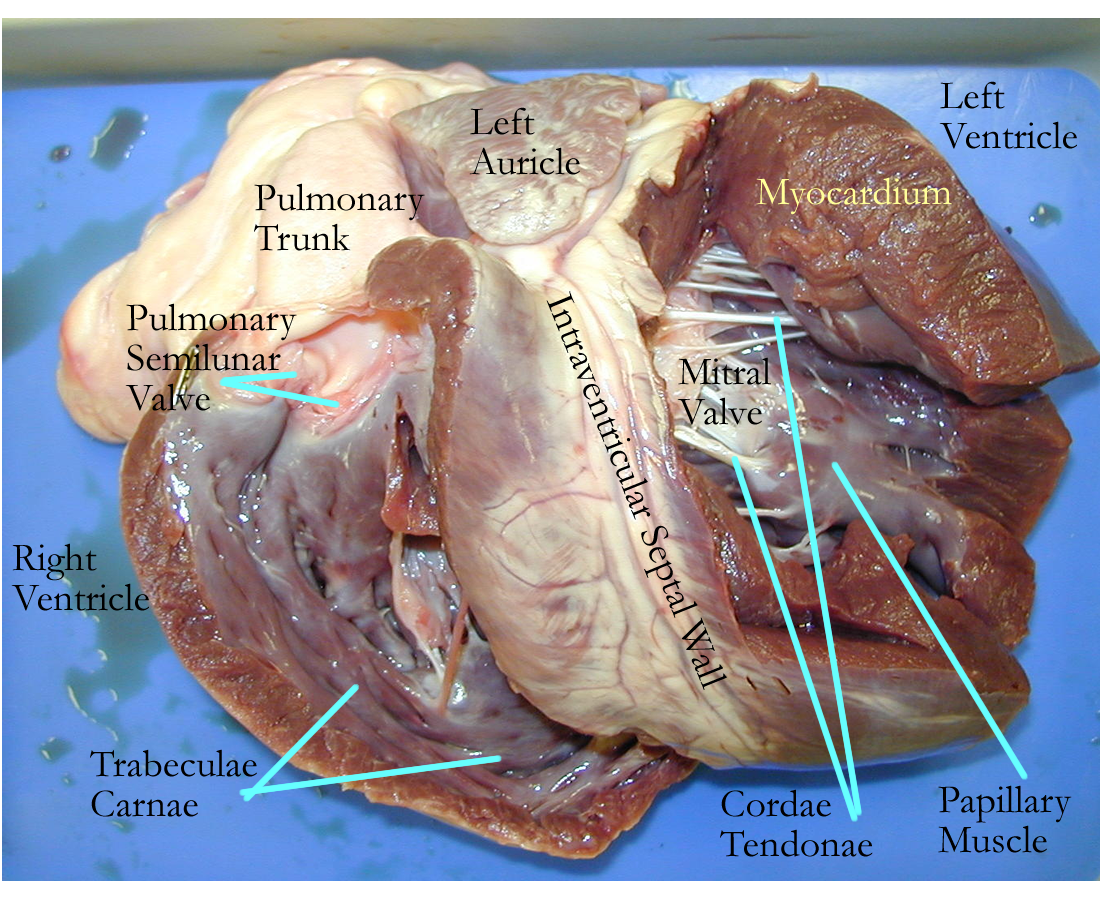
Ventricles
The ventricles have several associated structures common to both left and right sides. Their muscle bundles are called trabeculae carnae that help increase surface area and aid in contraction of the chamber. Finger like projections from the ventricle wall are called papillary muscles and are linked to the atrioventricular valves by connective tissue bands called chordae tendonae. The papillary muscle and chordae tendonae help anchor the valve leaflets and prevent them from inverting into the atria. The ventricles also each have a semilunar valve associated with the great vessel that leaves the chamber.
The differences in the right and left ventricular walls of each chamber are associated with their functions as pumps to generate the pressure needed to keep blood moving forward. The right side of the ventricle is thinner since the blood pumped from this region goes to the lungs for gas exchange. The left side of the ventricle is much thicker since blood pumped from this region has to go to all the body organs, above and below the heart.
The functions of the ventricles are:
a) collect blood from the atria
b) to pump blood to body organs
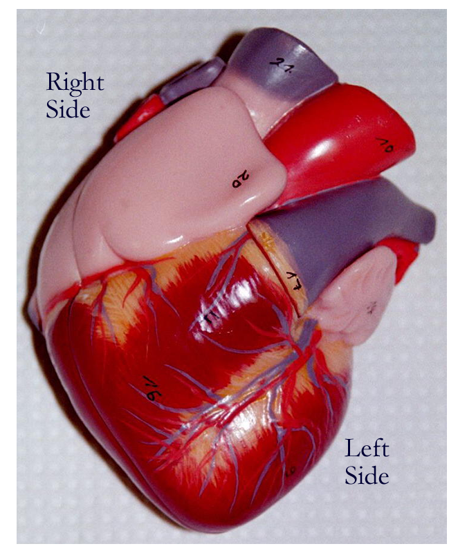
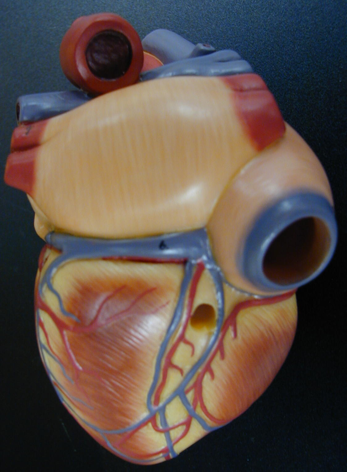
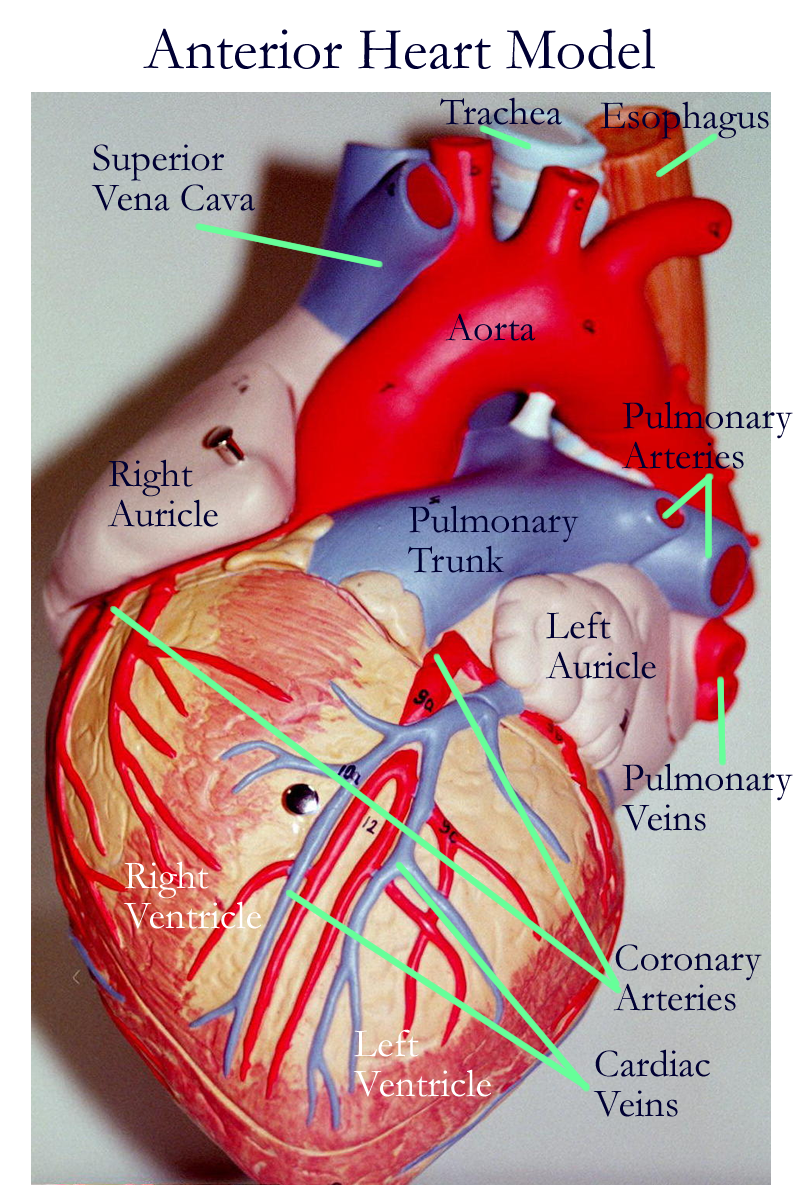
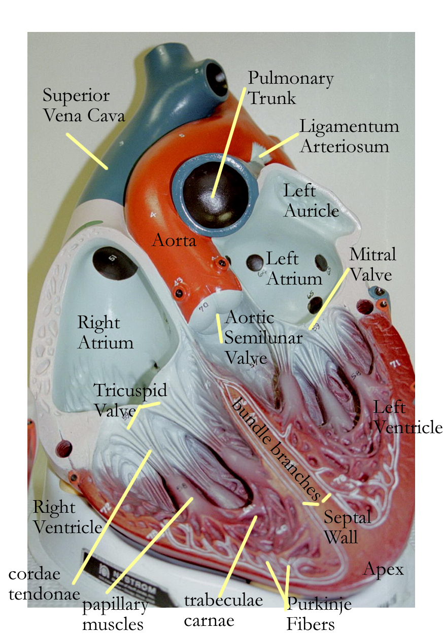
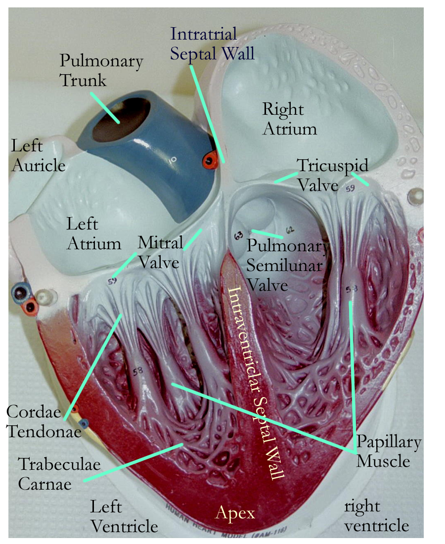
Heart model: Anterior, Posterior
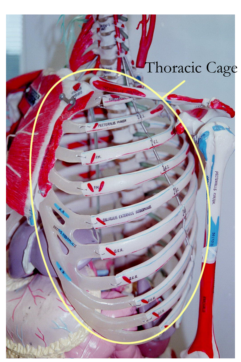
Organman: Thoracic Cage, Thoracic Organs
Body Torso: Thoracic Cage, Transverse Sectioned
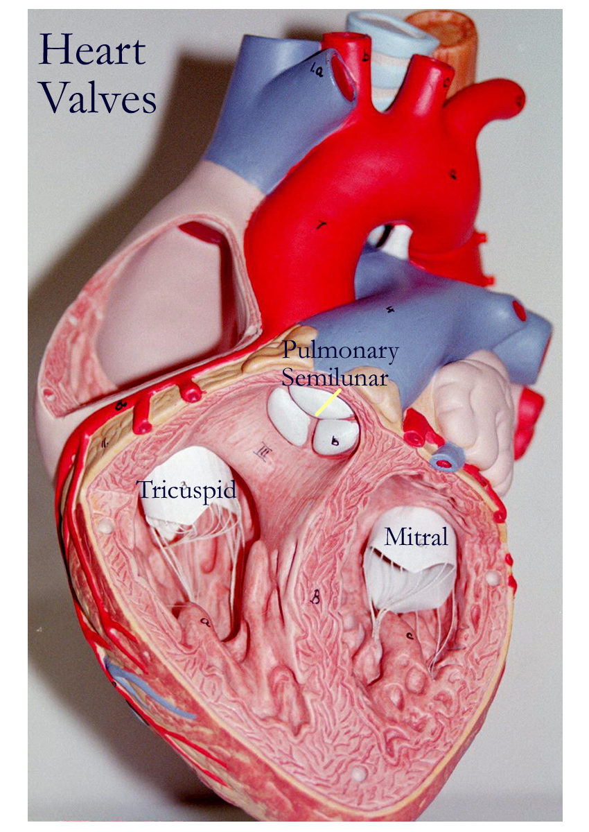
Heart valves
Heart valves are connective tissue flaps covered by the heart lining called the endocardium. Valves are passive in their movement, that is, they do not actively open and close since no muscle is present in their structure. Valves open and close based on pressure gradients created by the heart muscle pumping to move blood. All valves are designed to prevent backward flow of blood.
The four cardiac valves are:
a) Right atrioventricular (AV) valve or tricuspid valve, between the right atria and ventricle
b) Left atrioventricular (AV) valve or bicuspid or mitral valve, between the left atria and ventricle
c) Pulmonary semilunar valve, between the right ventricle and the pulmonary trunk
d) Aortic semilunar valve, between the left ventricle and the aorta
As seen in the above description, the atrioventricular valves have other names based on their anatomy. The right atrioventricular valve is also called the tricuspid valve since this valve is formed by three flaps or cusps. The left atrioventricular valve is known as the bicuspid valve since this valve is formed by two cusps or flaps. When these two flaps close, they resemble a mitre angle or mitre cap, so the clinical name for this valve is the mitral valve. It is under the greatest pressure and due to its structure and other related anatomical structures, it can fail to open (stenotic) or close (insufficient).
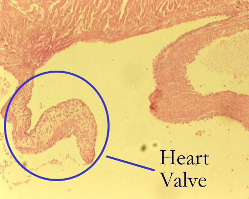
Heart valve
Pig specimen: Right Side Heart, Left Side Heart
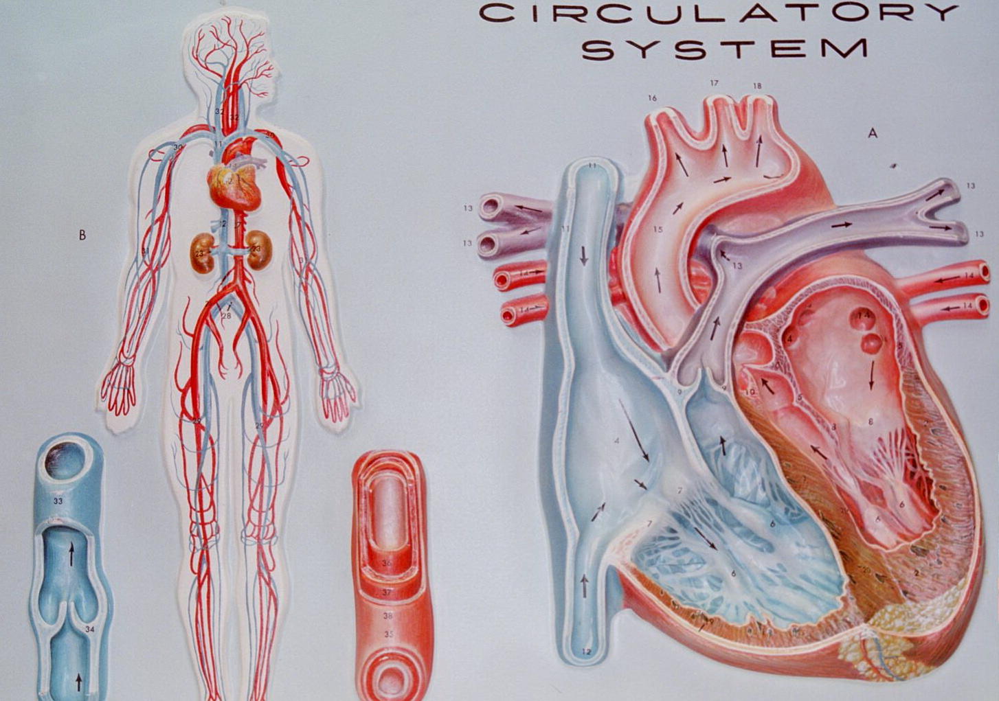
Blood flow through the heart
The heart muscle pumps blood through the series of vessels that form two major circuits: the Pulmonary circuit and the Systemic Circuit. Blood returning from the body enters the right atrium via the coronary sinus from the heart; itself the superior vena cava from the head, neck, and arms; and the inferior vena cava from the trunk and legs. Blood also is returned to the left atria via the right and left pulmonary veins from the lungs.
Blood is then allowed to enter the ventricles through the open AV valves. The atria contract simultaneously to pump any remaining blood into their respective ventricles.
In the pulmonary circuit, the right ventricle pumps blood through the pulmonary semilunar valve into the pulmonary trunk and pulmonary arteries to be delivered to the lungs for gas exchange. Blood returns to the heart through the pulmonary veins into the left atria. In the systemic circuit, the blood in the left ventricle is pumped through the aortic semilunar valve into the aorta for all the body organs. Blood then returns to the right atria to end the systemic circuit and begin the pulmonary pathways.
Heart model: Posterior Interior, Anterior Interior
Heart Physiology
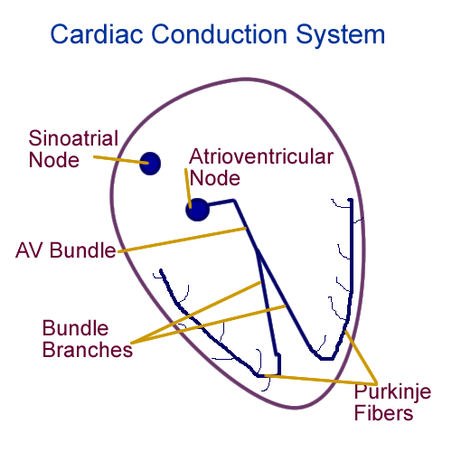
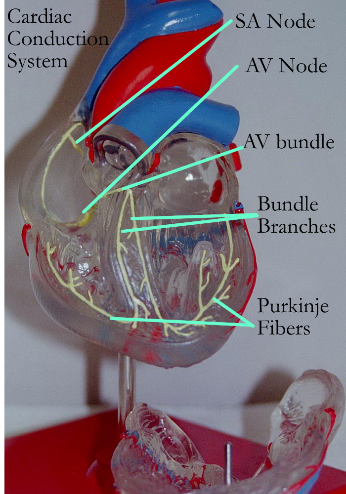
The heart has its own intrinsic (internal) electrical cells. These are modified cardiac muscle cells that are specialized to initiate and respond to electrical (ionic) changes. These cells can be regulated and modified by the ANS. The cardiac conduction system consists of the:
A) Sinoatrial (SA) node in the right atria near the SVC
B) Atrioventricular (AV) node in the right atria near the Rt AV valve
C) AV bundle & junction in the interventricular septum
D) Bundle Branches in the interventricular septum
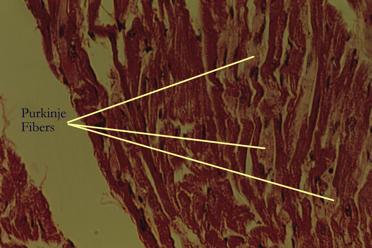
E) Purkinje Fibers in
the ventricular free walls
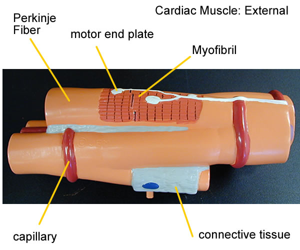
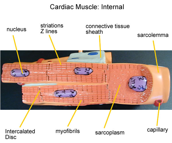
Heart Histology Model
The conduction system of the heart can be represented electrically by recording the changes in ion movement. This recording is called an electrocardiogram or ECG.
Wave deflections on the tracing represent ion movement and reflect how the conduction system is working. The first wave deflection is the p wave and it represents atrial depolarization triggered by the SA node firing. The second deflection wave is a group of three waves called the QRS complex. It represents ventricular depolarization triggered by the firing of the AV nodal system: the AV node, AV bundle-junction, Bundle Branches, and Purkinje fibers. ECG abnormalities are generally termed arrhythmias and can represent problems in the conduction system, ionic concentrations, heart muscle response, and ANS control.
Drawing of Cardiac Conduction System
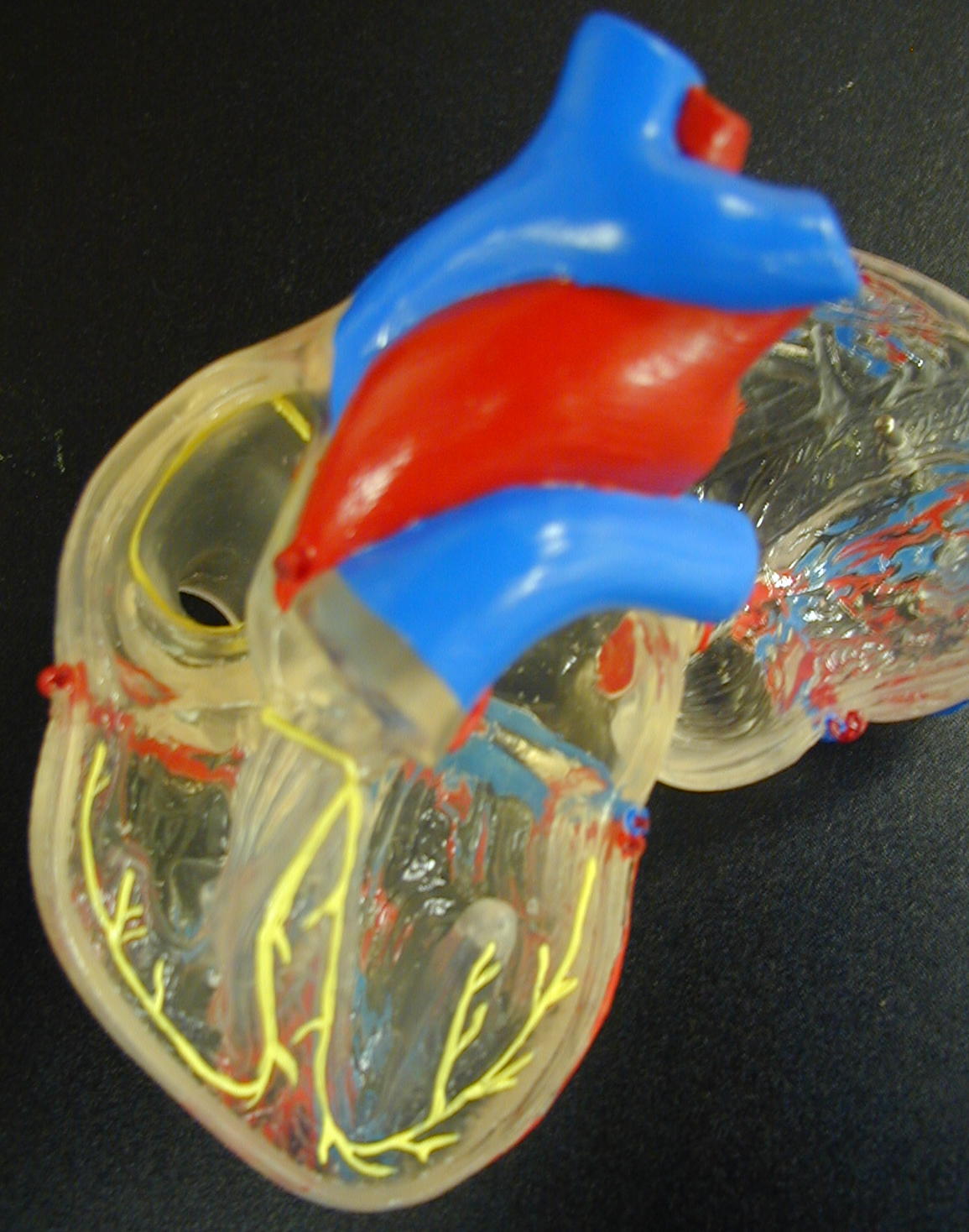
Heart Model with Conduction System
Clear Heart Model with Conduction System
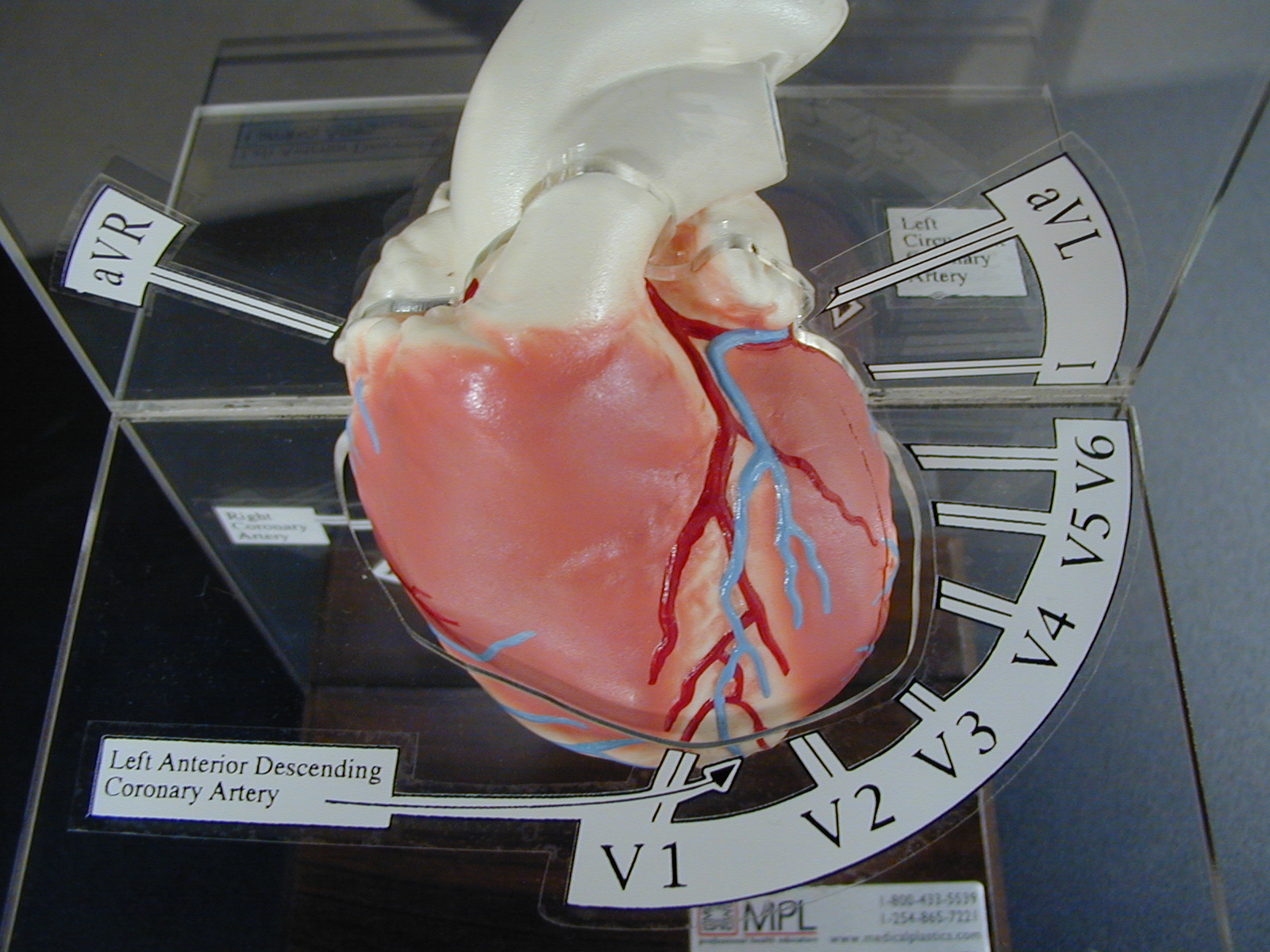
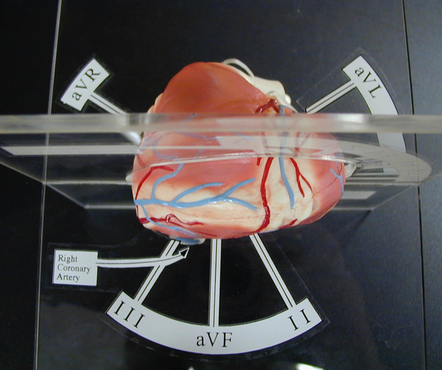
ECG Heart with Leads
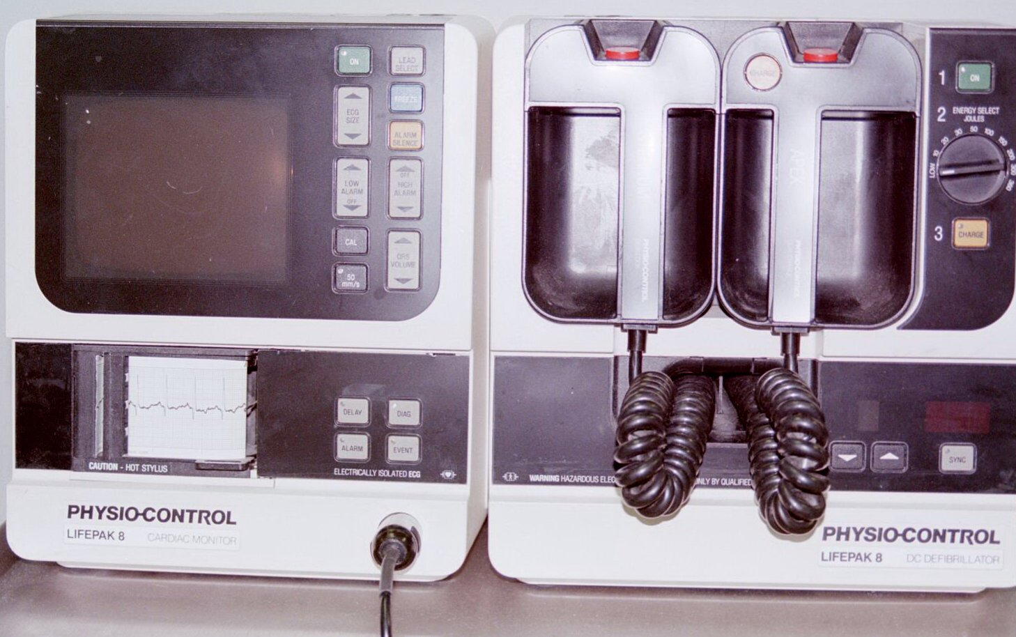
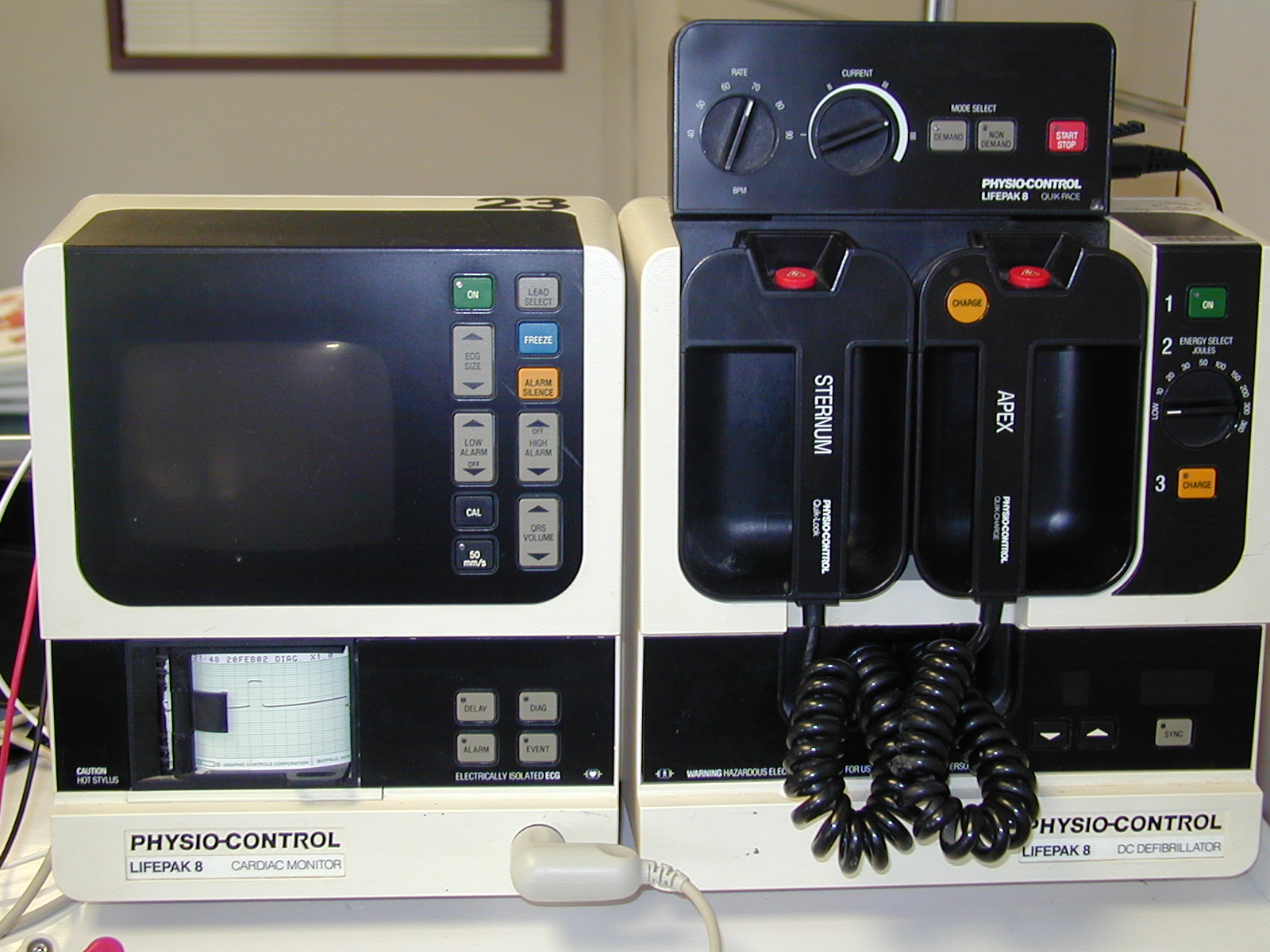
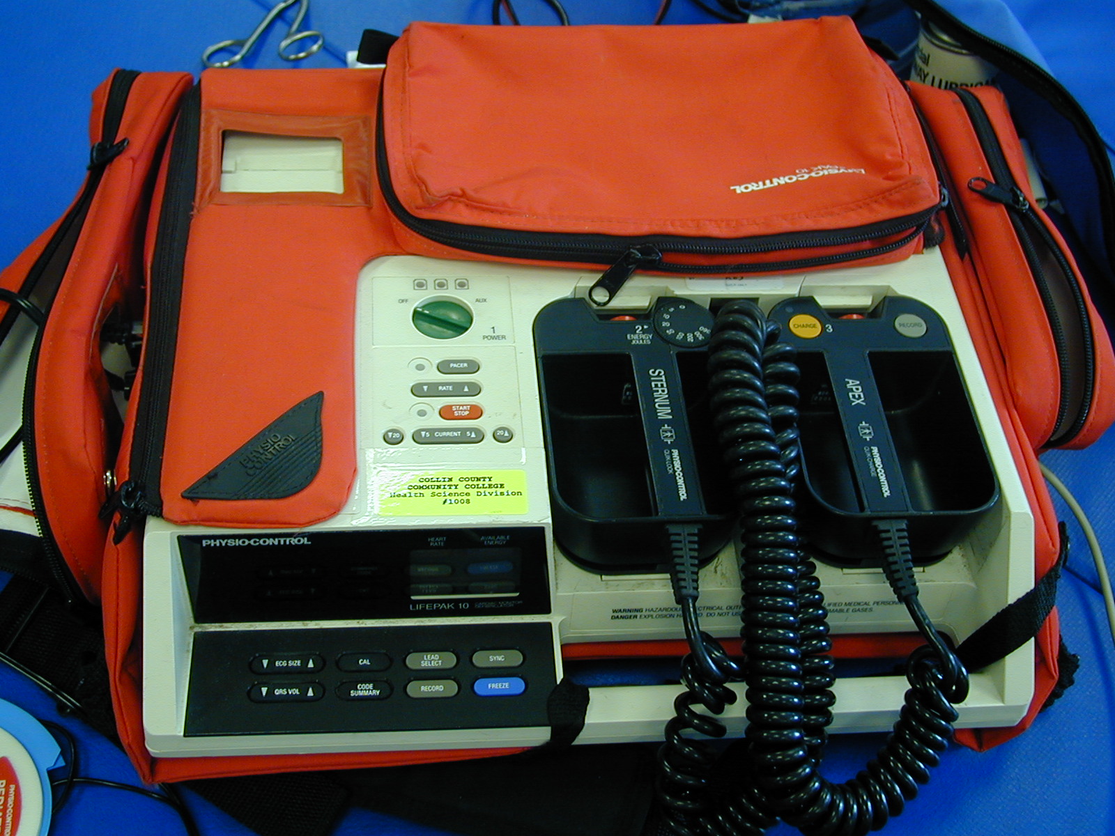
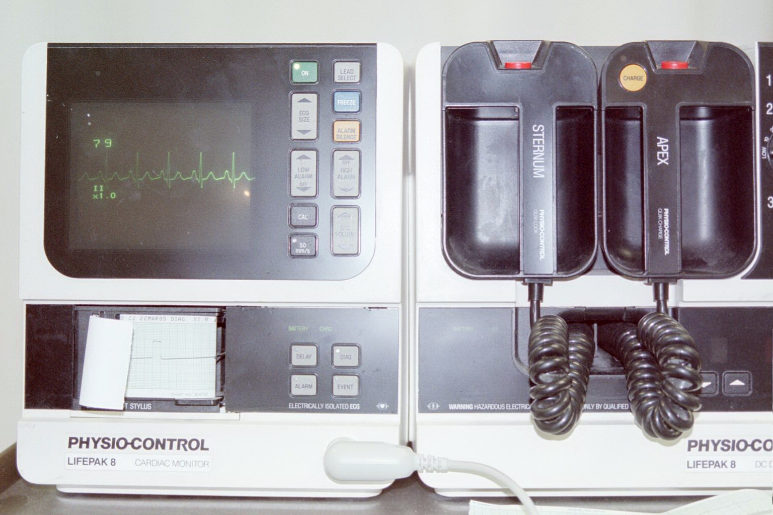
ECG machines: Field, Lifepack 8, Lifepack 10
ECG tracing: Drawing of wave forms
ECG tracing, Lead II on Machine
ECG tracing, 12 leads
Cardiac Cycle
The heart’s electrical events tie to mechanical events in a sequence marked from the beginning of one heart beat to the next. This cycle, known as the cardiac cycle, involves the contraction and relaxation of the heart. During contraction or systole, the heart muscle pumps blood and during relaxation or diastole, the heart chambers fill with blood. Since the heart muscle contracts and relaxes, it pushes blood creating pressure changes that open and close the heart valves. When each set of heart valves close in relation to the heart movements, there are two heart sounds that are heard.
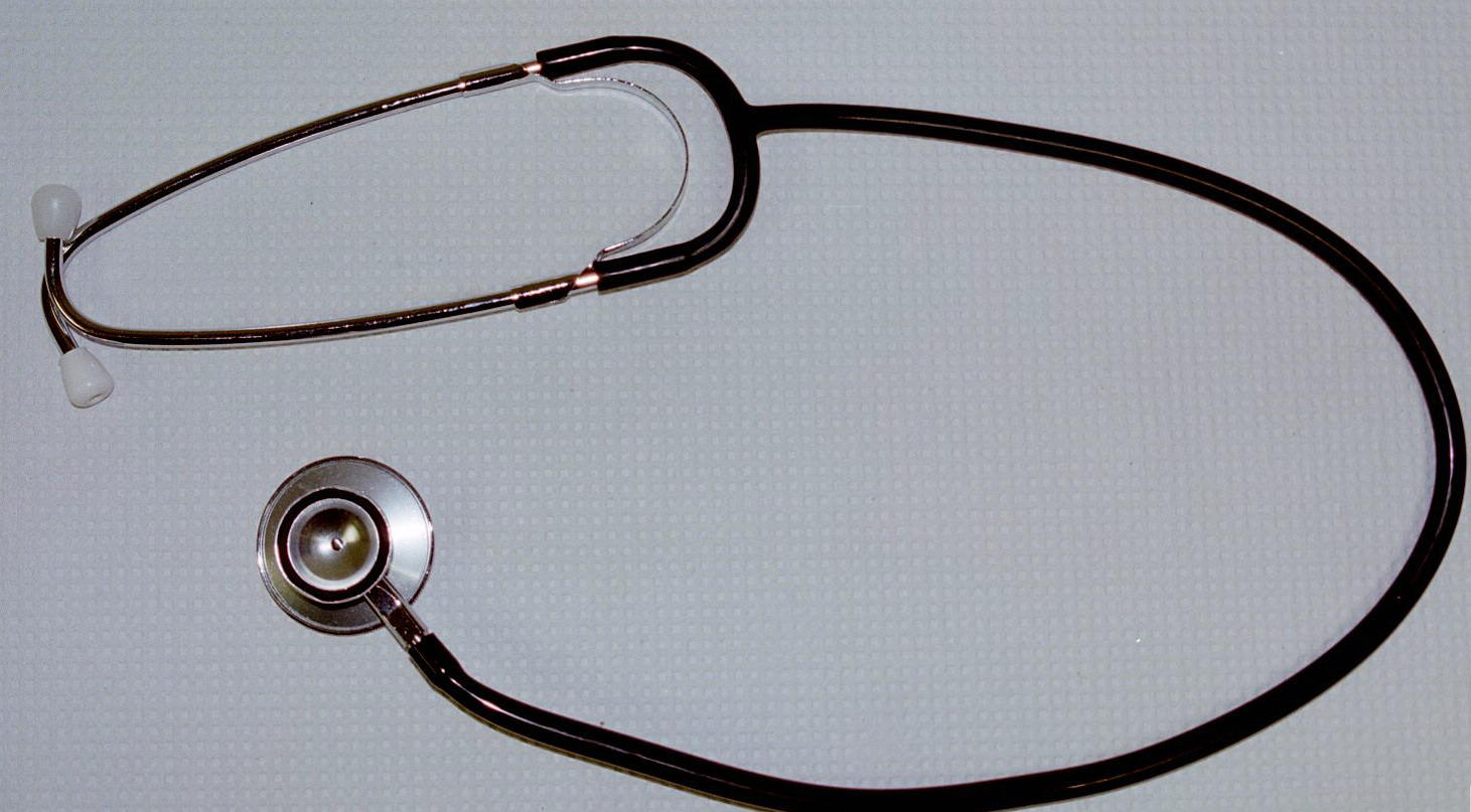
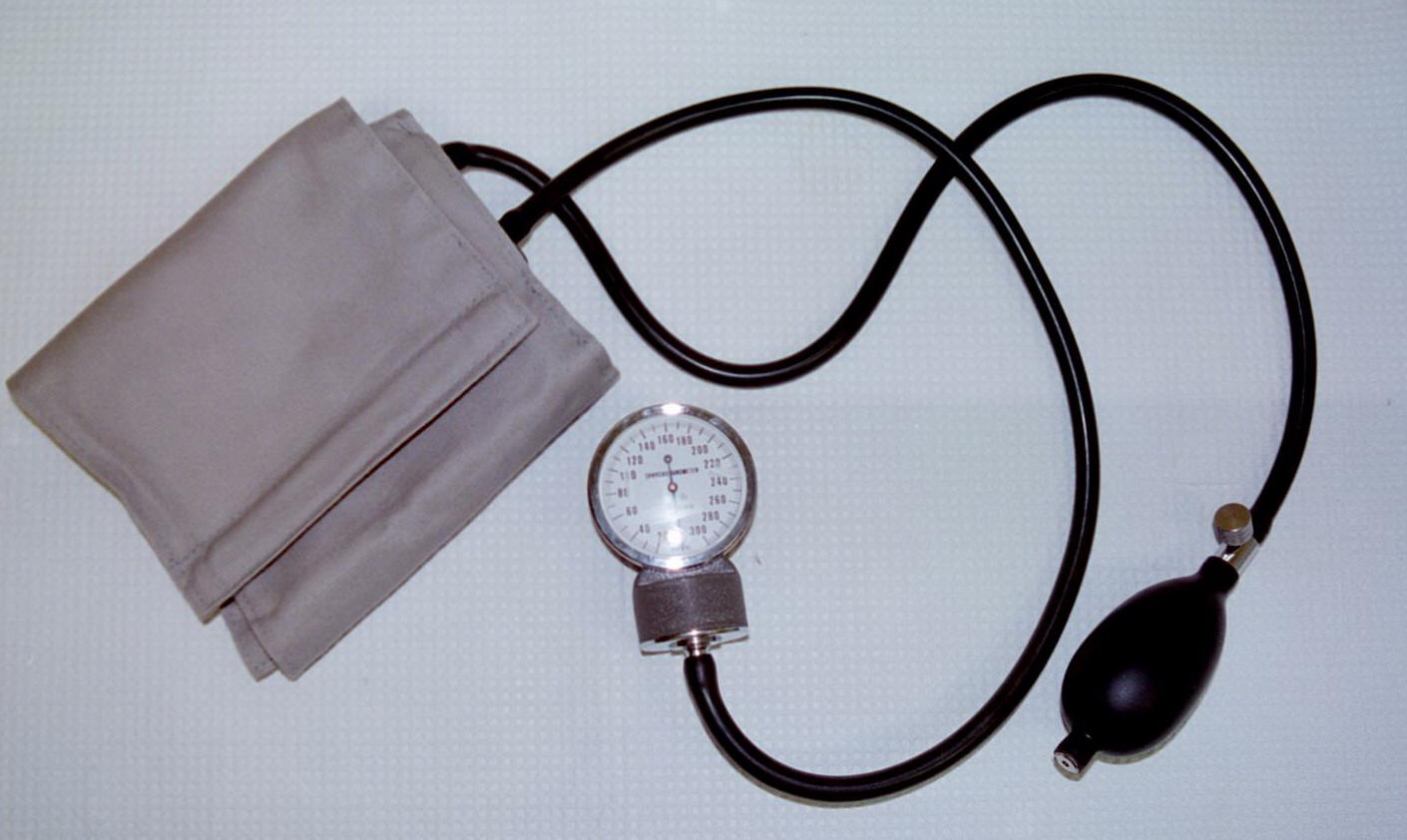
The first heart sound, “lub”, occurs when the left and right AV valves close at the beginning of the ventricular systole. The second heart sound, “dup”, occurs when the pulmonary and aortic semilunar valves close during ventricular diastole. Heart sounds can best be heard by using a stethoscope over the chest wall at specific points between the 3rd and 5th intercostal spaces.
Cardiac Output
When the heart muscle contracts, a certain amount
of blood is pumped.
The amount of blood pumped by a ventricle in one minute
is called the cardiac output.
This amount of blood flow can also be represented mathematically as
CO = SV x HR.
There are other means to calculate cardiac output such as the Fick principle.
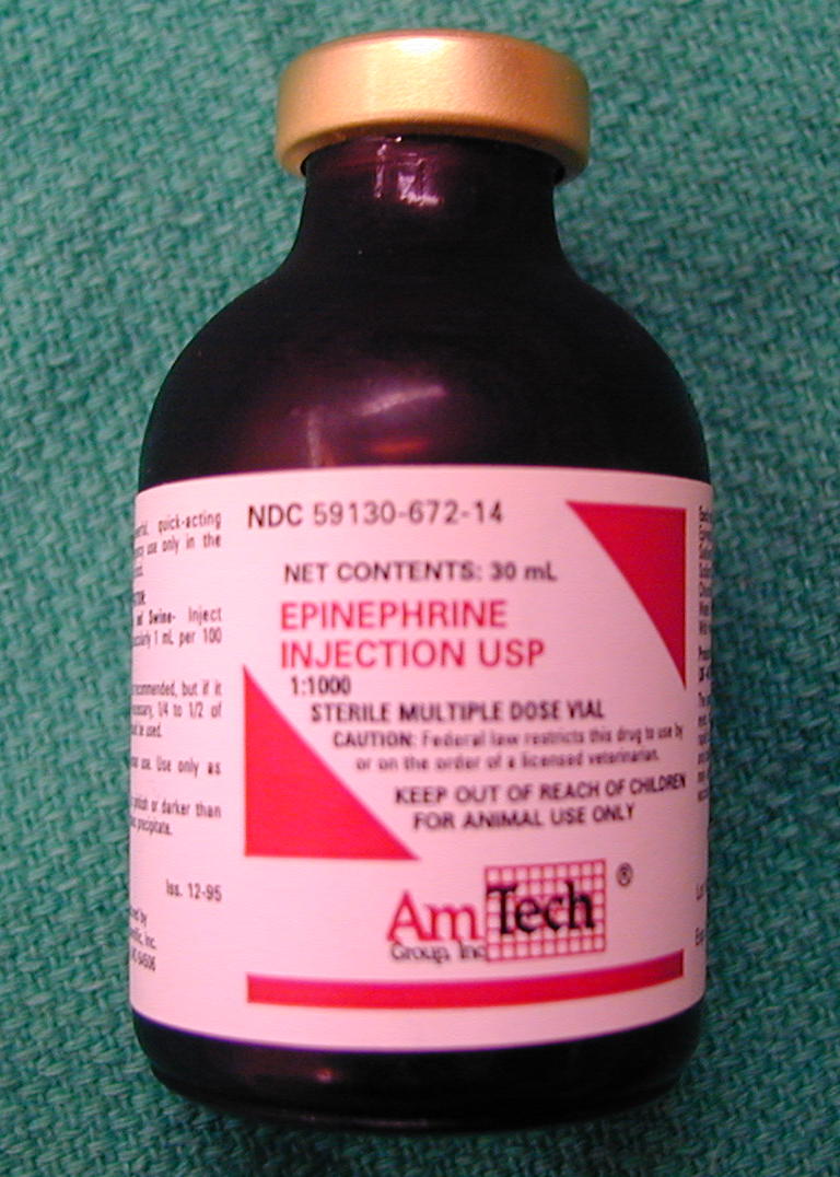
Heart rate (HR) is set by the interaction between the SA node and the AV node. The ANS modifies the response at these nodes to help keep the heart rate at ~ 72 beats per minute. Although the ANS is considered a peripheral control mechanism, it is tied centrally to the brainstem at the medulla. Higher brain centers at the diencephalon and cerebrum can further modify and regulate HR. Other peripheral controls detect blood pressure changes as well as chemical changes by monitoring the respiratory gases O2 and CO2. These receptors are located in large elastic vessels like the aortic arch and carotid arteries. Other chemicals such as hormones like Epinephrine can affect dramatically affect HR.
Stroke volume (SV) is the difference in the volume given to the ventricle to pump and what remains after systole (contraction). Stroke volume is affected by how much blood is returned to the heart, known as venous return, and how hard the heart has to pump to keep blood moving forward, known as afterload. The ANS can also affect SV by changing heart rate and force of contraction.
Another way to represent heart function is to calculate the percentage of blood pumped by the heart. This percentage is called the ejection fraction and should normally be at 60%-70%. The heart muscle health is dependent on its own blood supply. Any changes in blood pressure, vessel size, and vessel lining can affect how efficient the heart can work and how HR, SV and therefore CO are affected.
CO calculation: Heart Rate x Stroke Volume = Cardiac Output
HR = 75 beats per minute and Stroke Volume = 70 mL per beat, the CO = 75 x 70 = 5.25 L / min
Heart Specimen: Right Side, Left Side
Cardiac Drugs: Procainimide, Epinephrine
Cardi/o- heart aur/o- ear
Papill/o- fingerlike atri/o- atrium
Peri- around ventricul/o- ventricle
-mural wall tachy- swift
brady- slow cor, coron/o- heart
thorac/o- chest inter- between
trans- across -gram recording
I. ID and Function of heart parts
A Model
B Drawing
II. Heart sounds: Obtain a stethoscope and listen for the heart sounds of the cardiac cycle. Explain what you are hearing.
III. Name and define the major events of the cardiac cycle.
IV. Label the conduction pathway of the heart
VI. List the pathway of blood through the pulmonary circuit starting at the right ventricle.
VII. Calculate the Cardiac Output for the following parameters
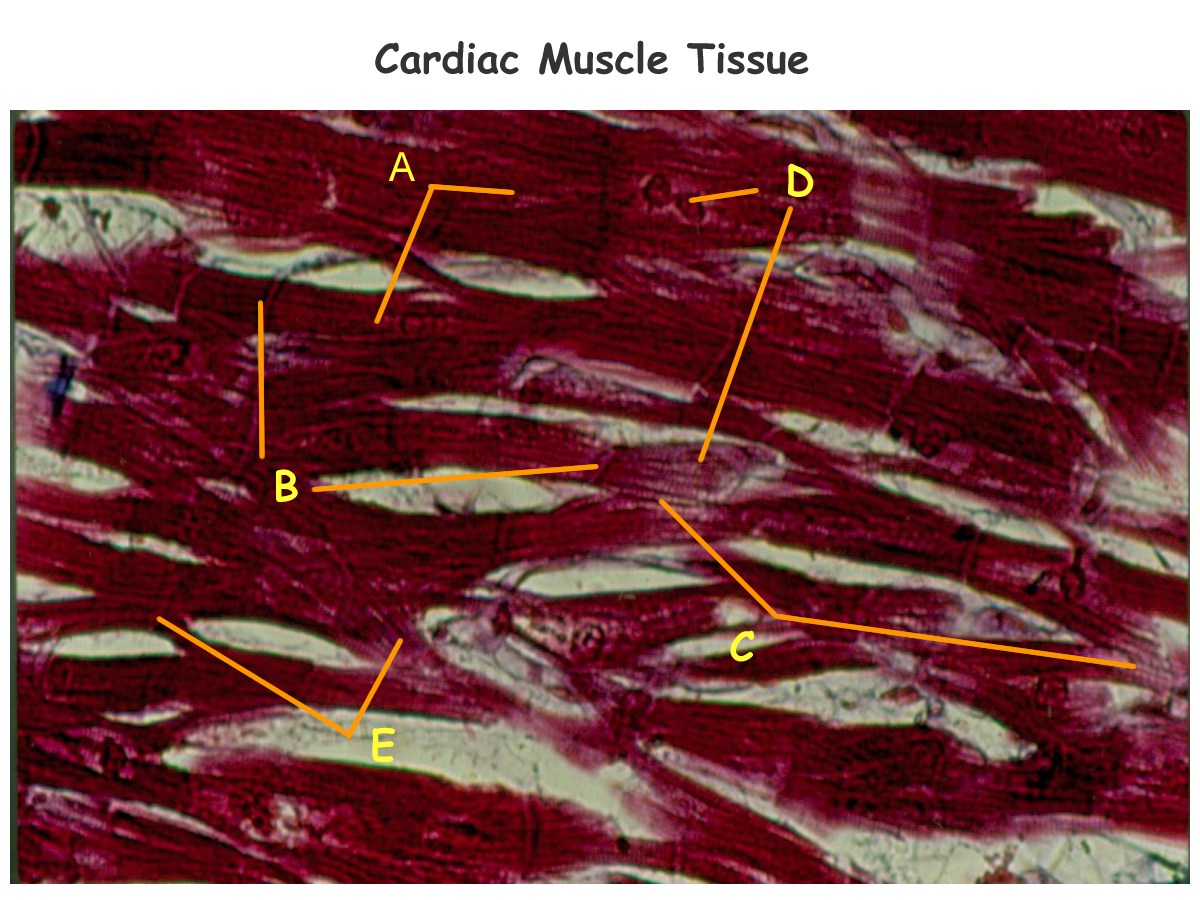
VIII. Label the histology of cardiac muscle
Concept
Map: Make a concept map of the heart (histo, gross) anatomy and physiological
function of conduction system as well as cardiac muscle in the atria and
ventricles.
Include this concept map in your LAR lab report (if selected)
as a document insert or as an addtional PDF scan of the map.
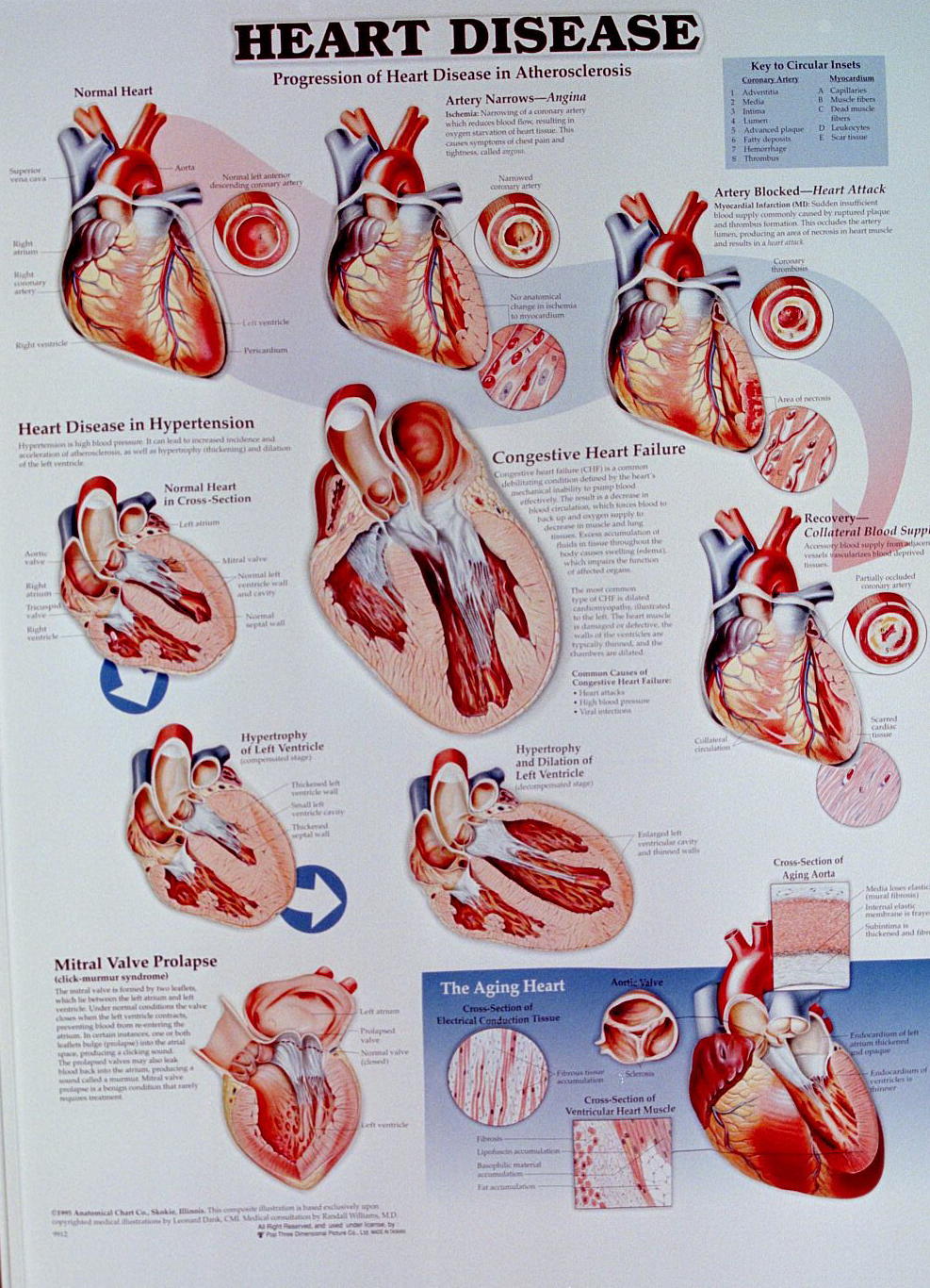
Myocardial ischemia Myocardial infarction (MI)
Atherosclerosis Coronary artery disease (CAD)
Valvular stenosis Cardiac Arrest
Bradycardia Tachycardia
Arrhythmias: flutter, fibrillation, heartblock, asystole
Rheumatic heart disease Congenital heart disease
Endocarditis Myocarditis
Pericarditis Angina Pectoris
Congestive Heart Failure (CHF)
Hypertensive Heart Disease Bypass surgery
Valvular Heart Disease: Insufficiency, Stenosis
Cardiologist
http://www.sciencedaily.com/news/health_medicine.htm
http://www.lumen.luc.edu/lumen/meded/histo/frames/histo_frames.html
http://americanheart.org/presenter.jhtml?identifier=10000056
http://www.americanheart.org (American Heart Disease Association)
http://sln.fi.edu/biosci/biosci.html
http://www.medtropolis.com/VBody.asp
http://www.kcmetro.cc.mo.us/maplewoods/Biology/Bio110/lab3.htm
http://endeavor.med.nyu.edu/courses/physiology/courseware/ekg_pt1/ekgmenu.html
http://calloso.med.mun.ca/%7Etscott/second.htm
http://www.track0.com/canteach/links/linkbodysystems.html
http://www.carr.lib.md.us/schs/science/anatomy/systems.html
http://www.stemnet.nf.ca/CITE/body.htm
http://sln.fi.edu/biosci/systems/circulation.html
http://www.fi.edu/biosci/preview/heartpreview.html
http://www.tmc.edu/thi/anatomy.html
http://www-medlib.med.utah.edu/WebPath/CVHTML/CVIDX.html
http://www.merck.com/disease/heart/coronary_health/anatomy/home.html
http://www.gen.umn.edu/faculty_staff/jensen/1135/webanatomy/wa_cvs/
http://www.andrews.edu/~murdick/write/fluid/system.htm
http://www.usouthal.edu/biology/shardo/bly152/cardiovascular.html
http://defiant.wbc.edu/wbc/jjohnson/pages/humphys/links/14cardiolinks.html
http://www.clt.astate.edu/radsci/pfgs311cvsimages.htm
http://www.student.loretto.org/anatomyphys/chp43.htm
http://www.crha-health.ab.ca/hlthconn/items/cvsystem.htm
http://cellbio.utmb.edu/microanatomy/cardiovascular/cardiovascular_system.htm
http://www.readingeagle.com/krt/health/cardio/cardio.htm
http://www.readingeagle.com/krt/health/cardio/cardio.htm
http://www.bmb.psu.edu/courses/bisci004a/cardio/41card3.htm
http://www.medem.com/MedLB/article_detaillb.cfm?article_ID=ZZZ8D8356JC&sub_cat=72
http://www.nlm.nih.gov/medlineplus/heartandcirculation.html
1. Give the location and function of the heart.
2. Name the cardiac chambers and associated valves.
3. Describe the histology of cardiac muscle.
4. Name the components of the cardiac conduction system.
5. Explain the electrical events represented by a lead II ECG tracing.
6. Define cardiac cycle.
7. What are heart sounds?
8. Define Cardiac output.
9. Follow the pathway of blood through the heart
10. How does the ANS affect the heart?