Biology 2404 A&P Basics Lab Exercise 15 Lymphatic System Dr. Weis
| Objectives | Background | Medical Terms | Activities | Applications | Careers | WWW | Review Questions |
Students should be able to
* define lymph regarding location, components, and function
* describe the circulation of lymph through lymphatics
* Give the location, function, and anatomy of the lymphoid organs:
lymph node, spleen, tonsils, thymus, and peyers patches
* Define related terms
Read related material in textbook
The lymphatic system is similarly composed as the cardiovascular system. There is a fluid called lymph, a system of vessels called lymphatics, and related organs such as lymph nodes, spleen, tonsils, peyers patches, and the thymus.
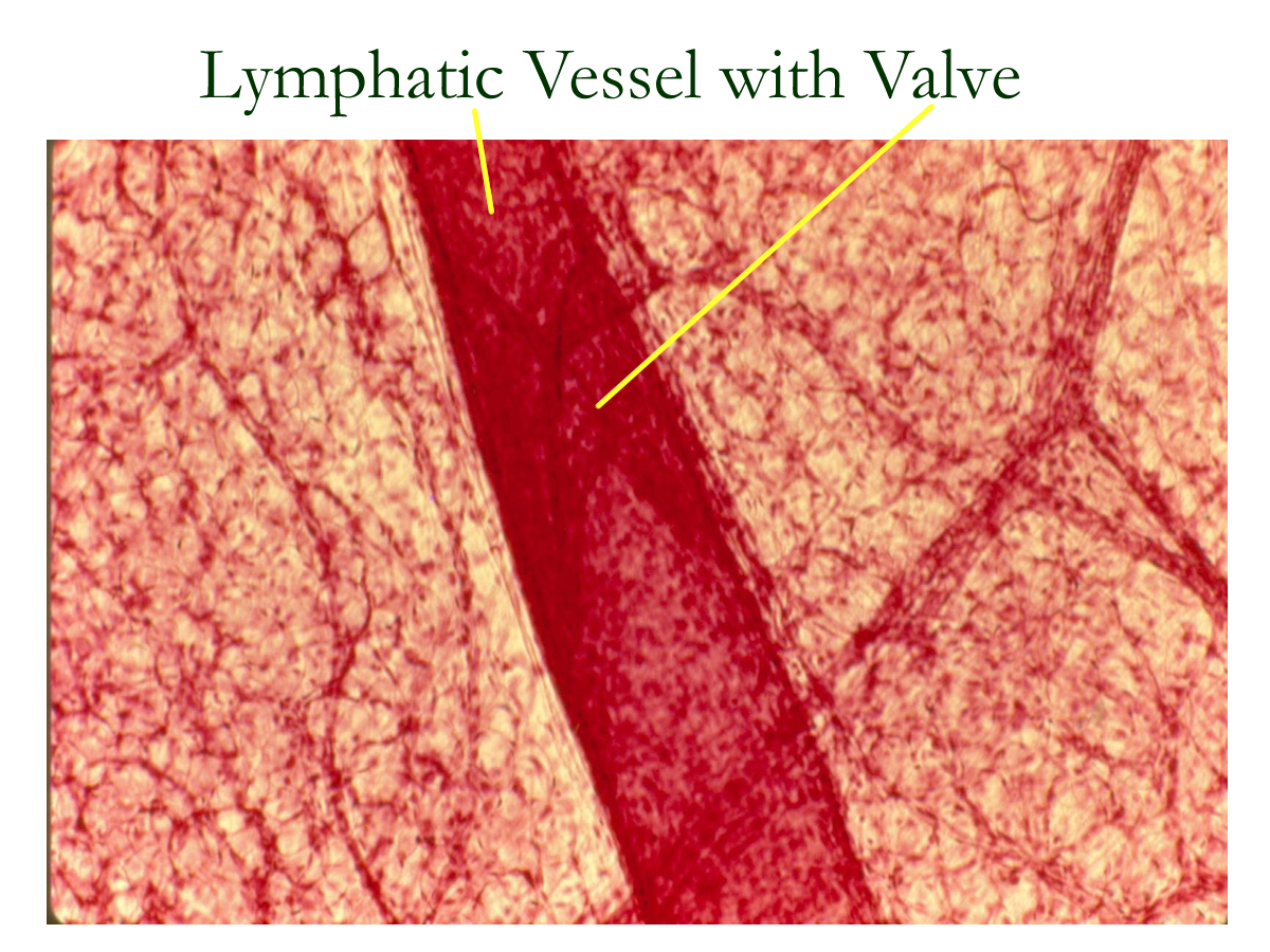
Lymph is the remaining 30% of tissue (interstitial) fluid that was collected by lymphatic vessels (lymphatics) after the blood capillary exchange. The lymph fluid resembles plasma in composition as it will contain water, ions, nutrients, and gases. However, lymph has a higher protein and cellular content than plasma. Lymph enters lymphatic vessels called lymphatic capillaries. These vessels resemble the venous structure in relation to valves and vessel walls and follow the venules and veins as they return to the heart. The lymphatic vessels drain into regional trunks that end in two major ducts: the right lymphatic duct and the thoracic duct. These ducts connect to the right and left brachiocephalic veins and allow the lymph to be returned back to the cardiovascular system. The process of returning lymph back to the cardiovascular system takes about 24 hours. If the excess interstitial fluid is not returned, pressures around the tissue increase causing swelling known as edema.
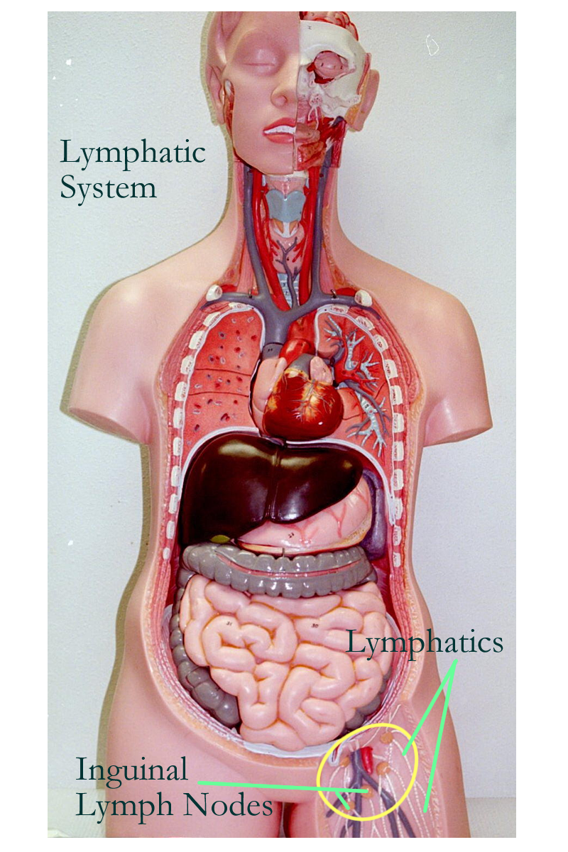
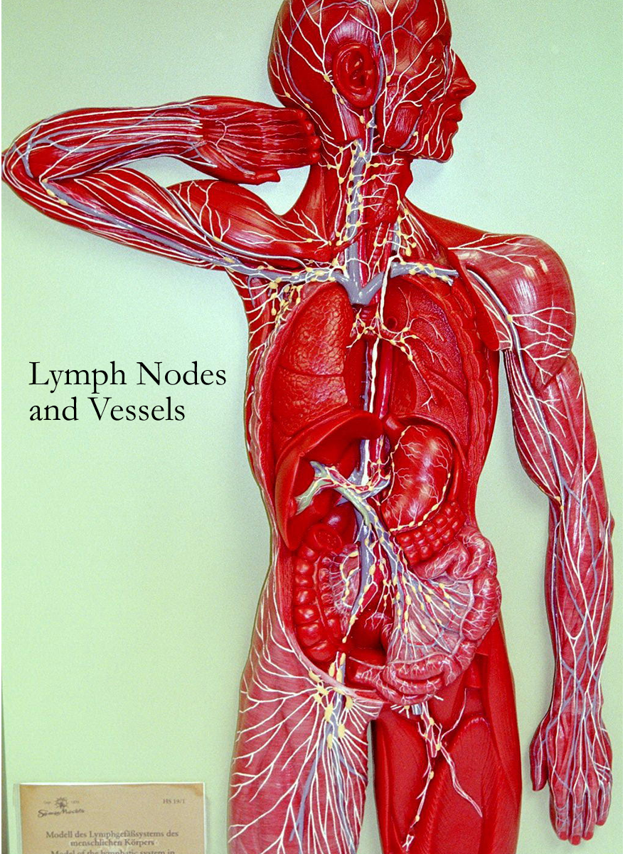
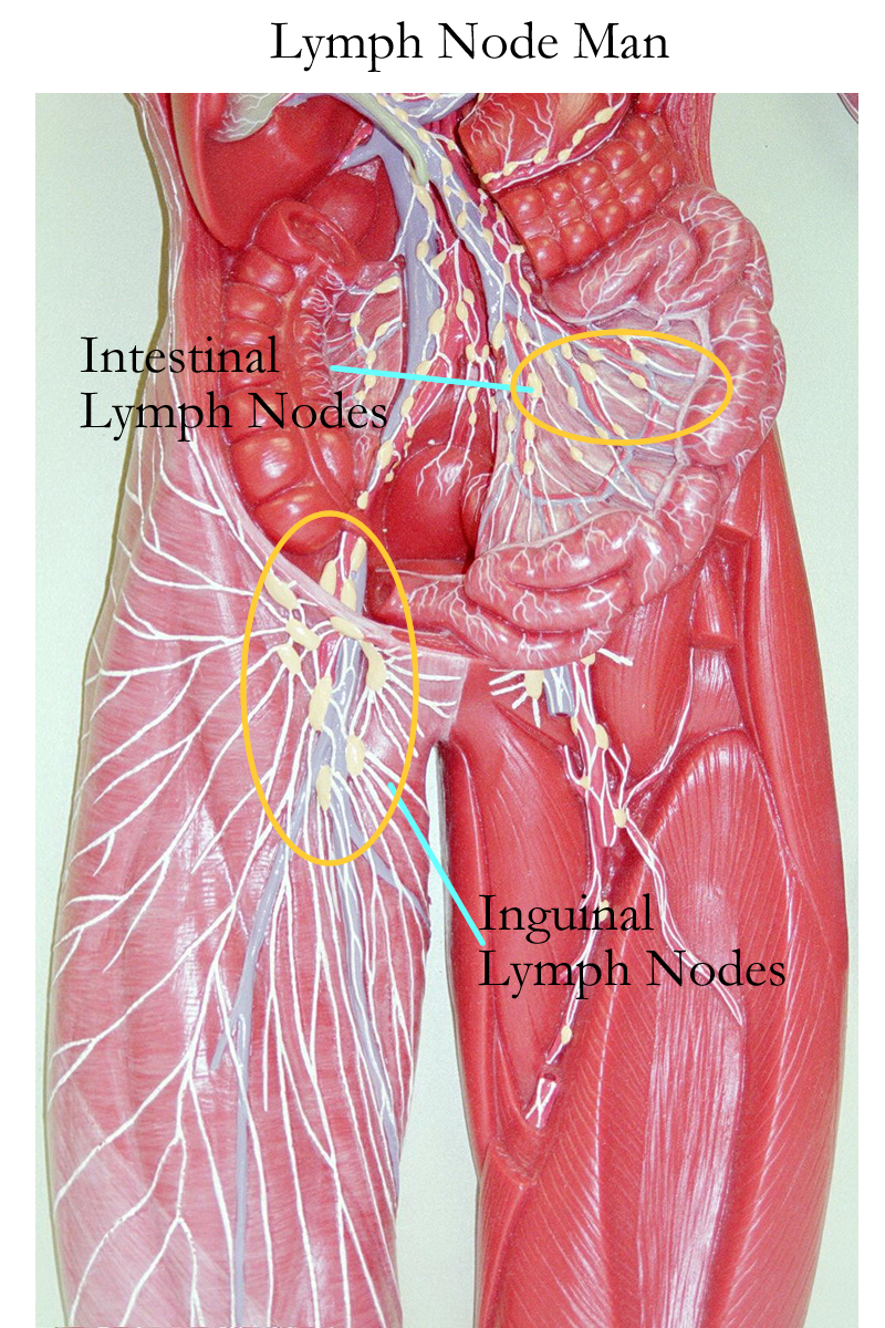
The lymphatic capillary lining consists of valve like flaps that open and close based on fluid pressure gradients. These vessels are passive in their collection of substances so that large proteins and even damaged or cancerous cells can enter lymphatic circulation. In order to clear and clean the lymphatic fluid before it is returned to the heart, the lymph vessels pass through lymph glands or nodes. These oval to bean shaped structures differ in respect to size and location. Their size can range from 1-25 mm and major locations are found in the inguinal, axillary, cervical, and intestinal regions.
Lymph Nodes
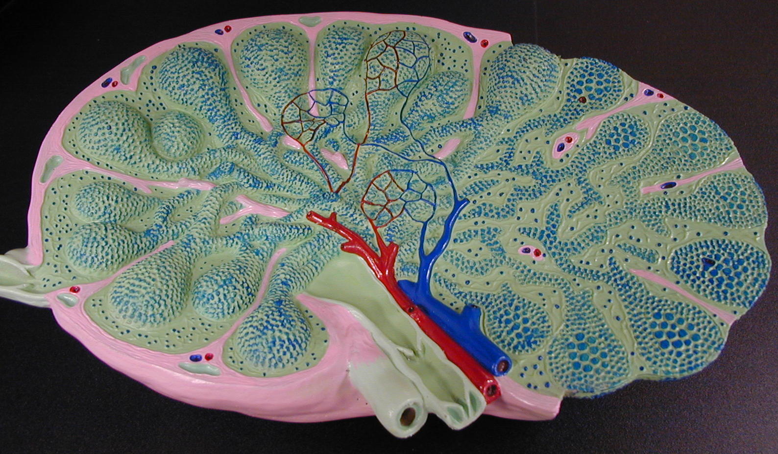
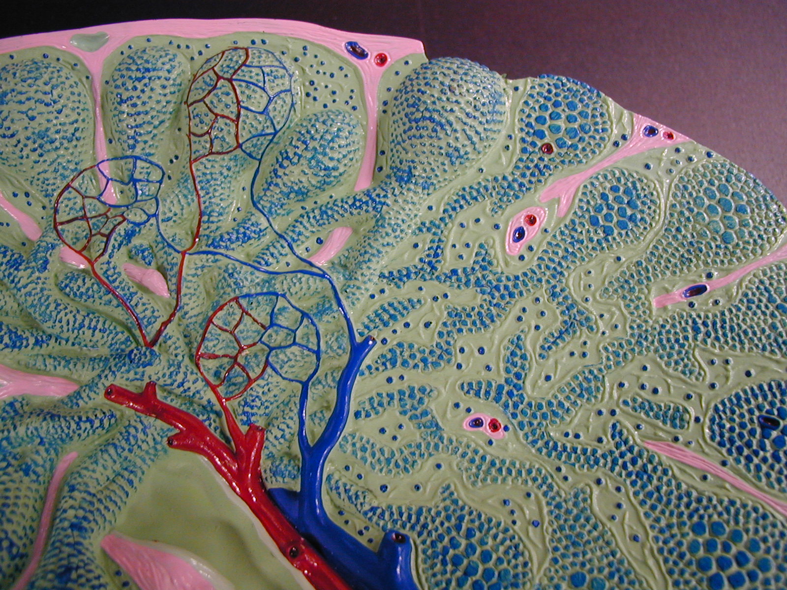
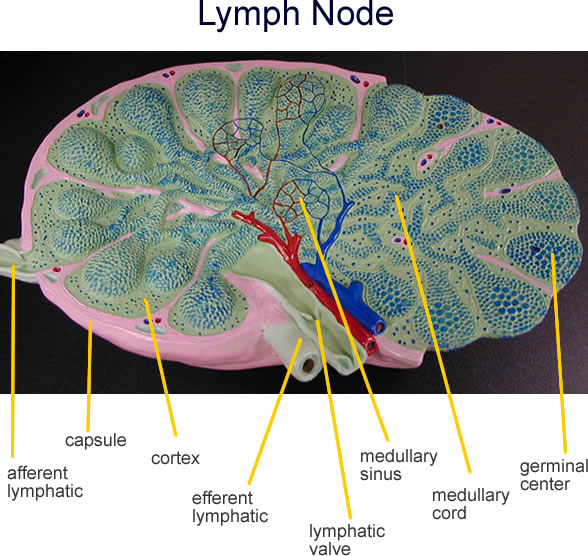
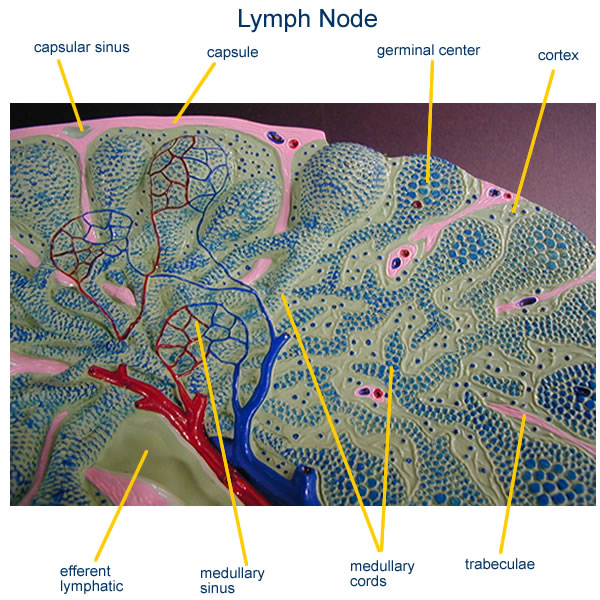
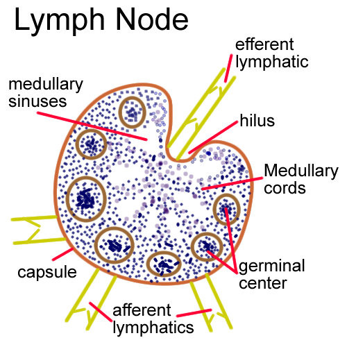
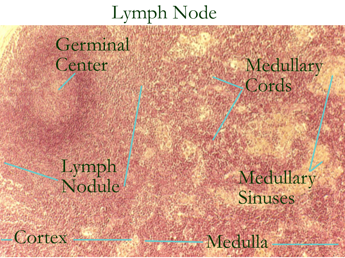
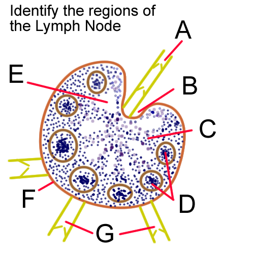
The structure of a lymph node consists of a capsule, cortex, and medulla.
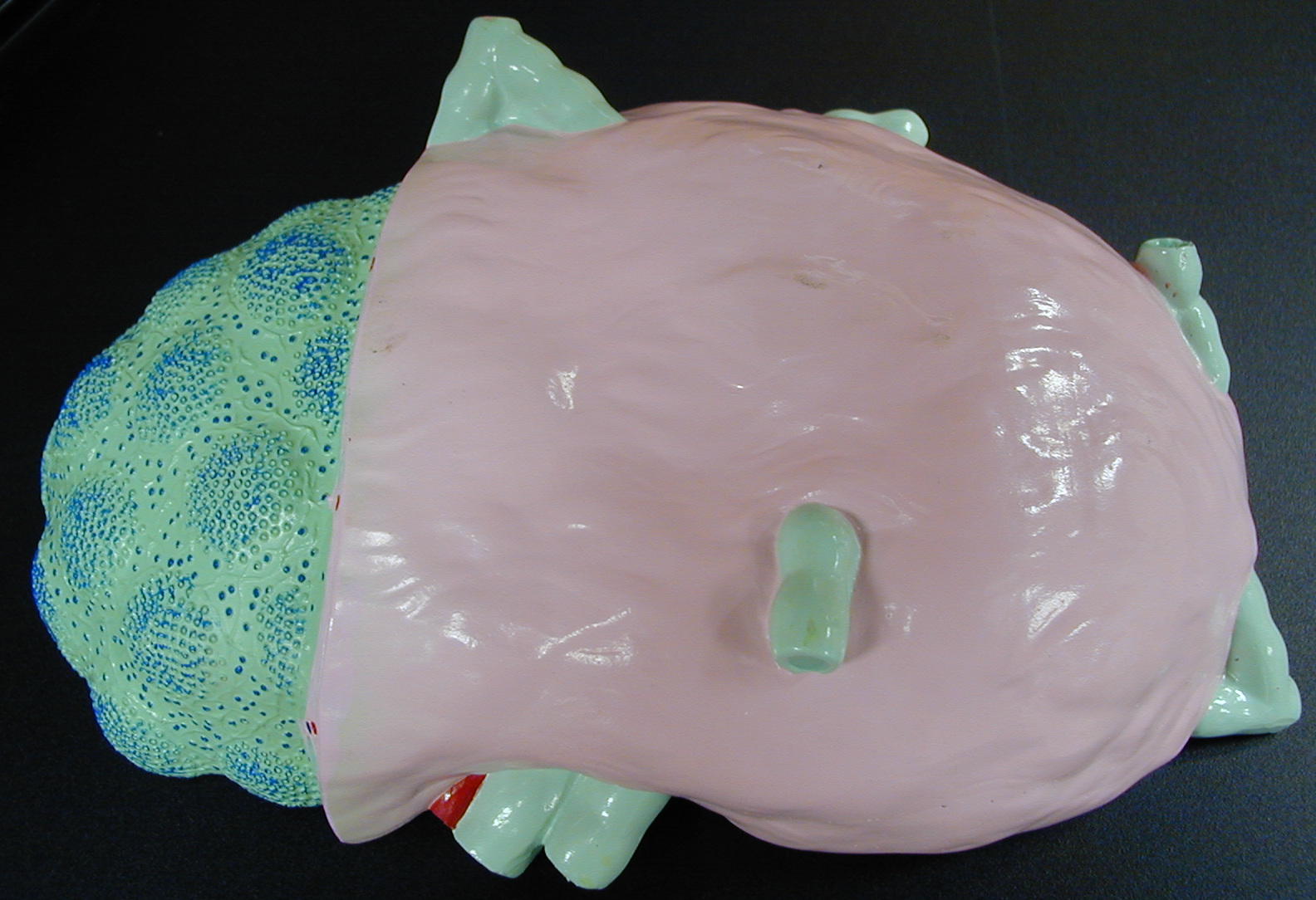
General Lymph Node Model Exterior
General Lymh Node Model Interior Transverse Section
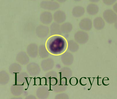
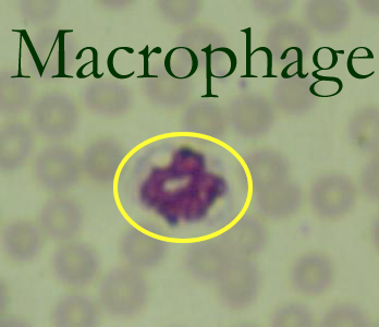
The connective tissue capsule forms a subcapsular space or sinus that allows for lymph collection from the afferent lymphatics. The lymph node cortex is arranged into lymph nodules(follicles) composed of B and T cells. The lighter germinal center contains naive B cells that will be sensitized and activated to divide in the outer regions of the follicle to produce daughter lymphocytes known as plasma cells. T lymphocytes are in the parafollicular region of the cortex and will work with macrophages to iniate cellular defenses. The lymph node medulla is reticular connective tissue with associated macrophages and lymphocytes forming the medullary cords.
Lymph Node Model: Cortex and Medulla
As lymph fluid flows through the cortex and medulla it is cleaned and filtered as it encounters the various WBCs that form the two major divisions of the immune system.
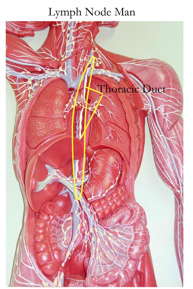
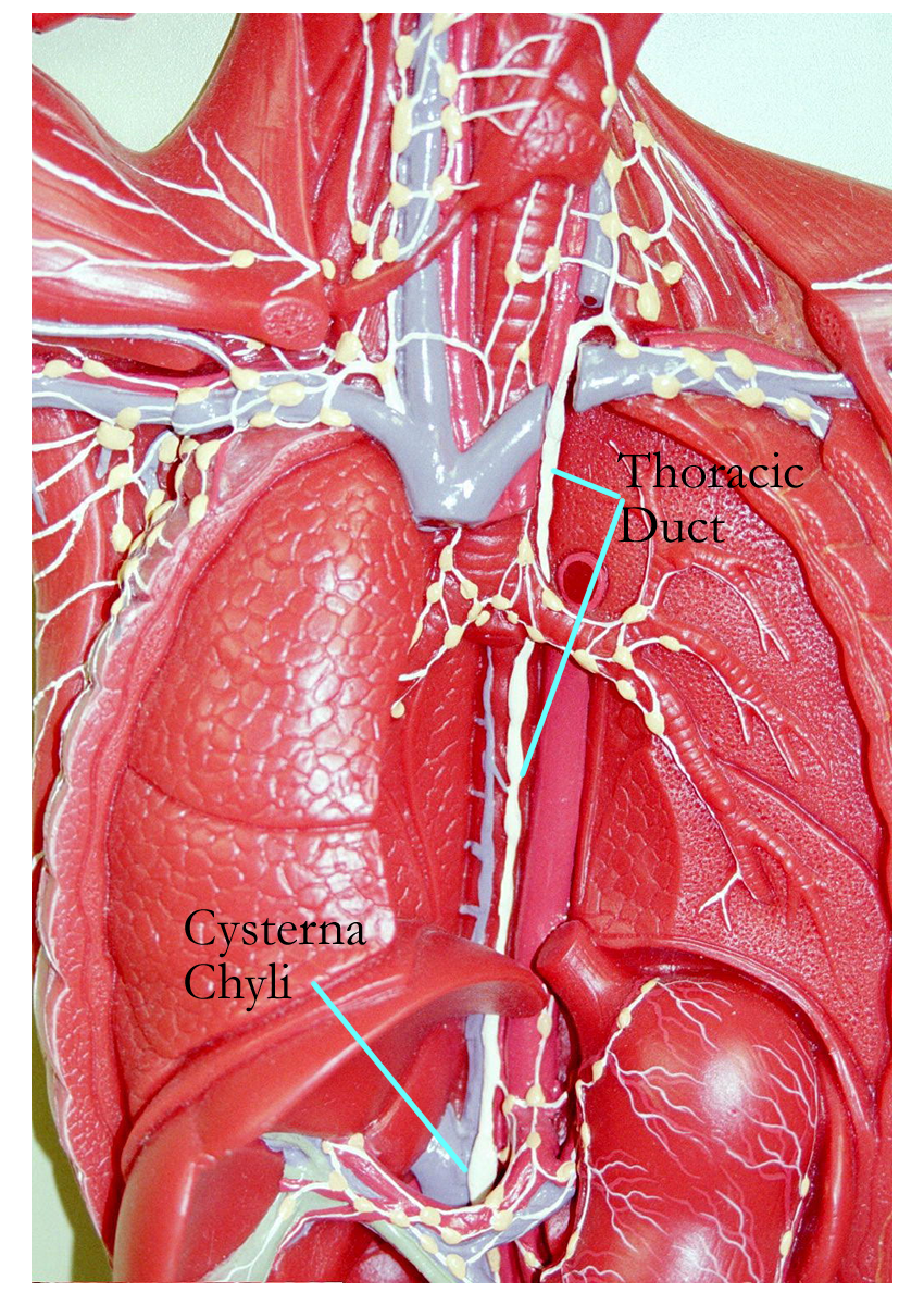
Specialized lymphatic vessels that occur in the small intestine of the gastrointestinal tract are called lacteals and function in fat absorption. Because of the milky appearance, the lymphatic fluid is called chyle. Chyle follows the normal flow of lymph and will be returned to the heart the same way as lymph is returned. Fats are then made available to the body organs for energy use or storage. Lymphatics return lymph back to the heart. Those from the lower legs combine to form the Cysterna Chyli. More lymph from the left trunk, left arm and head together with the cysterna chyli form the Thoracic Duct which empties cleansed lymph into the left brachiocephalic vein.
Lymph returning from the right side of the head, arm and trunk enters the
right lymphatic duct to be emptied into the right brachiocephalic vein. The
thoracic duct drains the entire left side of the body and the lower right side.
It empties into the left brachiocephalic vein.
Thus, all excess tissue fluid collected as lymph is processed by lymph nodes
and is then returned to the cardiovascular system.
Lymph Node Model (above) Labeled
Regional Lymph Nodes: Cervical, Inguinal, Intestinal
Other lymphatic organs / tissues
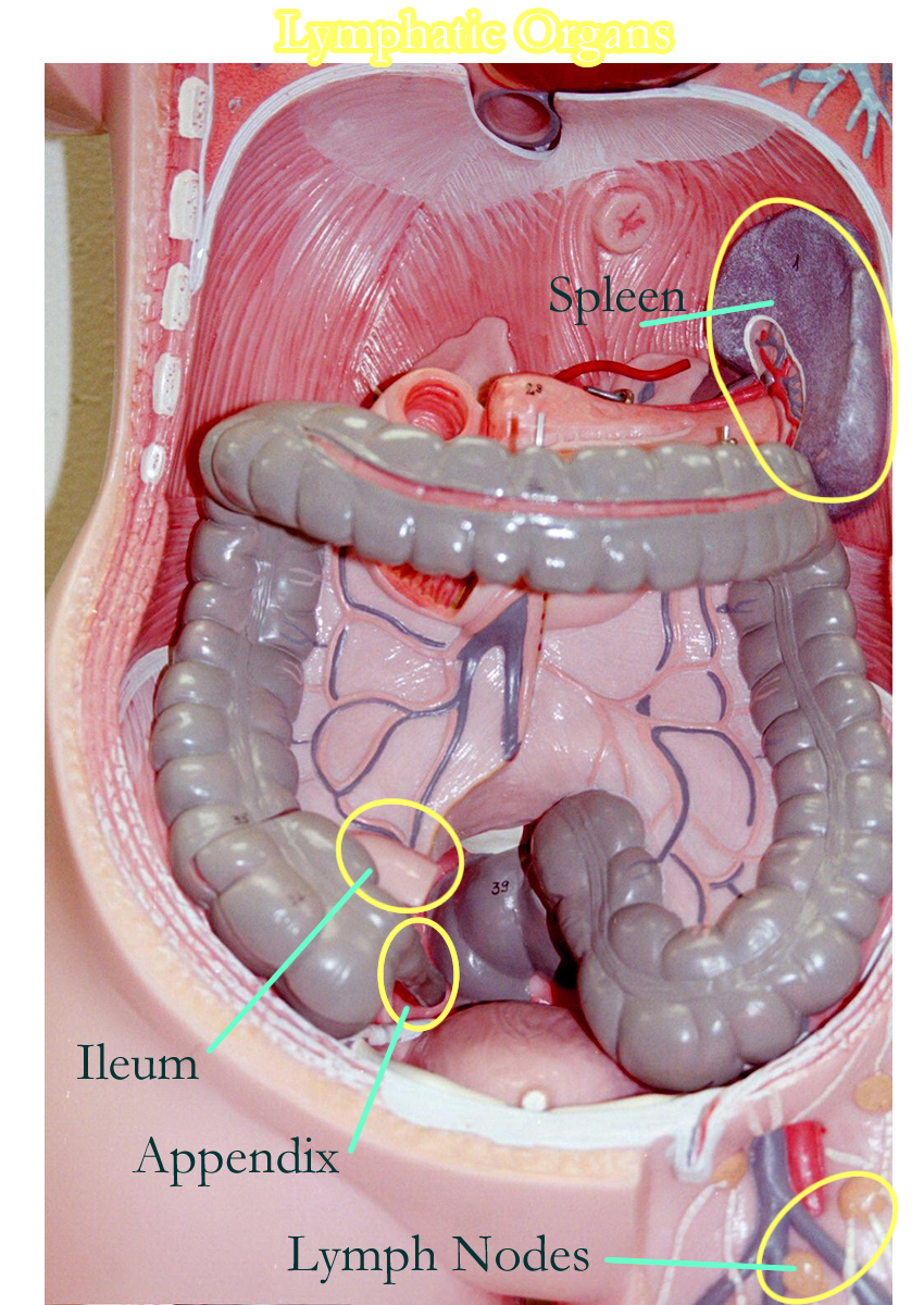
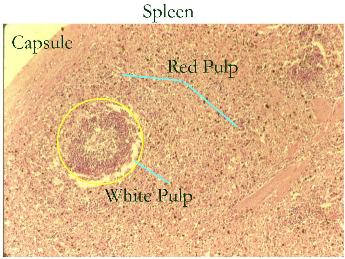
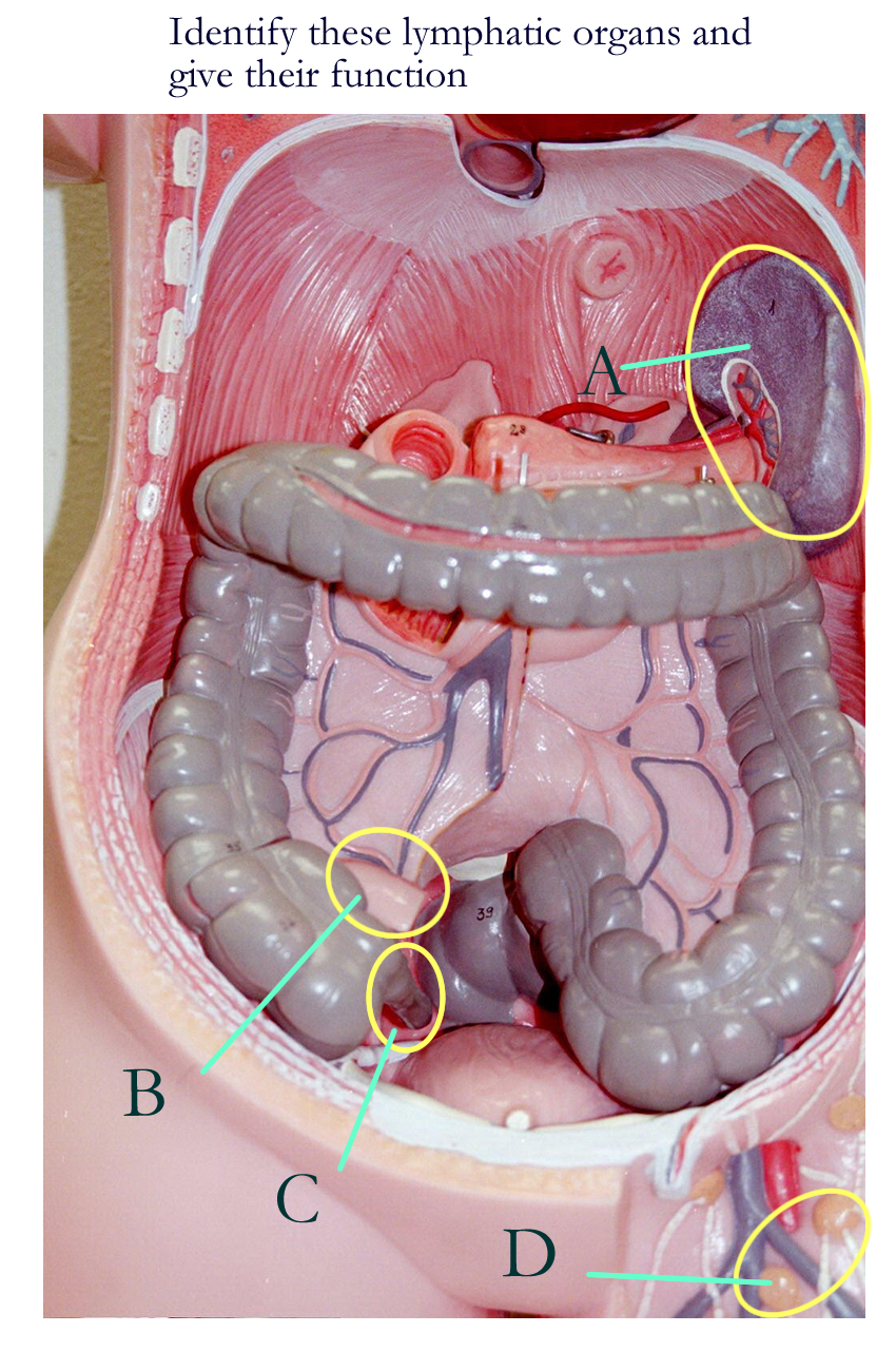
Spleen located in the left upper abdominal quadrant
Histologically has a capsule and red and white pulp
Red pulp is composed of RBCs
White pulp is composed of WBCs around arterioles
Function to recycle red blood cells and immune surveillance of blood entering this organ.
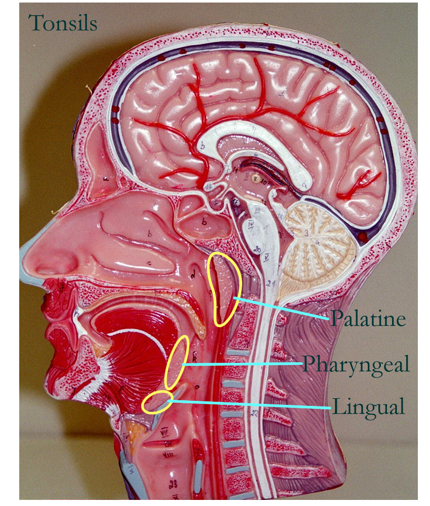
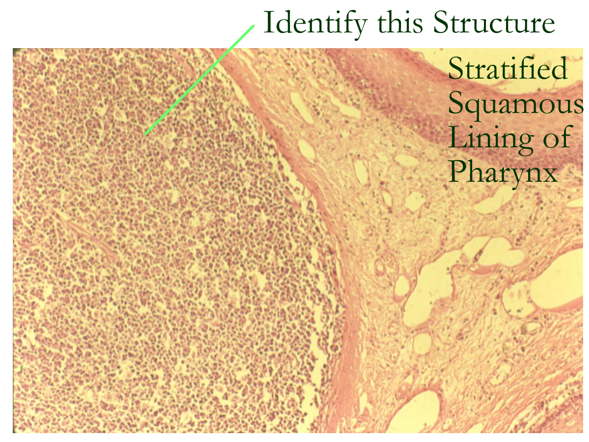
Tonsils located in a circular ring in the pharyngeal regions
Names reflect location: Tubular, Pharyngeal, Palantine, Lingual
Histologically are lymphatic nodules embedded in the throat lining
Function in immune defenses related to respiratory and digestive organs
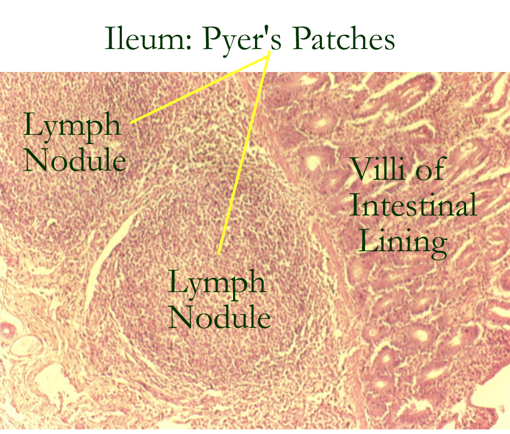
Peyer’s Patches located in the last portion of the small intestine called the ileum
Lymphatic nodules embedded under the lining of the digestive tract
Function to provide immune defense and alert for the digestive tract
Part of the GI lymphatic tissue known as GALT.
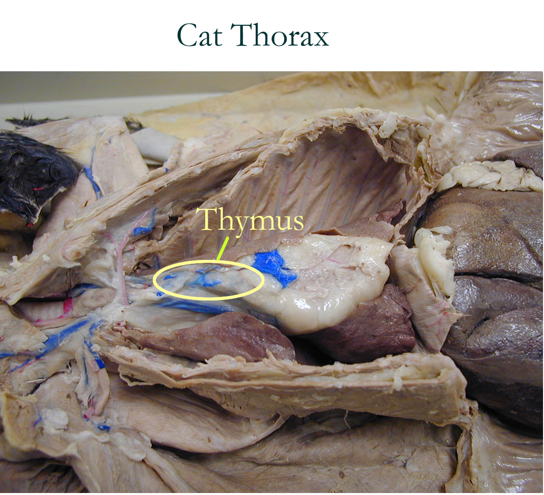
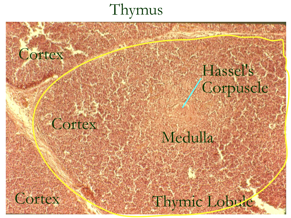
Thymus located in young mammals in the mediastinum, dorsal to the heart
Histologically is composed of a capsule, cortex, and medulla
Bilobed in humans, elongated triangle shape
Functions to program lymphocytes to become various T-cells for the Specific immune defenses.
Torso model: Spleen, Ileum, LN, Appendix
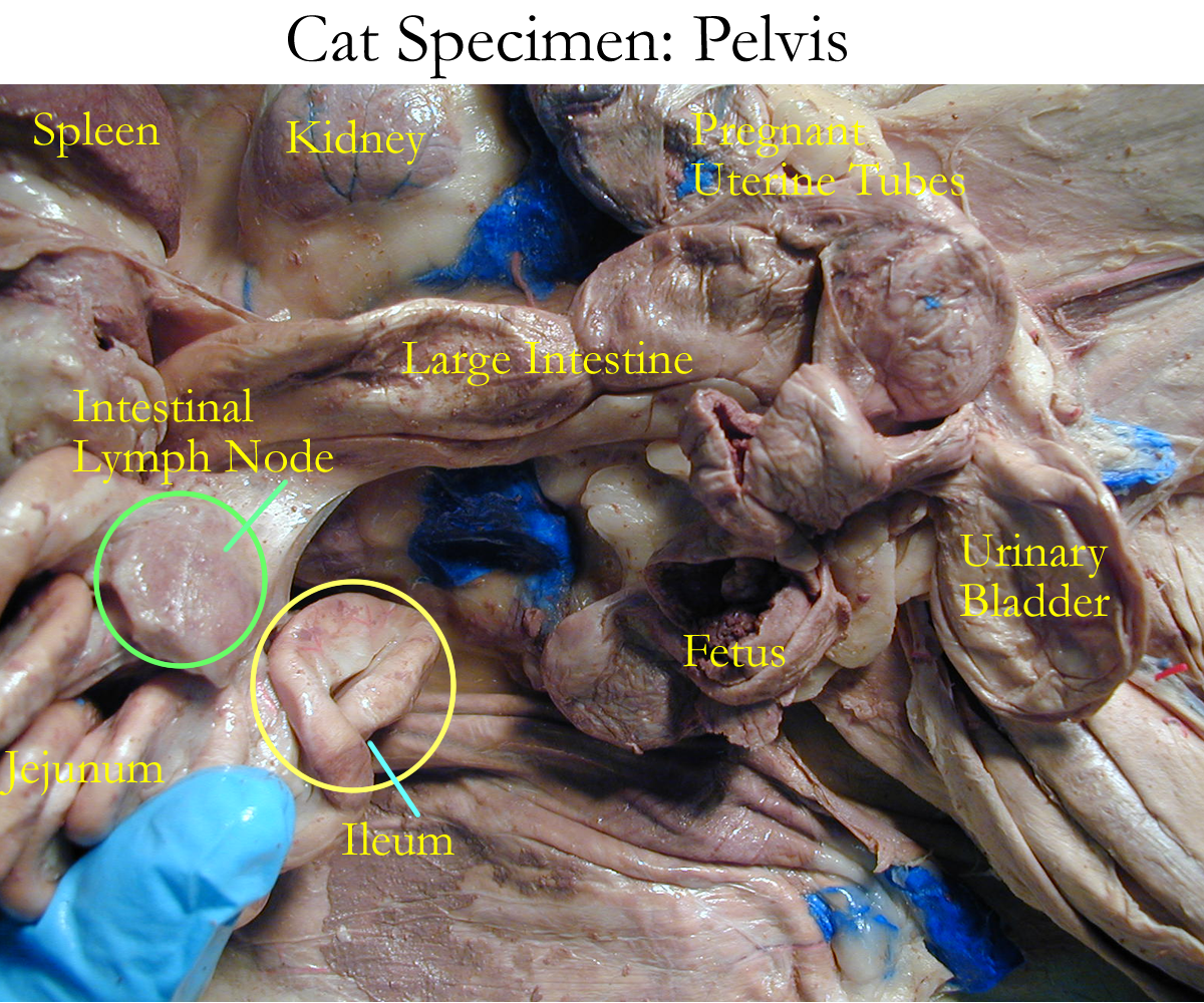
Cat specimen: Spleen, Thymus, Ileum
Histology
Lymph/o- lymph splen/o- spleen
Tonsil/o- tonsils -asis condition, state
Lien- spleen lymphaden/o- lymph gland
I. Palpate lymph nodes : Locate regional lymph nodes on drawings or models and see if you can palpate these organs. List the locations where you palpated a lymph node. What would a painful, enlarge lymphnode found on palpation possibly indicate?
II. ID and functions of the lymphatic organs
A Drawing
B Model
III. How is lymph collected, processed, and returned to the cardiovascular system? Name the structures involved and a problem or condition that can obstruct this flow.
IV. Fill in the following table
Organ |
Location |
Cell Types Present |
Function |
| a) Spleen | |||
| b) Tonsils | |||
| c) GALT | |||
| d) Lymph Node | |||
| e) Thymus |
Concept Map: Make a concept map of the lymphatic system (gross and histo) anatomy, location, and physiological function. Include this map in your LAR lab report (if selected) as a document insert or as an additional PDF document scan.
Lymphadenitis
Tonsillitis
Lymphadenopathy
Lymphedema
Lymphangitis
Splenectomy
Tonsillectomy
Elephantitis
Lymphoma
Lymphosarcoma
Hodgkin’s Disease
Bubonic Plague
Chylothorax
Immunologist
http://www.nlm.nih.gov/medlineplus/healthtopics.html
http://www.lumen.luc.edu/lumen/meded/histo/frames/histo_frames.html
http://www.track0.com/canteach/links/linkbodysystems.html
http://www.medem.com/medlb/article_detaillb.cfm?article_ID=/ZZZG0S6CGJC&sub_cat=198
http://www.nlm.nih.gov/medlineplus/bloodlymphaticsystem.html
1. Define lymph
2. Explain how lymphatic capillaries reclaim excess interstitial fluid
3. Name the two lymphatic ducts that return blood back to the venous circulation
4. Give the location, anatomy, and function of the lymph node.
5. Give the location, anatomy, and function of the spleen
6. Give the location, anatomy, and function of the tonsils
7. Give the location, anatomy, and function of the thymus
8. Give the location and function of the peyers patches
9. Define edema
10. Define chyle