Biology 2404 A&P Basics Lab Exercise 18 Digestive System Dr. Weis
| Objectives | Background | Medical Terms | Activities | Applications | Careers | WWW | Review Questions |
Students should be able to:
* Name the organs involved with the alimentary canal and their location
* Give the functions of the organs of the alimentary canal
* define related terms such as digestion, absorption, ingestion, segmentation, peristalsis
Read related material in text book
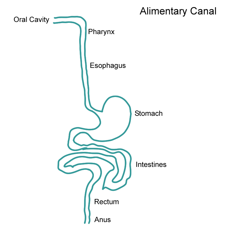
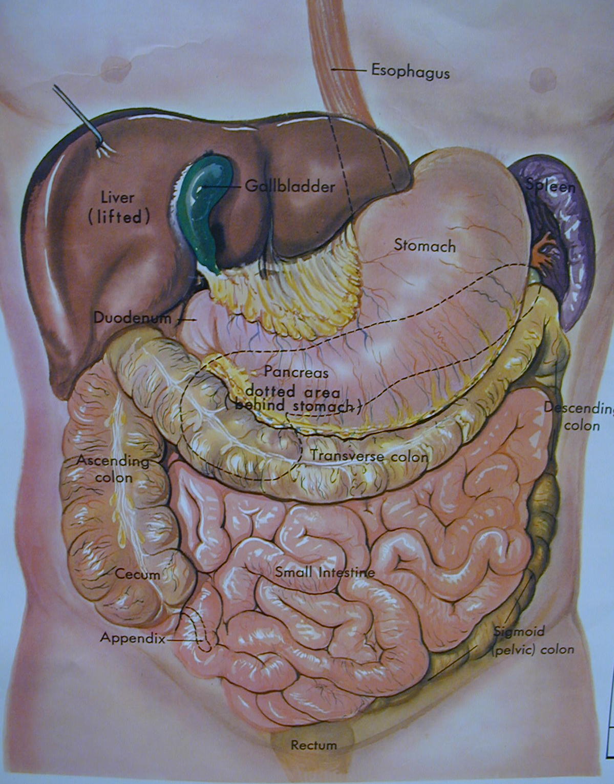
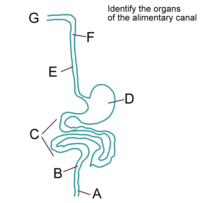
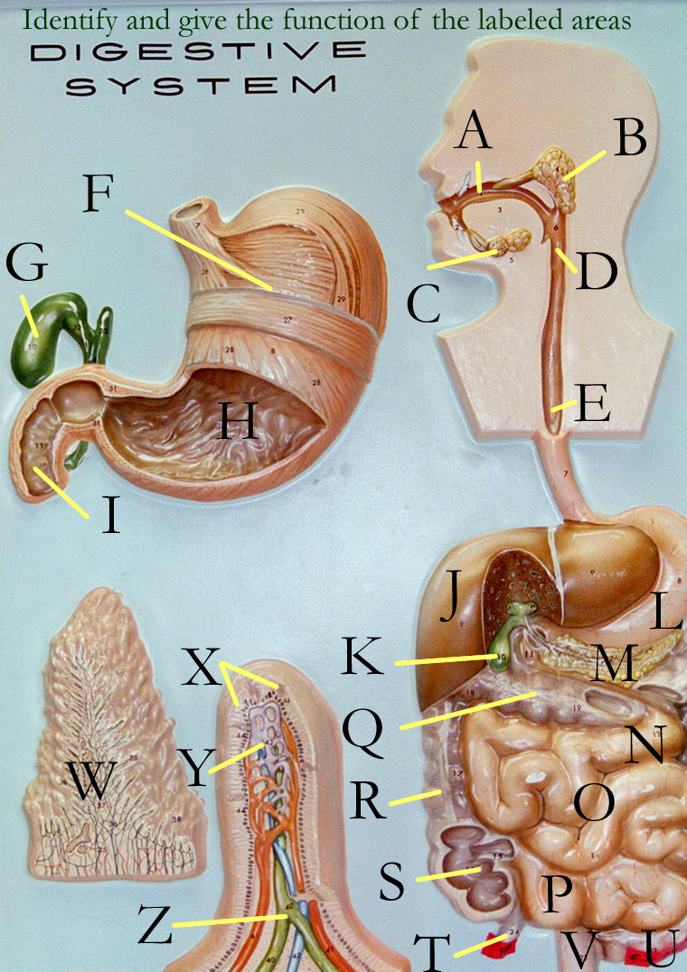
The alimentary canal or gastrointestinal GIi) tract is a 30 foot body tube that is an open cavity extending from the mouth to the anus. Parts of the GI tract are the oral cavity, pharynx, esophagus, stomach, small intestine, large intestine, rectum, and anus.
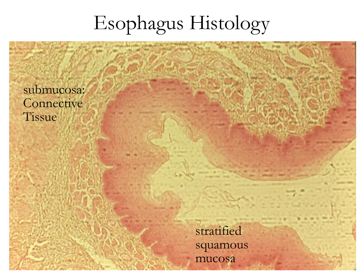
Histology of this tube consists of a mucosal lining that is stratified squamous or simple columnar epithelium, a connective tissue submucosa, a smooth muscle muscularis in at least 2 layers in different planes, and an outer connective tissue adventitia or serosa.
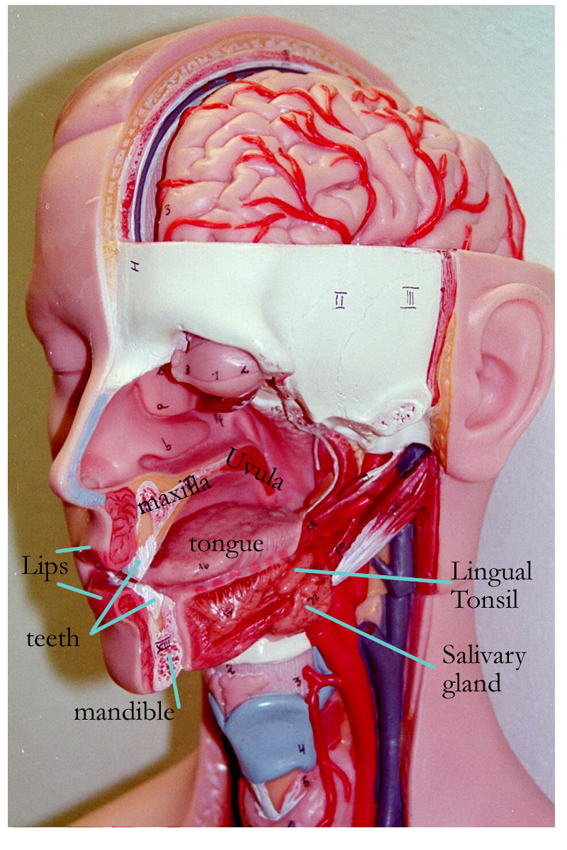
Oral Cavity
The oral or buccal cavity is the stratified squamous lined opening of the GI tract formed by the bones of the skull and soft tissue of the cheeks and lips. The oral cavity contains several accessory organs: the tongue, teeth, and salivary glands.
The function of this region is:
Ingestion: taking in, by the way of the mouth, food and liquids into the body.
Digestion: mechanical digestion = physical breakdown of food by the tongue, teeth, and palates
chemical digestion = enzymatic breakdown of food by the salivary enzymes in the saliva.
Pharynx
The pharynx is a stratified squamous lined muscular tube that allows for passageway of food, liquid, and air. The three regions are the nasopharynx, oropharynx, and laryngopharynx. For digestion, the oropharynx aids in the voluntary stage of swallowing or deglutition. Movement of the food bolus triggers closure of the nasopharynx by the uvula of the soft palate in order to prevent food from entering the nasal cavity. Extrinsic muscles of the larynx elevate to allow passage of the food bolus and the movement of food into the laryngopharynx triggers the closure of the epiglottis over the glottis to prevent food from entering into the respiratory tract. The continuation of the laryngopharynx is the esophagus.
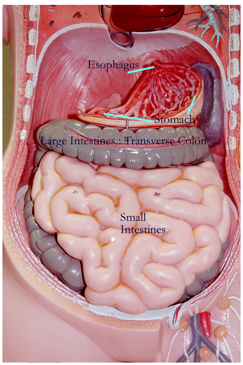
Esophagus
The esophagus is a collapsible, stratified squamous lined muscular tube for the transport of food from the pharynx to the stomach. The esophagus passes through the thoracic cavity in the region of the mediastinum and then through the diaphragm at the esophageal hiatus. The muscular wall of the esophagus changes from voluntary skeletal muscle near the laryngopharynx to involuntary smooth muscle 2/3 of the way down the tube. Muscle contractions in the wall create peristaltic waves that help propel food to the stomach. Movement of food into the stomach is controlled by a lower esophageal sphincter, a thickened region of circular smooth muscle that relaxes to allow food entry and contracts to prevent backward movement of food from the stomach to the esophagus.
Stomach
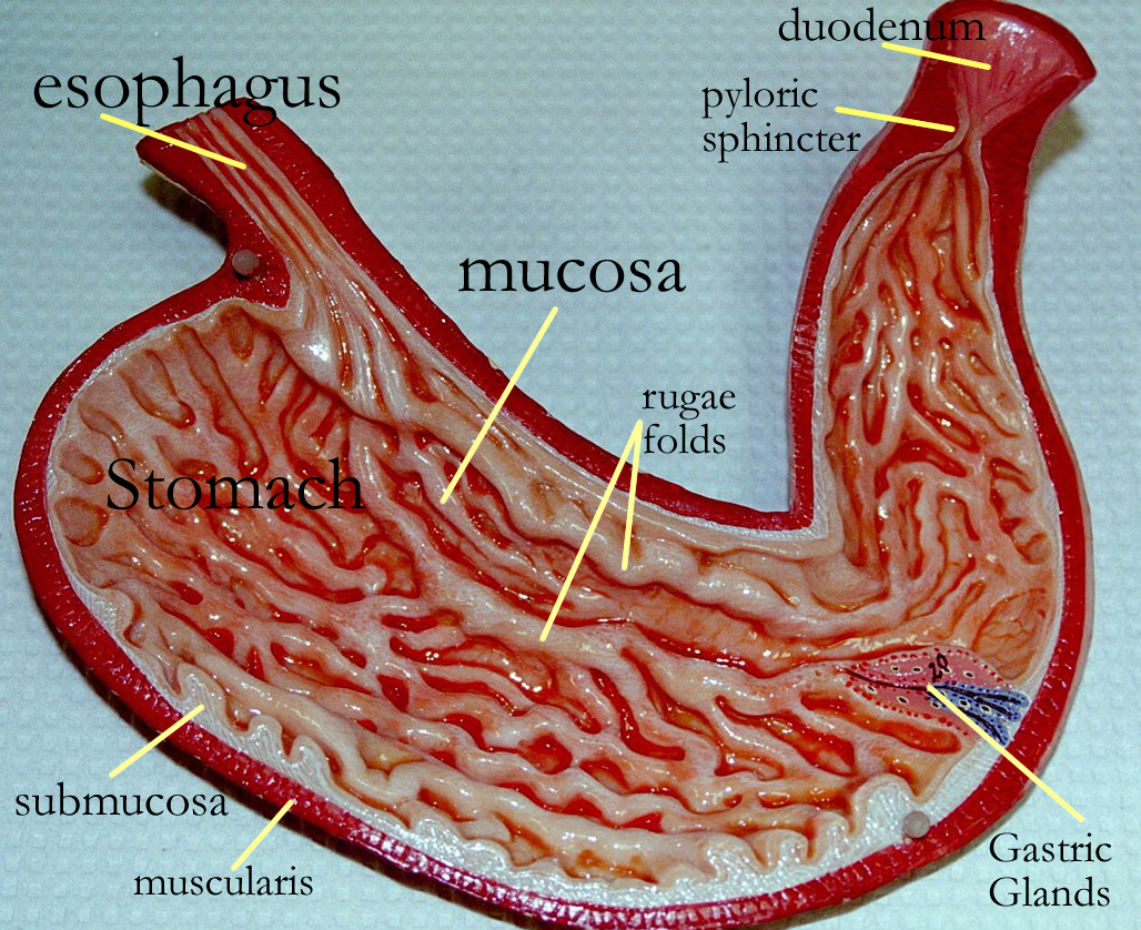
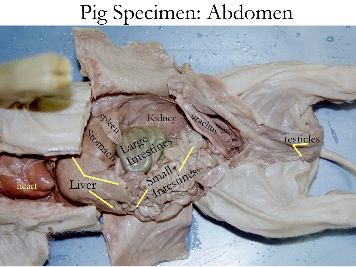
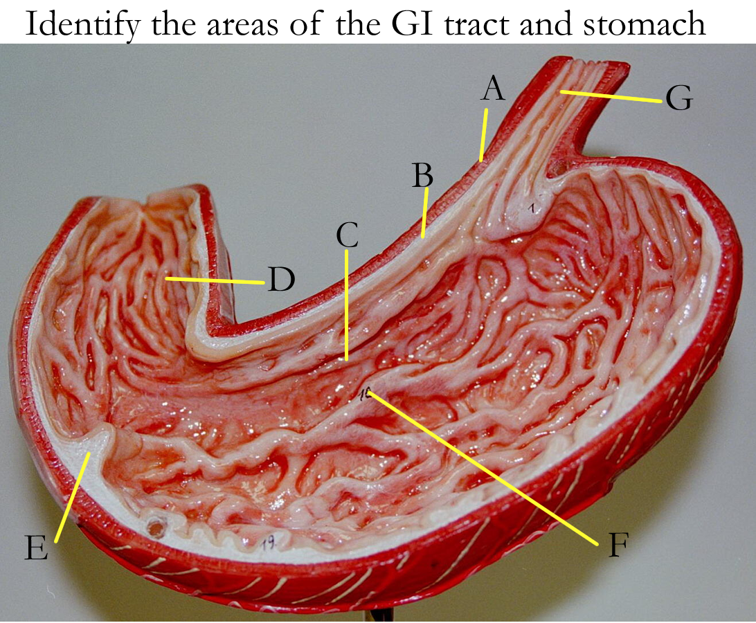
The stomach is an outpocketing of the GI tract and is located in the upper mid to left quadrant of the abdominal cavity. Grossly, the stomach is divided into four regions: the cardia, fundus, body, and pylorus. The cardia of the stomach is the region where the esophagus joins the stomach, known as the gastroesophageal junction. The fundus is the upper domed region of the stomach and is non-glandular in most mammals, except humans. The body of the stomach is the main glandular portion of the stomach and the pylorus is the lower, narrow portion that joins the stomach to the small intestine.
The stomach externally has a greater and lesser curvature. Along these curvatures is an extra fold of parietal peritoneum called the mesentery of the stomach or the omentum. The lesser omentum is found along the lesser curvature of the stomach and helps to anchor the stomach to the liver. The greater omentum is found along the greater curvature of the stomach and helps to anchor the stomach to the intestines. The purpose of anchoring the stomach is to prevent rotation along the long axis known as volvulus or torsion that would create a blockage or obstruction to outflow.
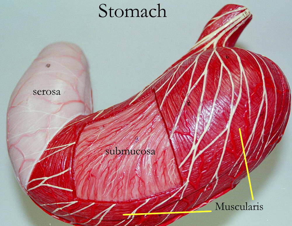
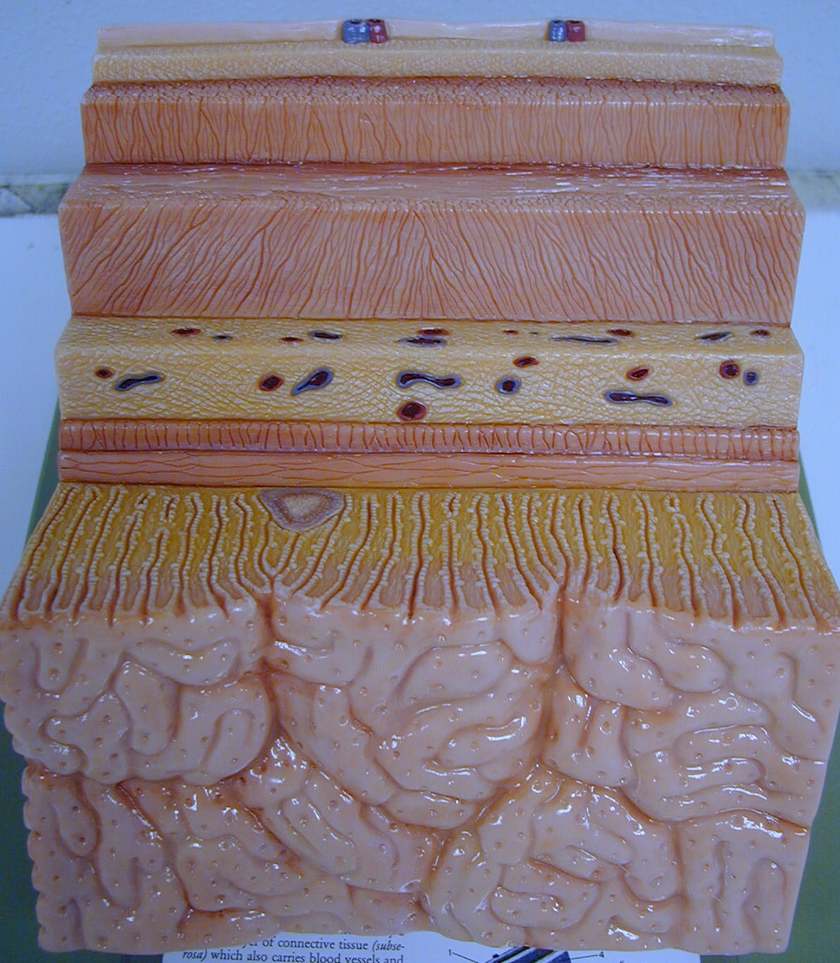
Histologically, the stomach has a simple columnar mucosal lining, a submucosa, a muscularis mucosa, and a serosa. When the stomach is empty, the submucosa’s connective tissue throws the lining of the stomach into large visible folds called rugae.
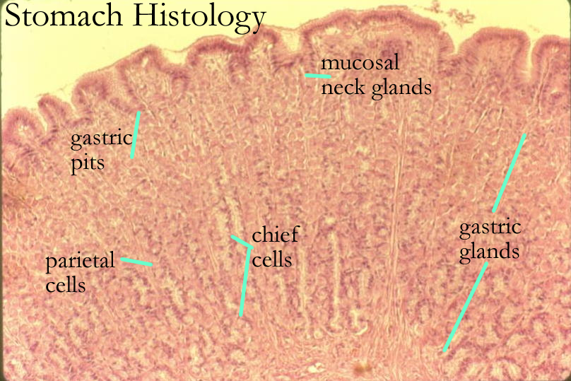
The mucosal lining of simple columnar cells is folded inward to create gastric glands and their openings, the gastric pits. Secretions from these gastric glands are collectively called gastric juices. The gastric glands, their location and function are:
Mucosal Neck Cell upper region of the lining secrete mucus for protection
Enteroendocrine Cells mid region of the lining secretes hormones for the GI tract
Chief Cell lower region of the lining secretes pepsinogen
Parietal Cells lower region of the lining secretes HCl and Intrinsic factor
HCl from the parietal cells is necessary to activate pepsinogen (from the chief cells) to pepsin. Pepsin then begins the chemical digestion of proteins eaten in the diet. Intrinsic factor is a hormone that signals the manufacturing and absorption of many B complex vitamins in the large intestine. An example of such signaling involves an important B vitamin, B12, that is needed for proper RBC development.
Gastric secretions are triggered by sight, smell, hormones, and neurotransmitters such as ACh.
Functions of the stomach include:
Mechanical Digestion : due to smooth muscle movements in the muscularis
Chemical Digestion: due to pepsin digestion of proteins into peptides
Intrinsic Factor: aid in signaling large intestine bacteria to make B complex and in the absorption of
these vitamins
Absorption: certain drugs such as aspirin and alcohol
Once the food has been liquefied in the stomach, it is called chyme. Movement of chyme from the stomach to the small intestine is controlled by the pyloric sphincter muscle of the pyloric region. The sphincter will open and close in response to duodenal signals.
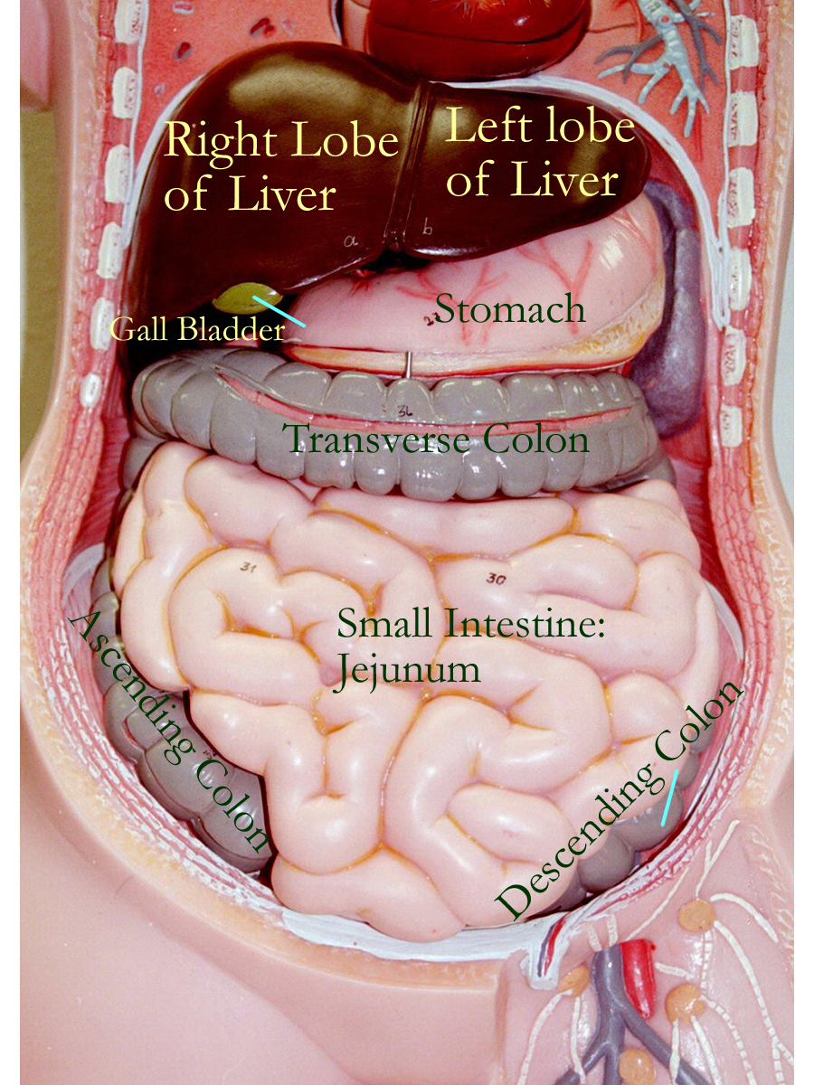
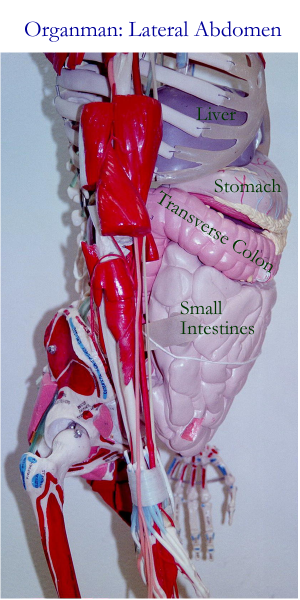
Organman Model : Anterior, Lateral
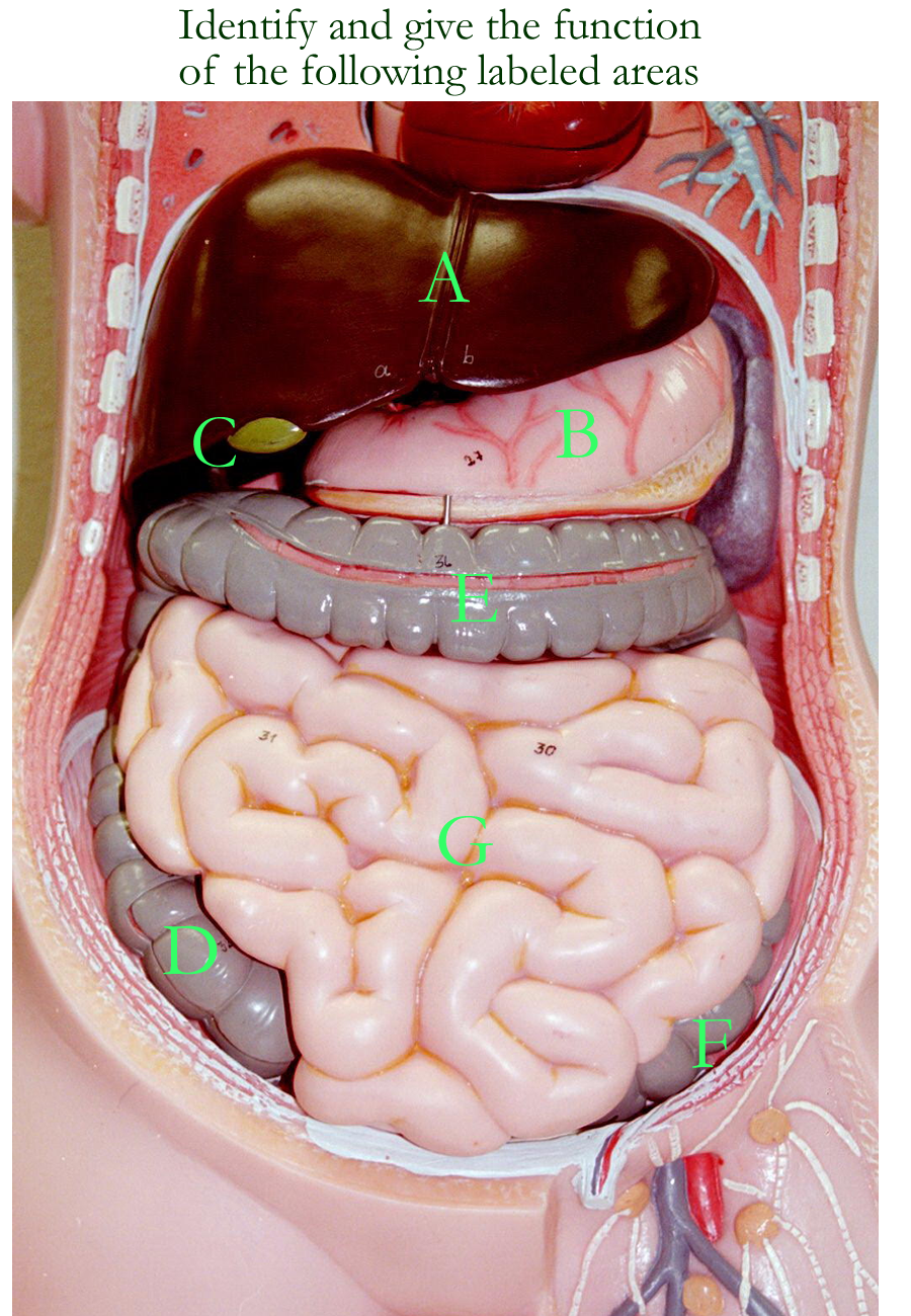
Small Intestine
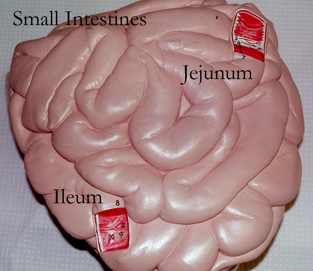
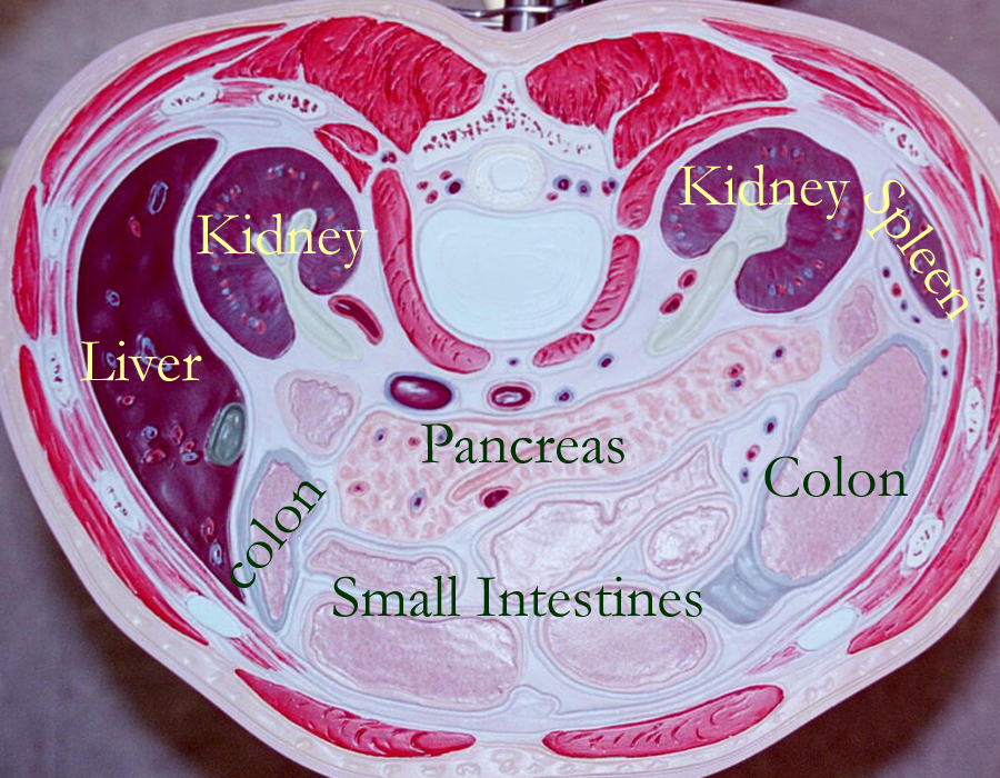
The small intestine is located in the abdominal cavity and is anchored by an extra fold of parietal peritoneum called the mesentery proper. The small intestine is divided into three regions: the duodenum, the jejunum, and the ileum.
The duodenum is the first 12 inches of the small intestine and functions primarily to receive and chemically adjust the stomach chyme. Ducts from the pancreas and liver to the duodenum allow for transport of secretions that aid in digestion. Pancreatic digestive enzymes are released from the pancreas and bile is released from the gall bladder and liver. Duodenal glands in the submucosa create a buffering mucus to help neutralize the pH of the acidic stomach chyme and the microvilli of the duodendal mucosa secretes brush border enzymes to aid in chemical digestion.
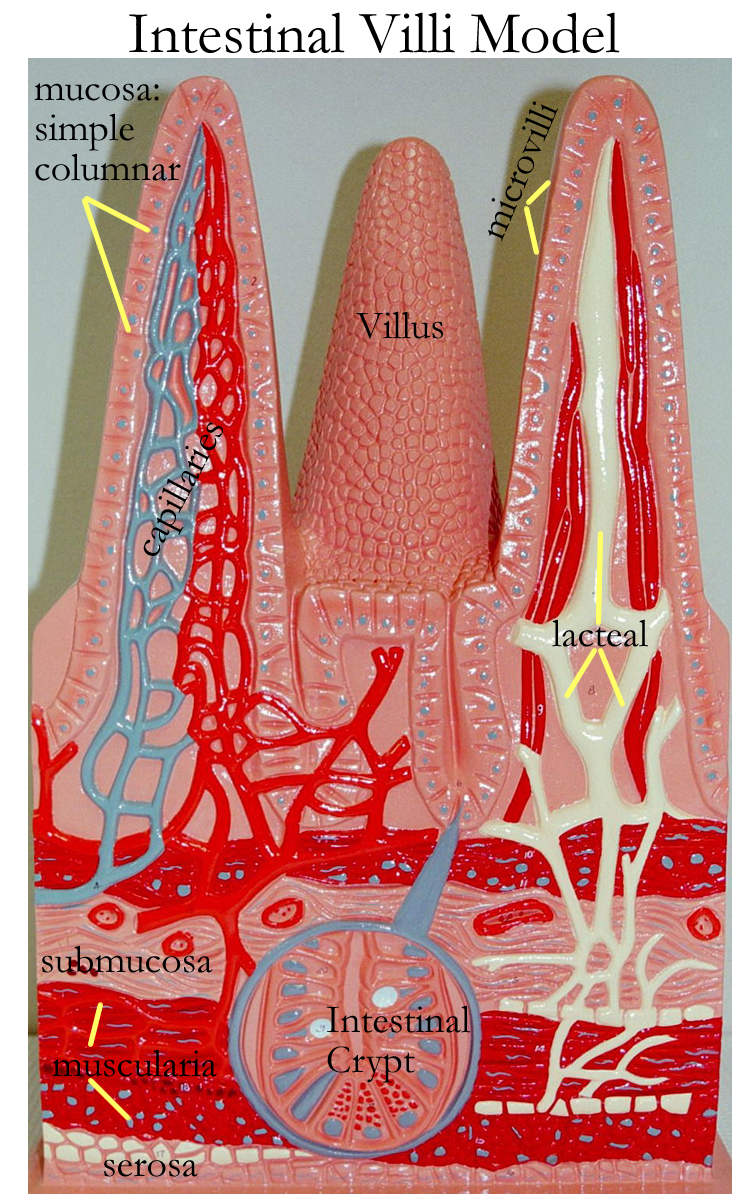
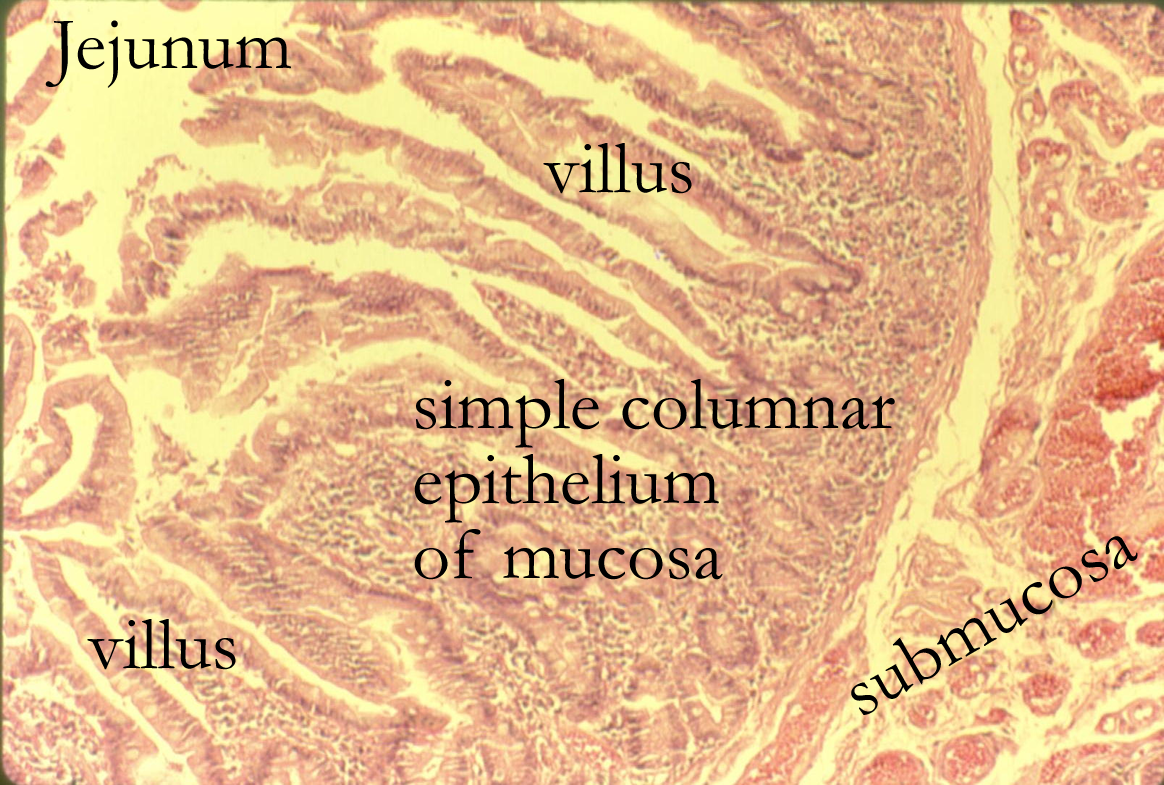
The jejunum is the mid section of the small intestine and most of the absorption happens in this region. Histologically, the submucosa creates small folds called plica that help to increase surface area. The mucosa has folds called villi and they are the longest of any seen in the small intestine. Each simple columnar cell of the mucosal lining also has folds called microvilli and they too secrete brush border enzymes that aid in digestion.
The folds of the plica, villi, and microvilli all help to increase surface area for absorption.
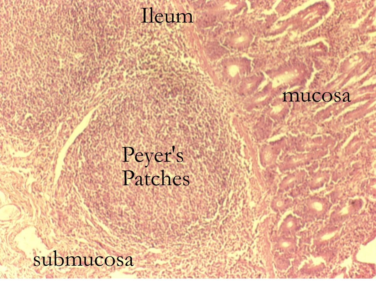
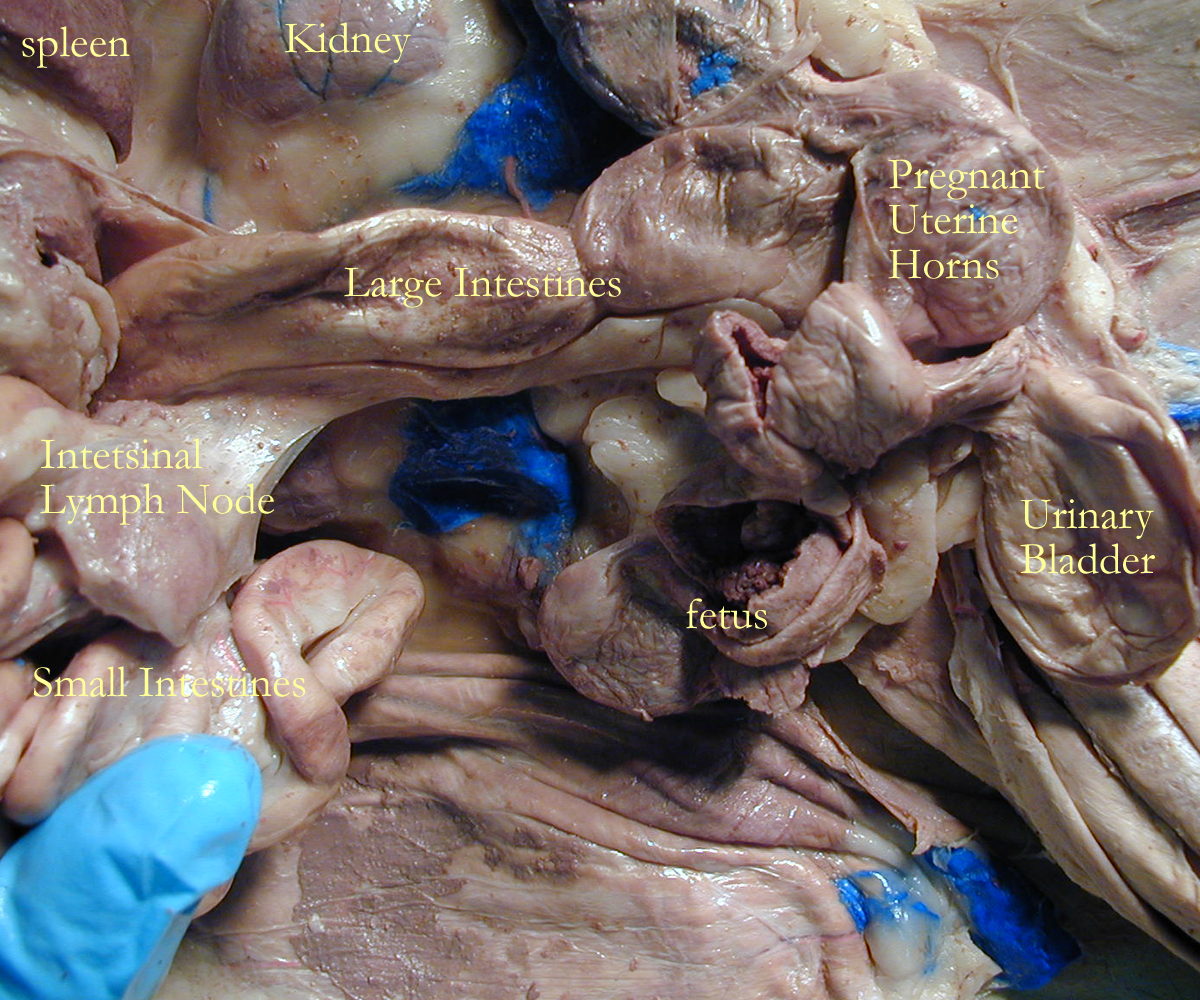
The ileum is the terminal portion of the small intestine. Its villi are shorter and function in fat absorption into the lymphatics called lacteals. In the submucosa, there are lymphatic nodules called peyer’s patches that aid in immune function for the digestive system. Movement of ingesta from the small intestine to the large intestine is controlled by the ileocecal sphincter muscle.
Histology: Duodenum, Jejunum, Ileum
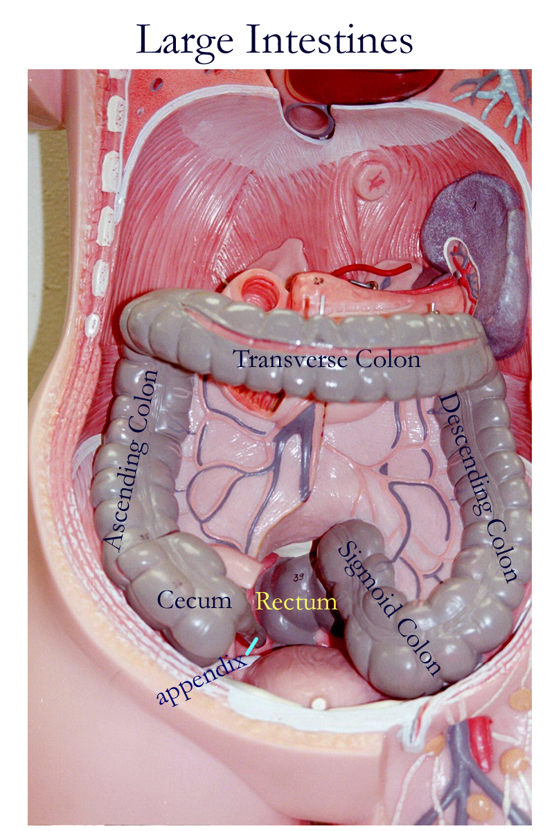
Large Intestines
The large intestine is located laterally around the small intestine in the abdominal cavity. The major regions are the cecum, colon, and rectum. The colon is further divided into the ascending colon, the transverse colon, the descending colon, and the sigmoid colon. These regions of the colon bend at 90 ° angles as they turn to create each of the regions of the colon. On the right side there is a right colic flexure also called the hepatic flexure from the ascending to the transverse colon. On the left side there is a left colic flexure also called the splenic flexure from the transverse to the descending colon. The cecum has an appendix, a blind appendage lined with lymphatic nodules. The rectum terminates into the anal canal and its opening, the anus.
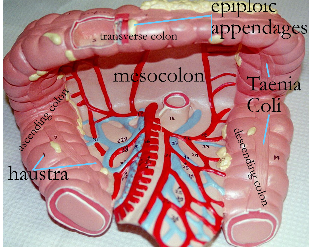
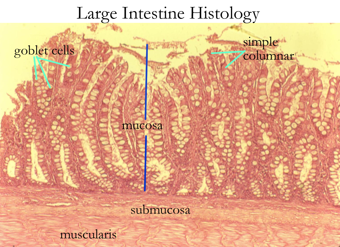
The large intestine is held in place by an extra fold of parietal peritoneum called the mesentery of the colon or mesocolon. It is anchored to the muscle wall of the lumbar regions. The epithelial lining of the mucosa is simple columnar with goblet cells for mucus production. The outer muscular layer of the colon is incomplete and forms bands called taenia coli. The complete smooth muscle layer then creates pouches called haustrae and movements in this area are called haustrations.
The functions of the large intestine are water absorption, production and absorption of Vitamin K and B complex, and storage & elimination of the fecal waste matter.
Strong peristaltic waves in the large intestine are called mass movements. They begin in the middle of the transverse colon and move the feces to the rectum. In the rectum, the muscle layers are complete and the lining forms internal valves in the anal canal called the anal columns. The opening of the anal canal is called the anus and the mucosal lining changes from simple columnar to stratified squamous to form a protective mucus membrane barrier for immune defenses. During the defecation reflex, stretch receptors signal the movement of feces into the rectum and anal canal. Internal smooth muscle sphincters relax and open and the external skeletal muscle sphincters are then signaled to relax and open. Because skeletal muscles are under voluntary, somatic control, the external anal sphincter can be closed until there is an appropriate time to deficate.
Model: Large Intestine GrossHisto
Vitamins produced by the large intestine vitamins : B complex, Vitamin K
Colo, colon/o- colon duoden/o- duodenum
Ile/o- ileum enter/o- intestine
Jejun/o- jejunum, hungry proct/o- anus, rectum
Rect/o- rectum, straight or/o-, stomat/o- mouth
Sigmoid/o- sigmoid colon cheil/o-, labi/o- lip
Esophag/o- esophagus gastr/o- stomach
Pharyng/o- pharynx pylor/o- pylorus
An/o- anus append/o- appendix
-emesis vomiting phagia swallowing
-scopy view de- down from
geni/o- chin cec/o- blind
chy- pour aliment/o- eat
typhl- blind ptyal/o- saliva
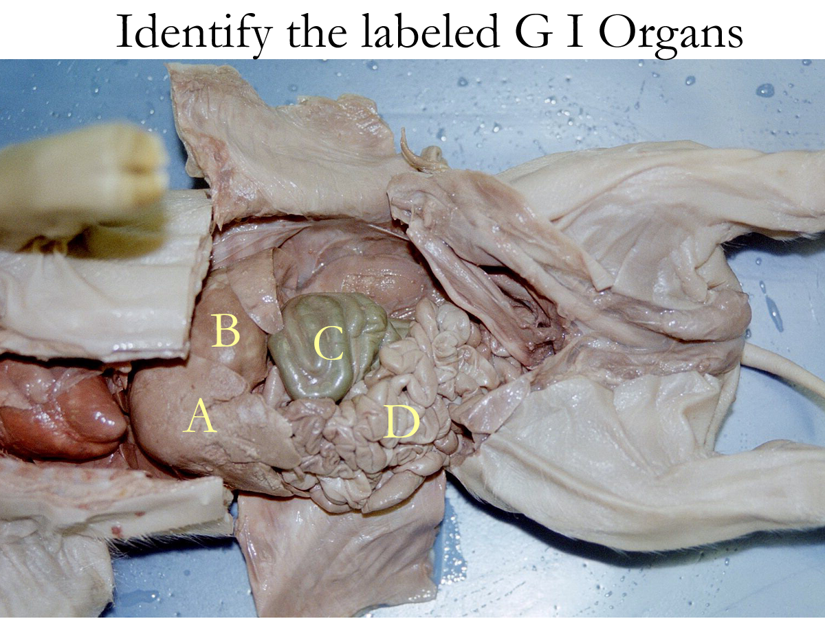
I. ID organs:
A Drawing
C Specimen
Concept Map: Make a concept map of the GI system (gross and histo) anatomy, location, and physiological function. Include this concept map in your LAR lab report (if selected) as a document insert or as an addtional PDF scanned document.
Vomiting Diarrhea
Pyloric Stenosis Ulcer: gastric, duodenal
Ileus Volvulus, torsion
Intussusception Diverticulitis
Hernias: hiatal, abdominal Crohn’s disease
Colitis Colorectal cancer
Appendicitis Endoscopy
Upper GI series, Lower GI series Enema
Stomach stapling Stomach cancer
Reflux esophagitis Stomatitis
Gastritis Gastroenteritis
Malabsorption/ Maldigestion H. pylori (stomach bacteria)
Irritable Bowel Disease (IBD) Hemorrhoids
Internal Medicine
http://www.nlm.nih.gov/medlineplus/healthtopics.html
http://www.lumen.luc.edu/lumen/meded/histo/frames/histo_frames.html
http://www.gen.umn.edu/faculty_staff/jensen/1135/webanatomy/
http://gened.emc.maricopa.edu/bio/bio181/BIOBK/BioBookTOC.html
http://www.kcmetro.cc.mo.us/maplewoods/Biology/Bio110/Labs.htm
http://calloso.med.mun.ca/%7Etscott/second.htm
http://www.track0.com/canteach/links/linkbodysystems.html
http://www.carr.lib.md.us/schs/science/anatomy/systems.html
http://www.leeds.ac.uk/chb/humbmods.html
http://www.stemnet.nf.ca/CITE/body.htm
http://www.gastro.org/public/yourdigest.html
http://medstat.med.utah.edu/kw/sol/sss/
http://arbl.cvmbs.colostate.edu/hbooks/pathphys/digestion/
http://biology.about.com/library/organs/blpathodigest2.htm
http://members.tripod.com/~rmoskowitz/digestive.html
http://w2.shorecrest.org/MSUS/C-Cruise/htmlpages/anatomy%2Fzoos%2Fgames_links.htm
http://biology.about.com/library/organs/bldigestoverview2.htm
http://www.teaching-biomed.man.ac.uk/mcwilliam/digest.htm
http://www.nlm.nih.gov/medlineplus/digestivediseasesgeneral.html
http://www.nlm.nih.gov/medlineplus/stomachdisorders.html
http://www.nlm.nih.gov/medlineplus/digestivesystem.html
http://www.niddk.nih.gov/health/digest/pubs/digesyst/newdiges.htm
http://www.medem.com/MedLb/article_detaillb.cfm?article_ID=ZZZ7C4T46JC&sub_cat=338 digestive system
http://www.medem.com/medlb/article_detaillb.cfm?article_ID=ZZZTD0TCGJC&sub_cat=50 teeth
http://www.nlm.nih.gov/medlineplus/mouthandteeth.html
1. Define digestion and absorption
2. Name the regions of the GI tract and give their functions.
3. Name the parietal peritoneal folds that anchor most of the GI tract.
4. Define ingestion and deglutition.
5. Define peristalsis and segmentation.
6. What is the purpose of rugae folds, plicae folds, villi, and microvilli and why.
7. Name the subdivisions of the small intestine and their function.
8. Name three hormones that affect the GI tract and give their specific functions.
9. Why is the GI tract considered an open body cavity?
l0. Name the regions of the large intestine and describe the defecation reflex.