Biology 2404 A&P Basics Urinary System Dr. Weis
| Objectives | Background | Medical Terms | Activities | Applications | Careers | WWW | Review Questions |
Students should be able to:
* Name, identify, and give the functions of the urinary system organs
Kidney
Ureters
Urinary Bladder
Urethra
* Define renal physiology terms such as
* Name the three hormones from the kidney JGA and their functions
* Define incontinence
* Define related terms
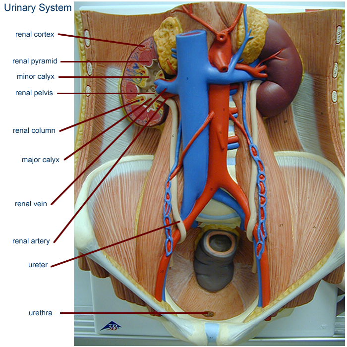
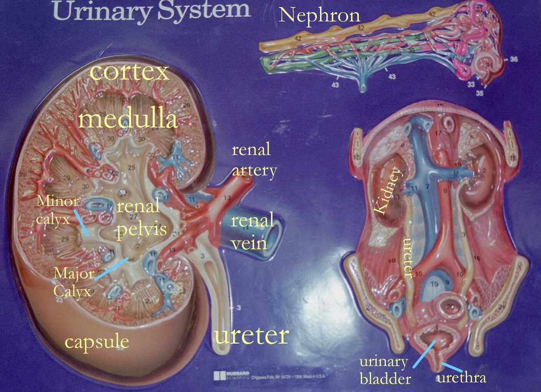
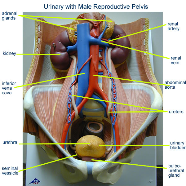
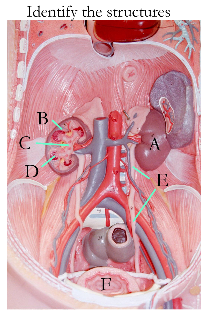
The urinary system is comprised of two kidneys, two ureters, a urinary bladder, and a urethra. The kidneys filter blood and the end product, urine, is then transported by the ureters, stored in the urinary bladder, and eliminated from the body by the urethra.
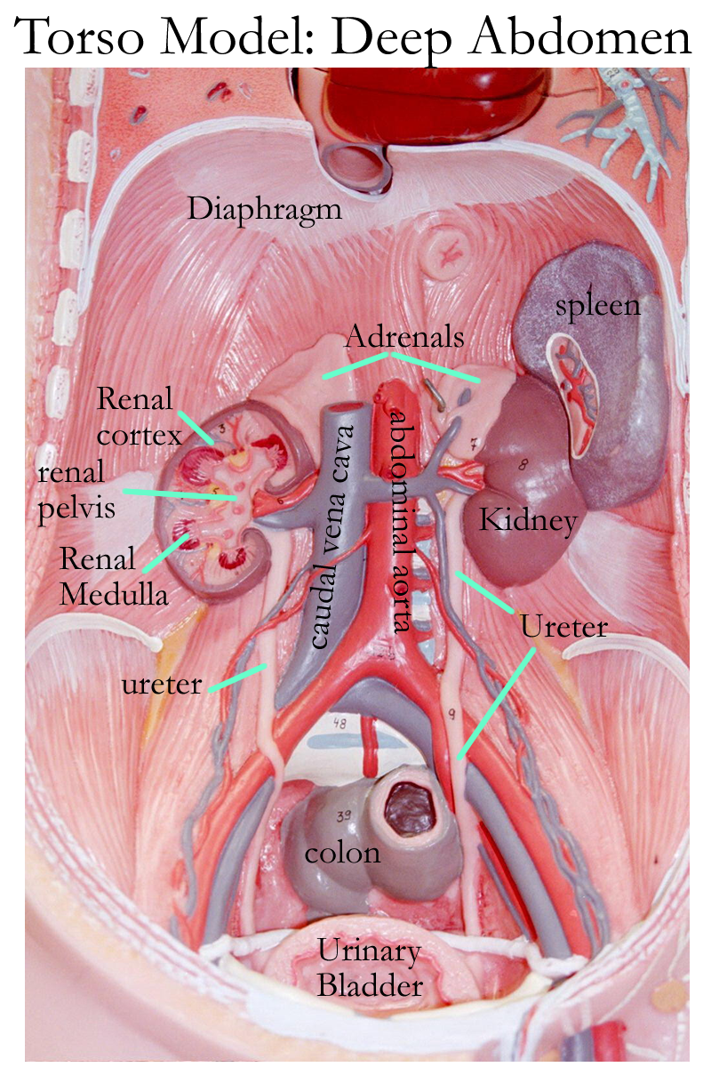
Urinary system wall mount (pig)
Kidneys
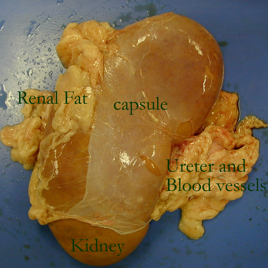
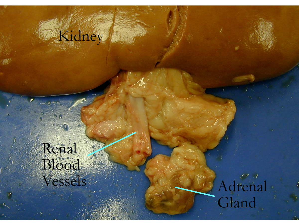
The kidneys are paired organs in the upper right and left abdominal quadrants on either side of the vertebral column. The kidneys sit retroperitoneally and are anchored and protected by two outer connective tissues: the adipose capsule and the renal fascia.
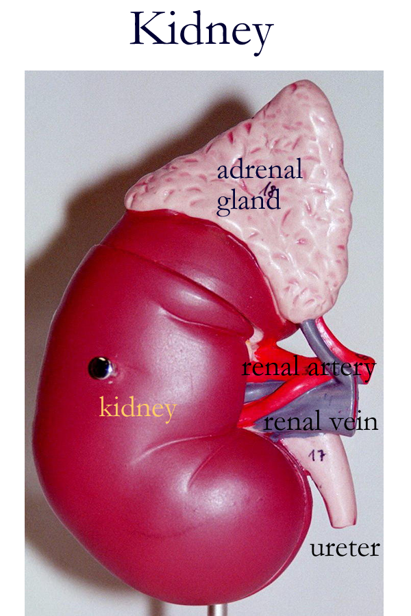
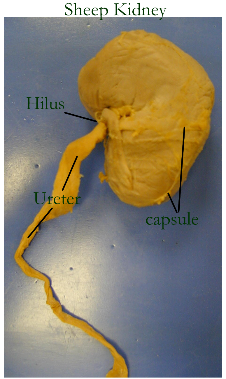
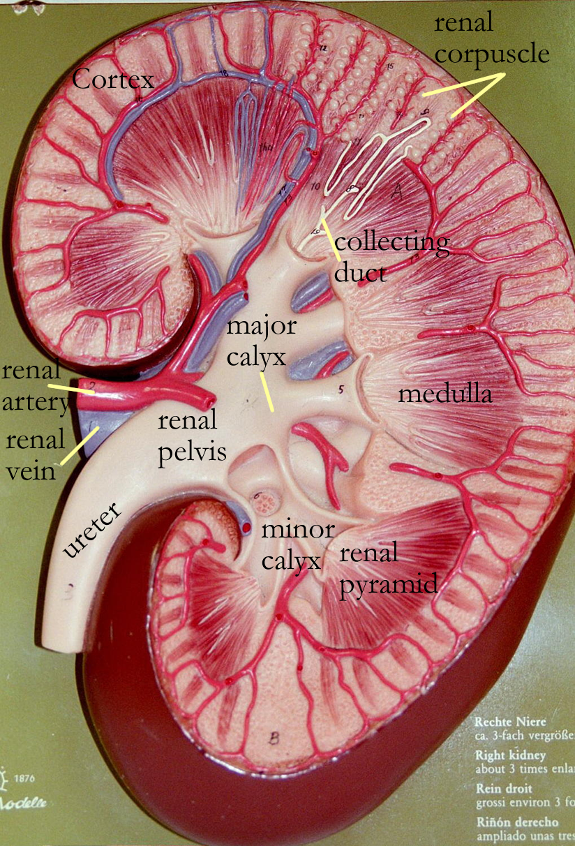
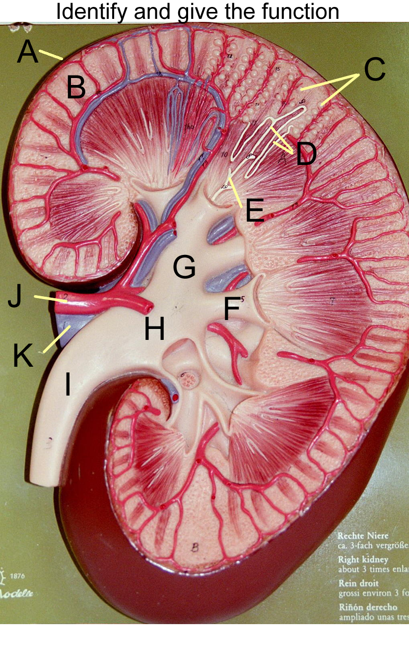
The kidney bean shaped organs are a little smaller than a closed fist, have a superior and inferior pole, and an indented region called the hilus where vessels and nerves enter and leave.
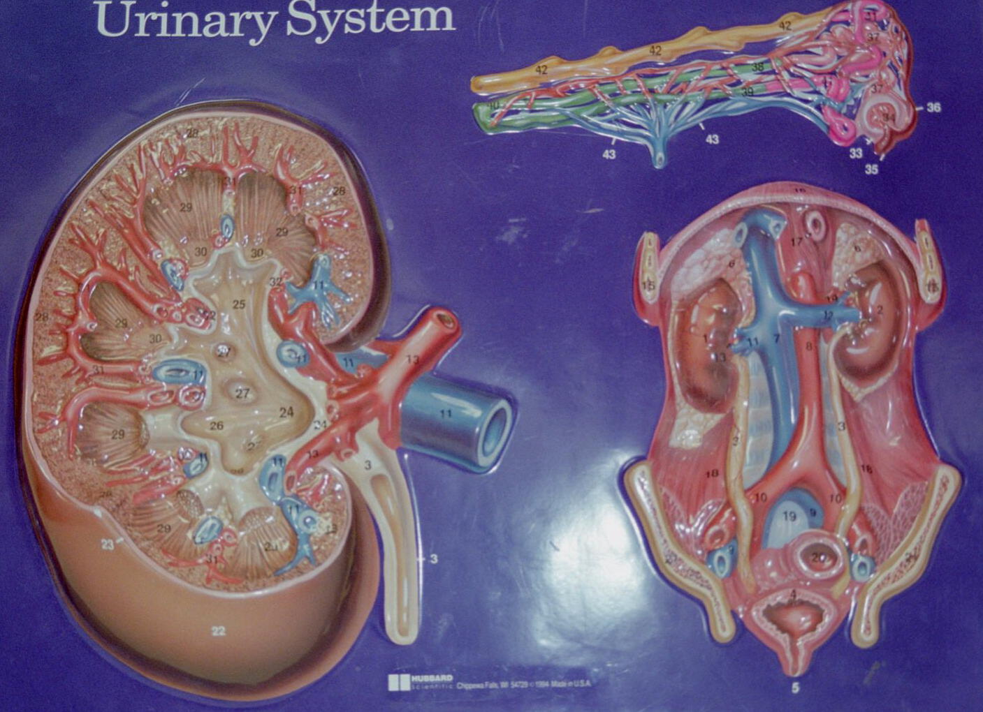
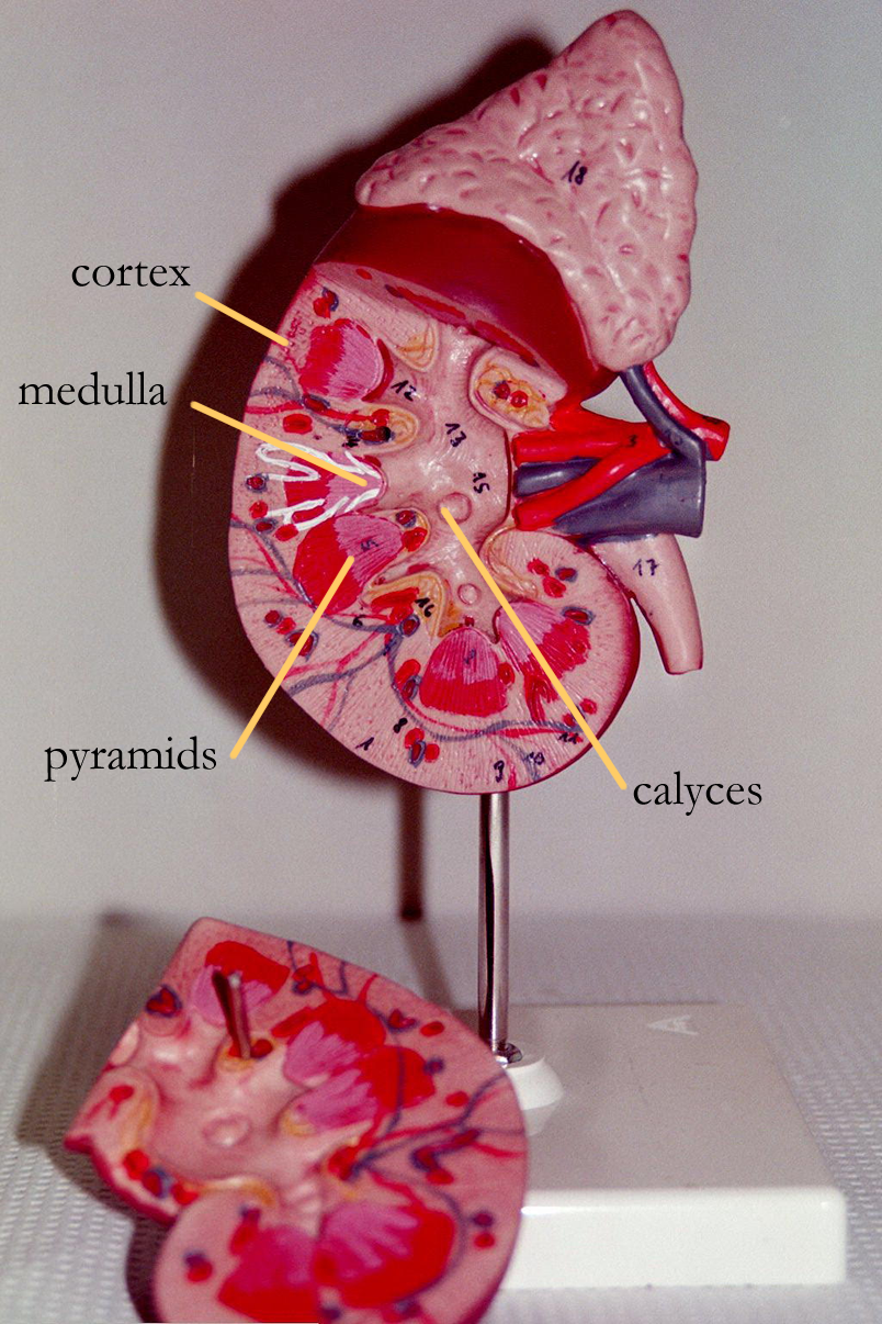
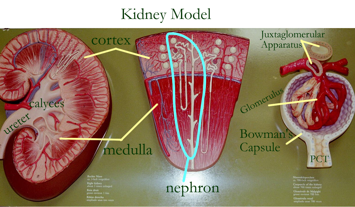
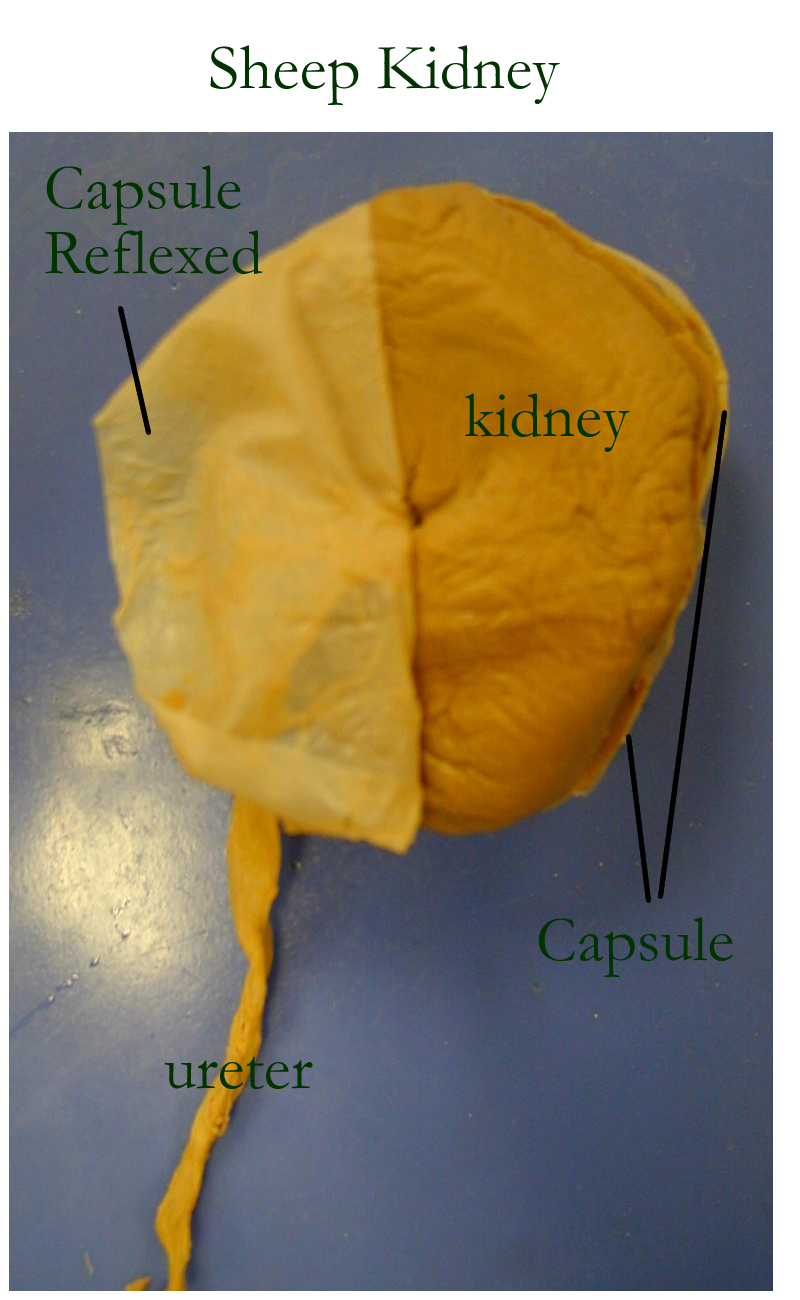
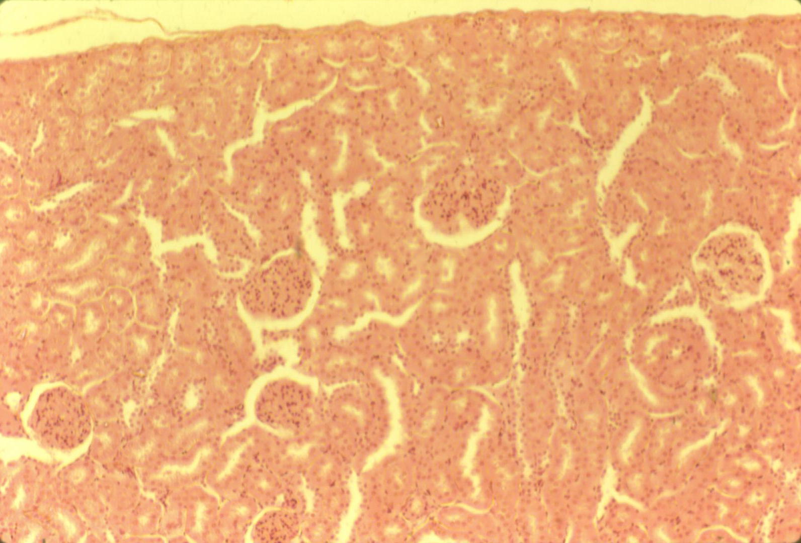
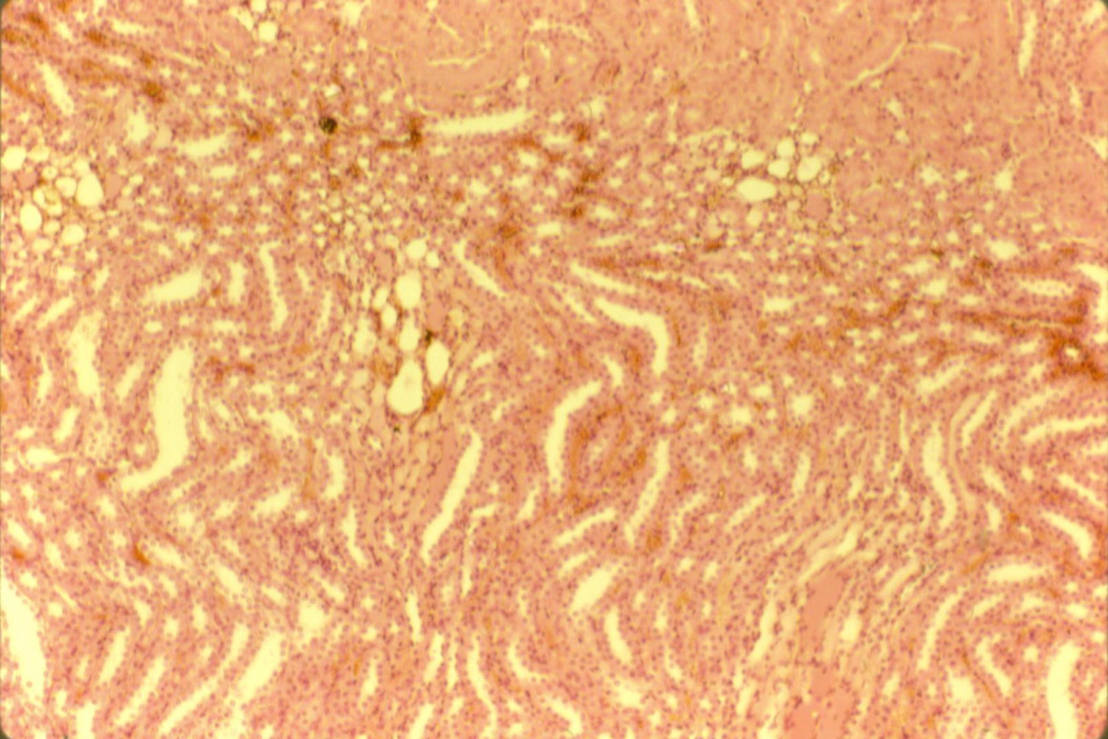
The kidneys grossly have a renal capsule, cortex, and medulla. The renal capsule also aids in anchoring the kidneys to the abdominal wall. The cortex of the kidney is comprised of nephrons with blood supply. Certain nephron renal tubules are for filtration and other tubules are for reabsorption and secretion. Parts of the renal cortex in humans and some other mammals dip down into the medullary area creating renal columns and dividing the medulla into sections.
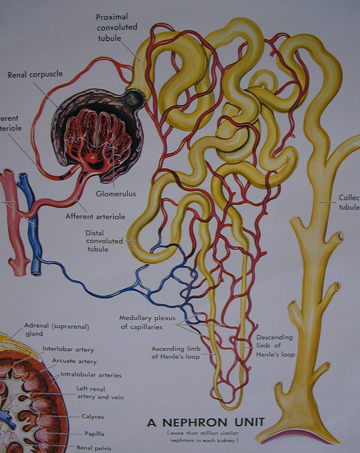
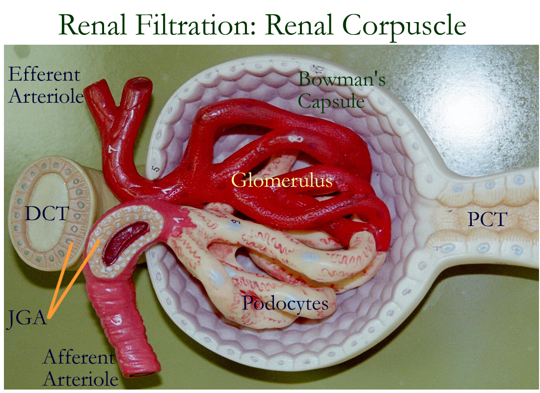
The juxtaglomerular apparatus is also located in the cortex and is the renal tubule and blood supply structure that produces the hormones that from the kidney.
The renal medulla is divided by the renal columns into triangular structures known as medullary pyramids. In the medulla, there are connective tissue tubules lined with cuboidal epithelium that adjust water and ions such as sodium in order to form a concentrated urine. The tip of the medullary pyramid called the renal papilla opens into the renal pelvis by means of calyces. The renal pelvis leaves the kidneys as the ureter at the hilus.
Kidney model: Anterior, Interior
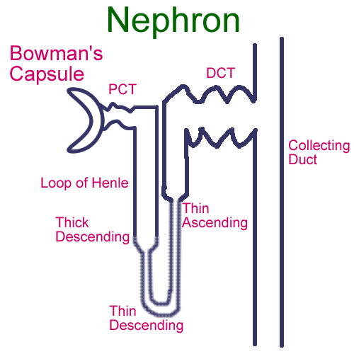
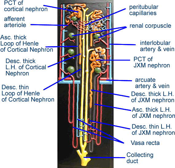
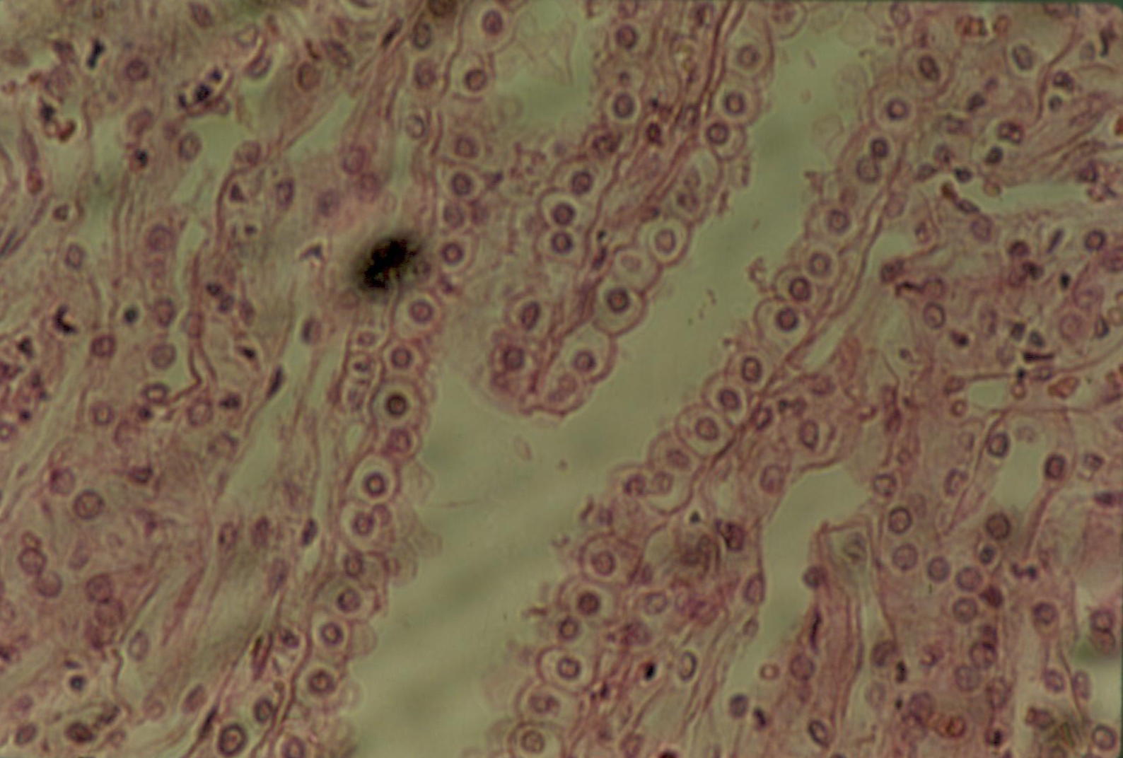
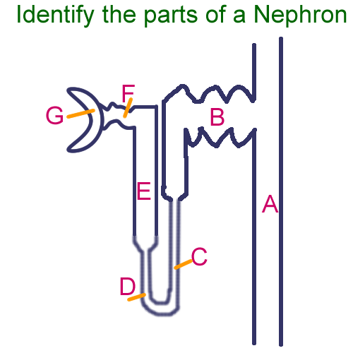
Microscopically, the kidneys are comprised of nephrons, the functional unit of the kidney. The nephron consists of simple cuboidal cells that line connective tissue tubules: Bowman’s capsule, proximal convoluted tubule, loop of Henle, distal convoluted tubule and collecting ducts. Each tubular region has special functions due to a corresponding blood supply from the renal artery divisions. The renal artery divides several times and follows the renal columns to the cortex to create the arcuate arteries, interlobular arteries, afferent arterioles and the renal capillaries called the glomerulus.
The Bowman’s capsule with the glomerulus together forms the Renal corpuscle. The function of the renal corpuscle is to filter blood. Proper blood pressure is needed to push plasma through the capillary fenestrations and the podocytes of Bowman’s capsule but still avoid pushing red blood cells and large proteins such as albumin through the renal filtration membrane. The fluid pushed through the renal filtration membrane that will then be collected in the Bowman’s capsule is called filtrate. Filtrate resembles plasma, except that there are no large proteins. The filtrate travels through the remaining tubules to be adjusted based on the body’s needs.
Bowman’s capsule opens into the proximal convoluted tubule and the glomerular capillaries continue as the efferent arterioles and then peritubular capillaries that surround the remaining tubes of the nephron. The proximal convoluted tubules (PCT) function primarily to reabsorb water, many ions, glucose, and amino acids. Reabsorption moves the substance from the PCT across and into the blood to be returned to the body.
In certain situations, the PCT can secrete ions, that is, move the ion from the blood into the filtrate to be excreted as urine. Examples of an ion that is secreted is hydrogen (H+) in order to adjust blood pH.
From the PCT, the filtrate continues into the Loop of Henle. Two types of loops exist and are identified by their location and length. The two different Loops of Henle give the nephron types their names. The shorter Loops of Henle are found in the cortical nephrons and comprise 75% of the nephrons found in the kidney. The longer loops of Henle dip down into the medulla and are found in the juxtamedullary nephrons.
The loops of Henle can only regulate water and salt (NaCl) concentrations. In the shorter loops of Henle, a dilute filtrate is formed, whereas in the longer loops of Henle, a more concentrated filtrate can be formed. The loops of Henle affect their surrounding tissue environments off the collecting ducts and so the filtrate concentration can be further regulated by response to hormones.
From the loop of Henle, the filtrate enters the distal convoluted tubule with it’s peritubular capillaries surrounding this section of the nephron. The early distal convoluted tubule (DCT) actually comes back around near the PCT and the afferent arteriole. The early DCT and the afferent arteriole form the endocrine structure known as the juxtaglomerular apparatus (JGA). The hormones secreted from this region are:
Erythropoietin (EPO) signal the red bone marrow to make RBCs
Calcitrol formed from Vitamin D, signals the Late DCT and Collecting Duct to reclaim (reabsorb)
Calcium and signals the digestive tract to get calcium from the diet
Renin signal the DCT and collecting duct to reclaim sodium by the renin-angiotensin system.
The rennin angiotensin system is as follows:
Renin (Juxtaglomerular apparatus) --> Angiotensin I (Liver) --> Angiotensin II (Lung) -->
Aldosterone (Adrenal Cortex, Zona Glomerulosa) --> Reabsorb Na+ (DCT, CD of kidneys)
The late DCT functions primarily in secretion, that is, movement of a substance from the peritubulary capillary (blood) to the tubule (DCT). Substances secreted are H+, ammonia, and certain drugs that have renal elimination. The late DCT can also reabsorb substances, but hormones are needed. The collecting duct (CD) functions the same as the late DCT due to the presence of similar epithelial linings.
Hormones that can trigger DCT / CD reabsorption are:
Renin Na+ reabsorption
Calcitrol Ca+ reabsorption
ADH water reabsorption
Aldosterone Na+ reabsorption
ANP blocks aldosterone’s affects on the kidney
In summary the functions of the kidneys are:
Regulate pressure of blood by reabsorption or elimination of water
Regulate blood concentration and O2 levels by signaling RBC production
Regulate pH and acid / base by secretion of H+ or reabsorption of HCO3-
Regulate concentration of minerals (electrolytes) such as Ca++, Na+, Mg++, K+
Activate Vitamin D
Hormone production
Formation of urine
Renal Physiology : Renal Corpuscle for Filtration
Ruminant Kidney: Capsule, Cortex/Medulla, Sagittal View, Physiology
Sheep Kidney: Capsule, Sagittal View
Kidney histology
Bloodwork Report : Renal Function Tests
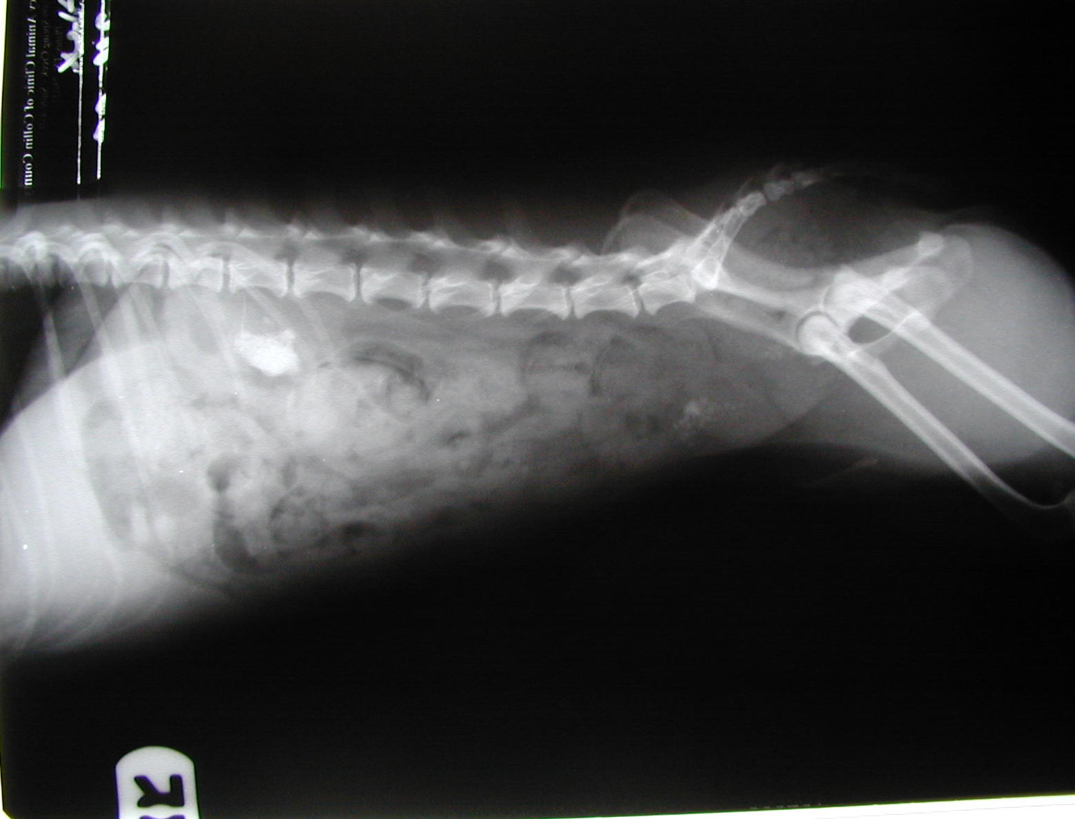
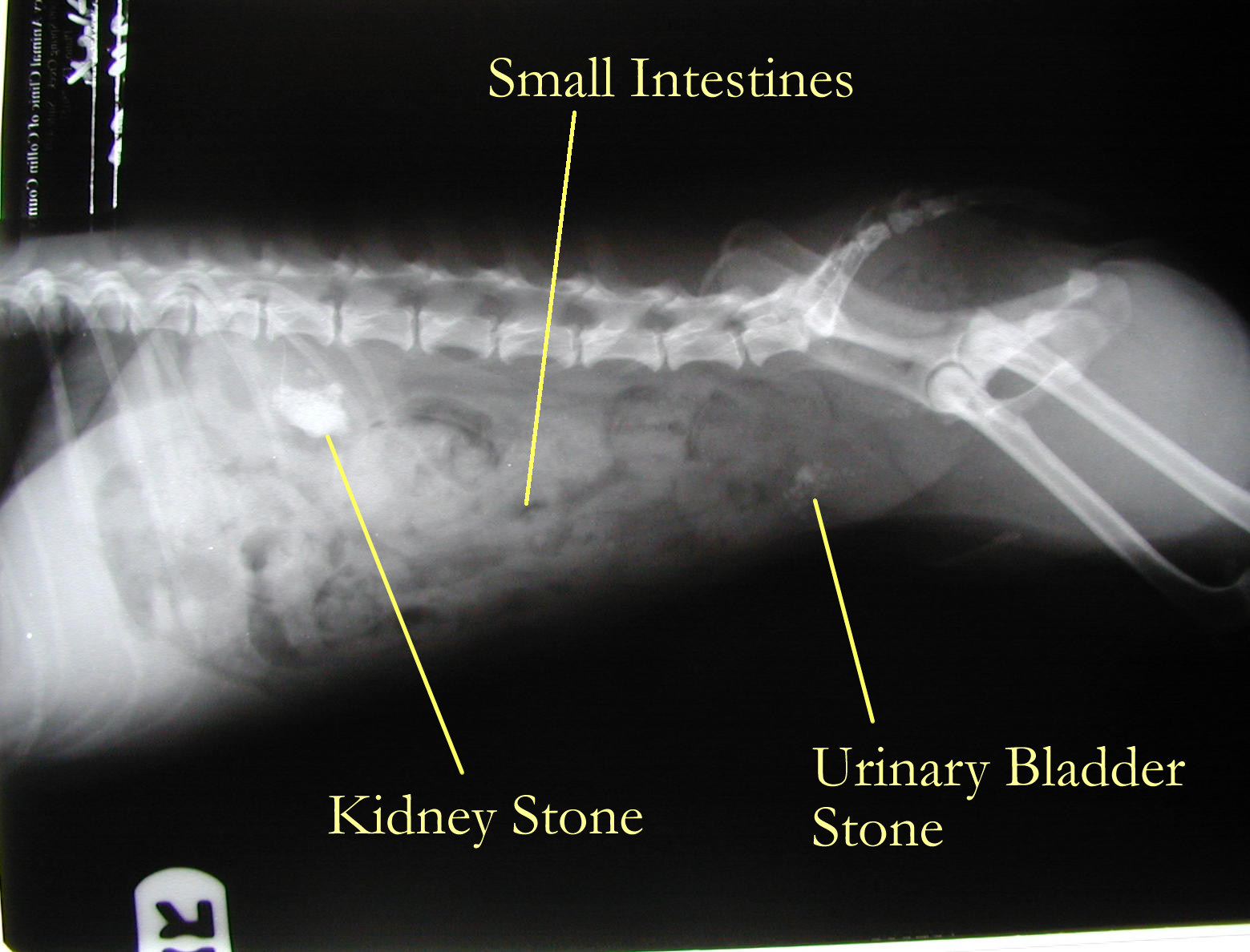
Kidney radiology : Stones in Renal Pelvis
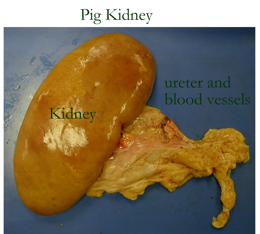
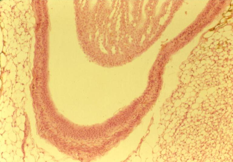
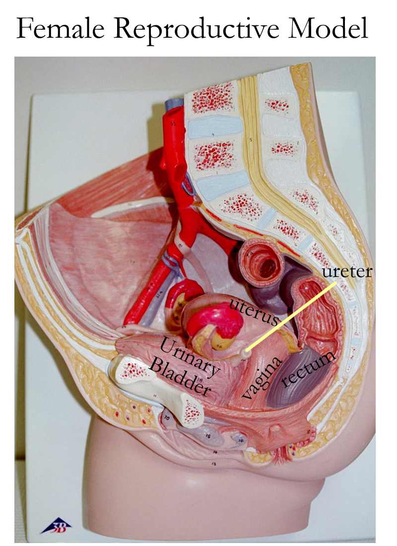
The filtrate that leaves the renal papilla of the renal medulla is collected as urine in the renal calyces and renal pelvis. The urine exits in the pelvis at the hilus and continues down the ureters. The cuboidal lining of the kidney tubules change to transitional epithelium in the renal pelvis and will be found through the rest of the urinary system until it changes to stratified squamous epithelium in the urethra. The ureters extend down to the urinary bladder posteriorly, enter obliquely, and function to transport urine to the urinary bladder.
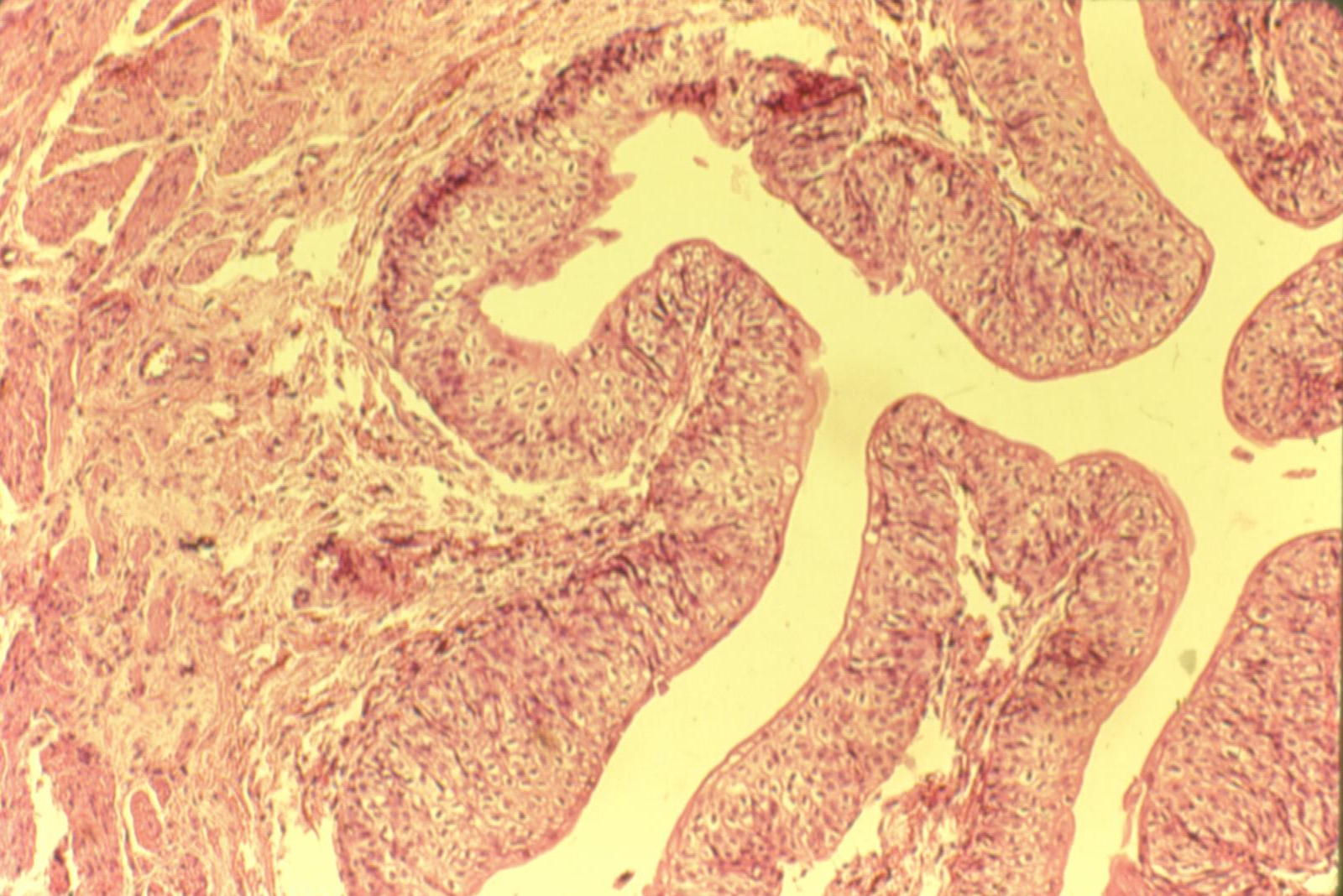
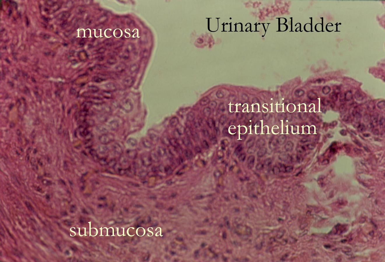
The urinary bladder is a sac like organ in the pelvic region that can change size due to expansion or contraction related to urine storage and elimination. The gross regions of the urinary bladder are the fundus, body, and neck. The histology of the urinary bladder is as follows:
Mucosa transitional epithelium
Submucosa connective tissue with blood supply
Muscularis 3 layers of smooth muscle termed the detrusor muscle
Serosa visceral peritoneum
The importance of this histology is found in the epithelial lining and the muscularis layer.
Transitional epithelial cells are linked together and will change shape from a cuboidal cell to a squamous shaped cell as the bladder expands. Prolonged expansion can damage the cell to cell junctions. The detrusor muscle enables contraction for urine expulsion.
Damage to the muscle or problems in the neuron signaling can lead to urinary retention or incontinence.
The urinary bladder’s three internal openings are formed by the two ureters and the urethra to create a triangular region known as the trigone. Bladder infections and cancers primarily occur in this area.
In the neck of the urinary bladder, there are two sphincter muscles. The internal urinary sphincter is smooth muscle and the external urinary sphincter is skeletal muscle. As the bladder expands to store the urine, the sympathetic nervous system is triggered to send signals to the brain to open the muscle sphincters. Urine is then expelled from the urinary bladder. This elimination of urine is also called micturition, urination, or voiding of urine.
Urine leaving the urinary bladder enters the urethra and is then eliminated outside the body when the external urethral sphincter, a skeletal muscle, is activated.
The differences in the urethra reflect the anatomy of the reproductive systems.
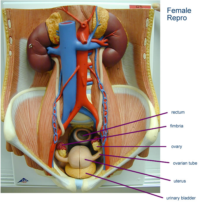
In females, the urethra is for urine elimination only and is very short, about 1.5 inches long. In males the urethra is also part of the male reproductive tract. The male urethra is about 8 inches long and functions in semen transport or urinary elimination.
Urinary Radiology: Stones in Urinary Bladder
Reproductive models
Urinary Model with Male Reproductive Organs
Urinary Model with Female Reproductive Organs
Urine production is the result of blood filtration and filtrate reabsorption & secretion.
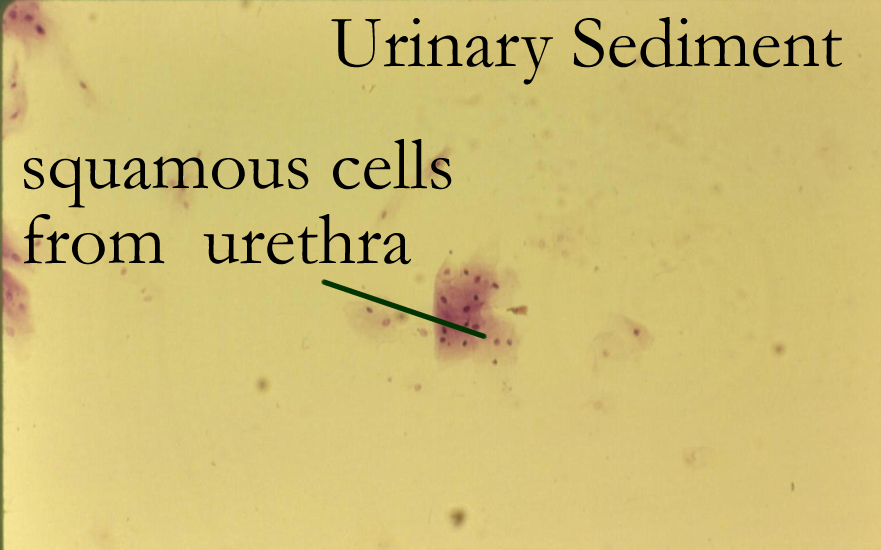
Urine is primarily water, with ions, nitrogen wastes (creatinine, urea, uric acid), drugs and toxins. Urine does not normally contain RBCs, protein, or glucose. Occasionally epithelial cells, casts, and crystals may be seen.
The urinalysis allows for identifying and testing substances eliminated by the urinary system. There are physical and chemical characteristics of urine that are identified during a urinalysis. See the chart below:
|
Color
|
straw, yellow, amber | abnormal if orange, red, brown |
|
Appearance
|
Clear, transparent | abnormal if cloudy |
|
Odor
|
no odor | older urine will smell ammonia odor |
|
Specific Gravity
|
Normal range is above 1.012 to 1.035 | concentration of urine based on solutes in water |
|
pH
|
diet dependent. Usually at 6.0 if on a protein diet | pH range is 4.50-8.0 |
|
protein
|
negative | abnormal values range from trace to 4+ |
|
glucose
|
negative | positive = abnormal |
|
blood
|
negative | positive= abnormal |
|
leukocytes
|
negative | positive = abnormal |
|
ketones
|
negative | positive = abnormal |
|
wastes
|
bilirubin, urobilinogen, creatinine, urea, uric acid | |
|
|
negative | unless older urine or pH changes |
|
Bacteria
|
depends on method of collection | Voided and catheter samples may have bacteria. Cystocentesis should be a sterile procedure |
|
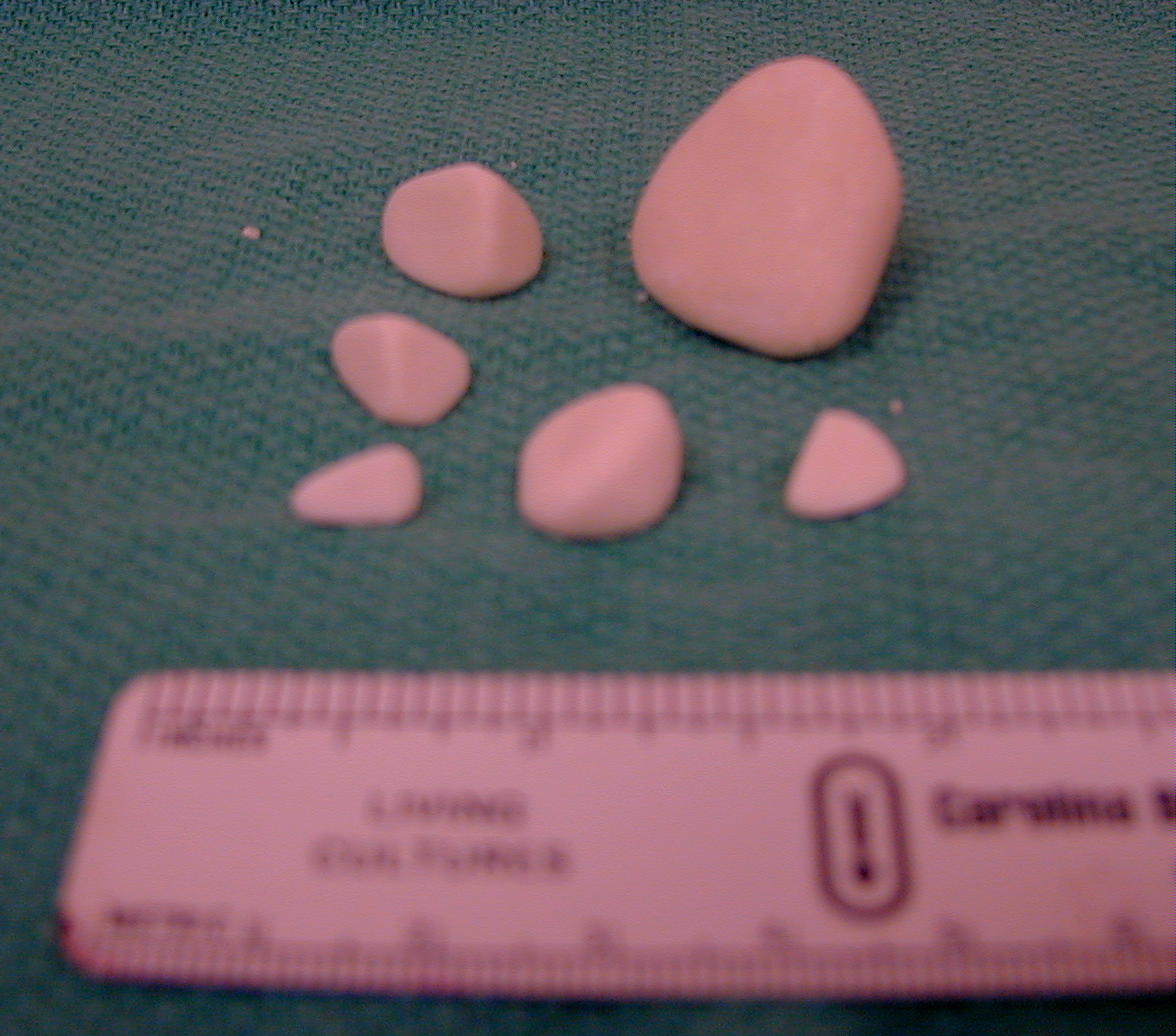
Stones
|
abnormal | Usually formed from mineral sedimentation |
|
Method collected
|
voided, catheter, cystocentesis | sterile sample is preferred |
|
amount
|
1-2 liters depending on water consumption | decreases (oligouria) or increases (polyuria) can indicate problems |
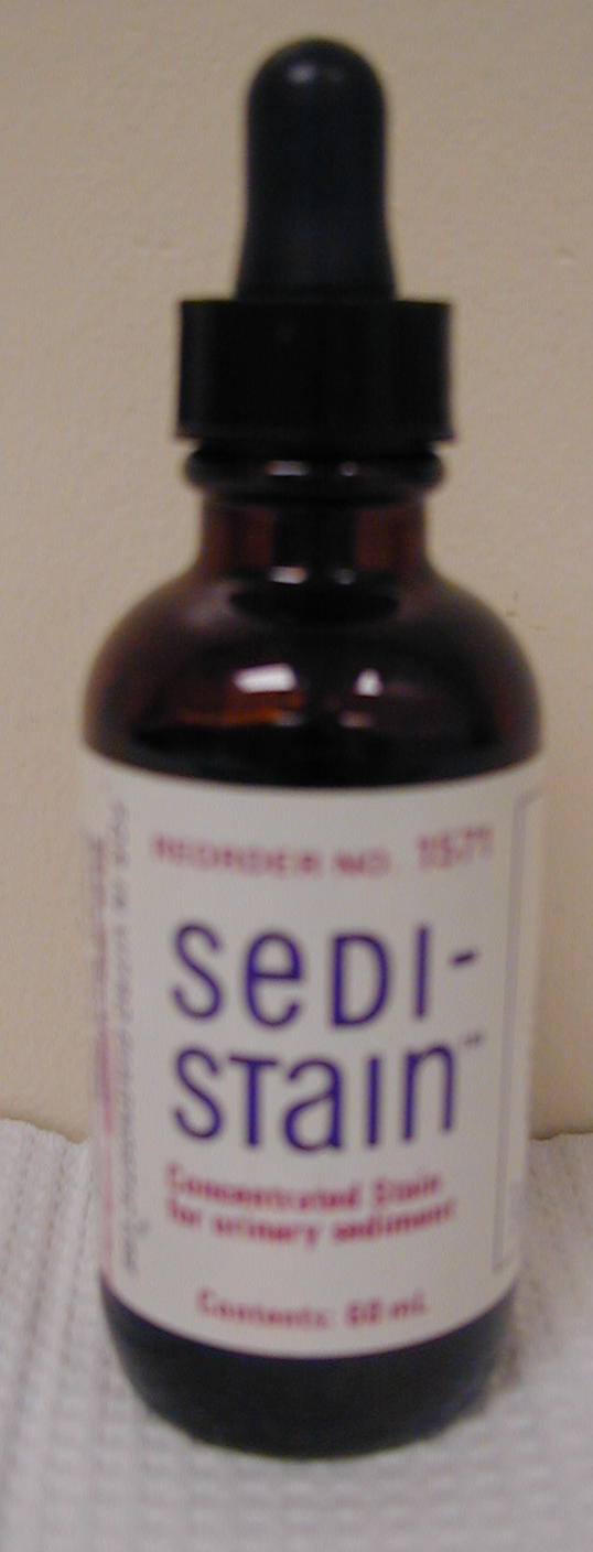
Urinalysis equipment: Sedistain, Dipsticks
Urinalysis and Sedimentation results
Picture of uroliths : Large, Small
Labeled Radiography of Kidney and Bladder stones
Cyst/o- sac diure- to urinate
-dynia pain dis- negative
dys- pain ecto- outside
nephr/o- kidney oligo- few, little
podo- foot pre- before
tri- three uro, -uria urine
calc- stone nocti- night
ren- kidneys vesic/o- bladder
glomerul/o- glomerulus ureter/o- ureter
urethr/o- urethra bacteri/o- bacteria
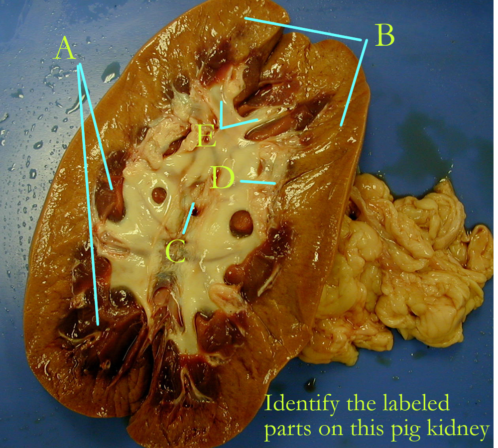
I. ID kidney parts
A Sheep
B Pig
II. ID organs
Urinary review and quiz : click on the urinary link on the main page of this web site
Concept Check:
Make a concept map of the urinary system (gross and histo) anatomy, location,
and physiological function. Tie this informatin to fluid and electrolyte (acid/base)
balance.
Include this map in your LAR lab report (if selected) as a document
insert
or as an additonal PDF document scan.
Renal calculi, nephrolithiasis (kidney stones)
Urinary tract infections (UTI)
Renal failure: acute and chronic
Polycystic Kidney Disease
Hypernephroma
Acute Tubular Necrosis (ATN)
Cystitis
Gout
Glomerulonephritis
Polyuria
Hemodialysis
Hematuria
Uremia
Stress Incontinence
Pyelonephritis
Nephrotic Syndrome
Hydronephrosis
Lithotripsy
Bladder neck obstruction
Anuria
Enuresis
Nephrotic syndrome
Nocturia
Cystoscopy
Retrograde pyelogram
Intravenous pyelogram
Cystourethrogram
Dialysis
Kidney Transplantation
Neurogenic Bladder
Urethritis
Urologist
http://www.nlm.nih.gov/medlineplus/healthtopics.html
http://www.lumen.luc.edu/lumen/meded/histo/frames/histo_frames.html
http://www.gen.umn.edu/faculty_staff/jensen/1135/webanatomy/
http://www.kcmetro.cc.mo.us/maplewoods/Biology/Bio110/Labs.htm
http://calloso.med.mun.ca/%7Etscott/second.htm
http://www.track0.com/canteach/links/linkbodysystems.html
http://www.carr.lib.md.us/schs/science/anatomy/systems.html
http://www.leeds.ac.uk/chb/lectures/anatomy7.html
http://www.stemnet.nf.ca/CITE/body.htm
http://www.med.virginia.edu/med-ed/phys/practice_board.html
http://www.niddk.nih.gov/health/kidney/pubs/yourkids/index.htm
http://www.niddk.nih.gov/health/urolog/pubs/yrurinar/index.htm
http://www.medem.com/MedLB/article_detaillb.cfm?article_ID=ZZZFQ0XCGJC&sub_cat=316
http://www.nlm.nih.gov/medlineplus/kidneysandurinarysystem.html
http://www.kidneyfund.org (American Kidney Fund)
1. Name the two types of nephrons
2. Define renal filtration
3. Define tubular reabsorption
4. Define tubular secretion
5. Name 5 functions of the kidneys
6. Define micturition
7. Give the location and function of the trigone
8. Give the normal physical and chemical composition of urine
9. Give the function of the urinary bladder
10. Describe the differences in the male and female urethra.
11. Name the three hormones from the kidneys and give their function.
12. Define incontinence.
13. Name two nitrogenous wastes eliminated by the urinary system.
14. Why doesn’t glucose and protein normally appear in the urine?
15. What is the detrusor muscle?
16. Be able to identify the organs of the urinary system.
17. Define diuretic.
18. What substances are commonly secreted?
19. How does glomerular filtrate compare to plasma?
20. Why should there not normally be RBCs in filtrate or urine?