Biology 2404 A&P Basics Lab Exercise 24 Development Dr. Weis
| Objectives | Background | Medical Terms | Activities | Applications | Careers | WWW | Review Questions |
Students should be able to:
* Define fertilization and where it occurs
* Define gestation and give the normal period for humans
* Name the stages of the zygote
* Identify the anatomy of a blastula and its function
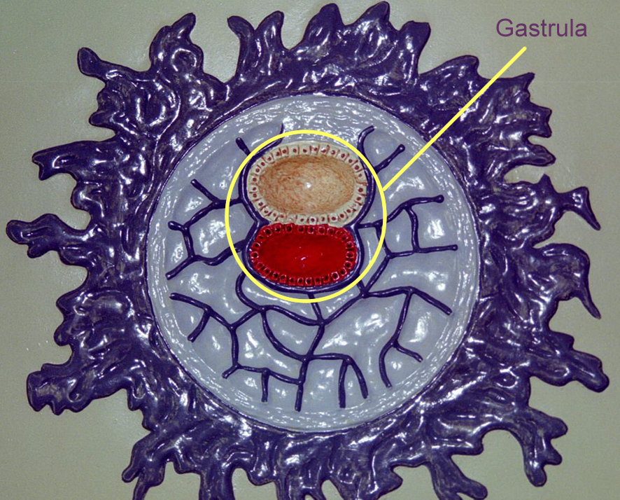
* Define gastrulation
* Name the embryonic tissues and their adult derivatives
* Name and describe the three stages of labor
* Define related terms
Read related material in textbook
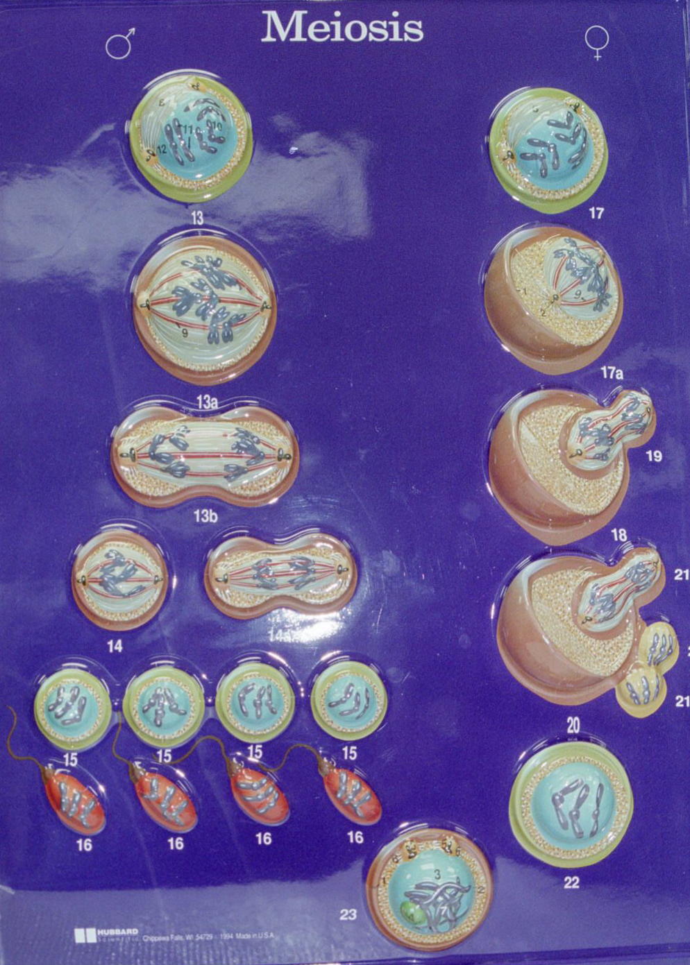
The gametes produced in the gonads undergo mitosis to create primary spermatogonia or primary oogonia and meiosis to each become haploid cells. Four haploid sperm are produced in the testes, while only one functional haploid oocyte is produced in the ovaries. Each has ½ the number of chromosomes once the process of meiosis is completed.
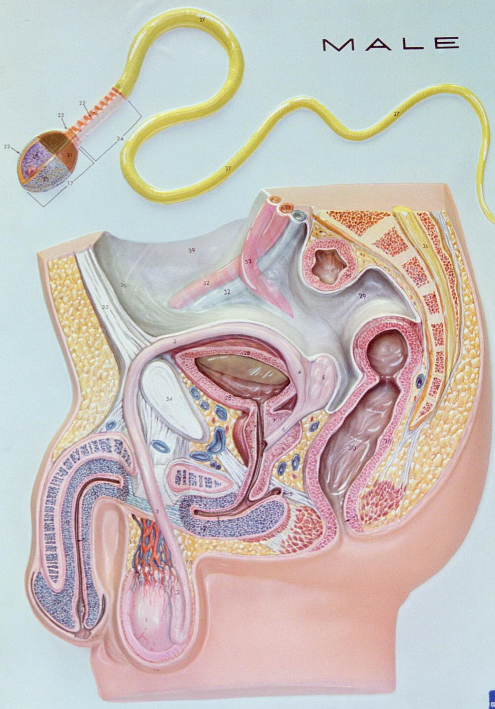
Sperm are stored and released through the male ducts and deposited into the female vagina during intercourse. Sperm have been activated and must be alive and motile to swim through the female ducts and reach the ampulla of the Fallopian tube.
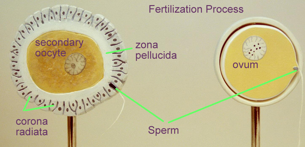
At ovulation, the ovaries release secondary oocytes into the peritoneum. The oocyte travels with some cuboidal cells that form the corona radiata which secretes a gelatinous material called the zona pellucida. The fimbria of the Fallopian tube sweep up the oocyte and it moves to the ampulla of this tube. The oocyte sends signals to the sperm moving up through the female reproductive tract. The oocyte stays in the ampulla for about 24 hours and the sperm are viable for about 48 hours.
During sperm movements through the female reproductive tract, they become capacitated and their acrosomal cap weakens. As the sperm encounters the corona radiata, the enzymes in the acrosomal cap are released and help to digest through the layers around the oocyte. When the sperm penetrates the cell membrane of the oocyte, the oocyte signals ionic changes that prevent other sperm from entering. The oocyte then completes its meiotic division to become a mature egg, or ovum.
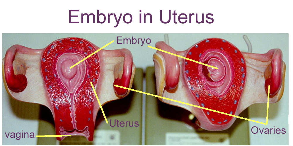
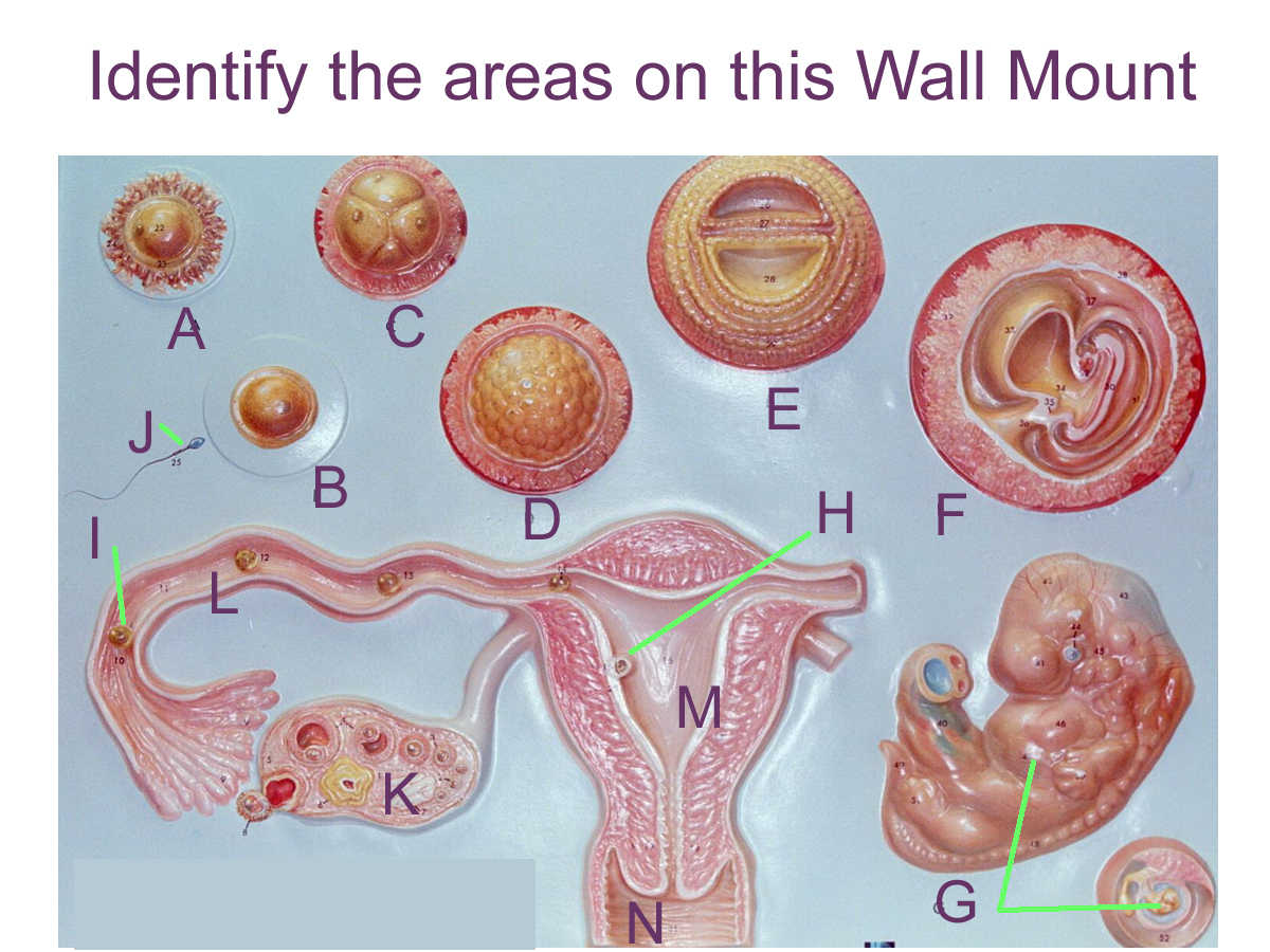
Female Reproductive wall mount with Oocyte
Male Reproductive wall mount with Sperm
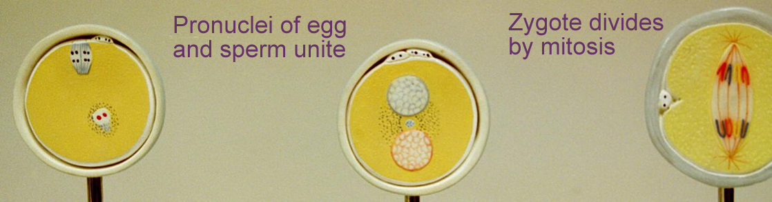
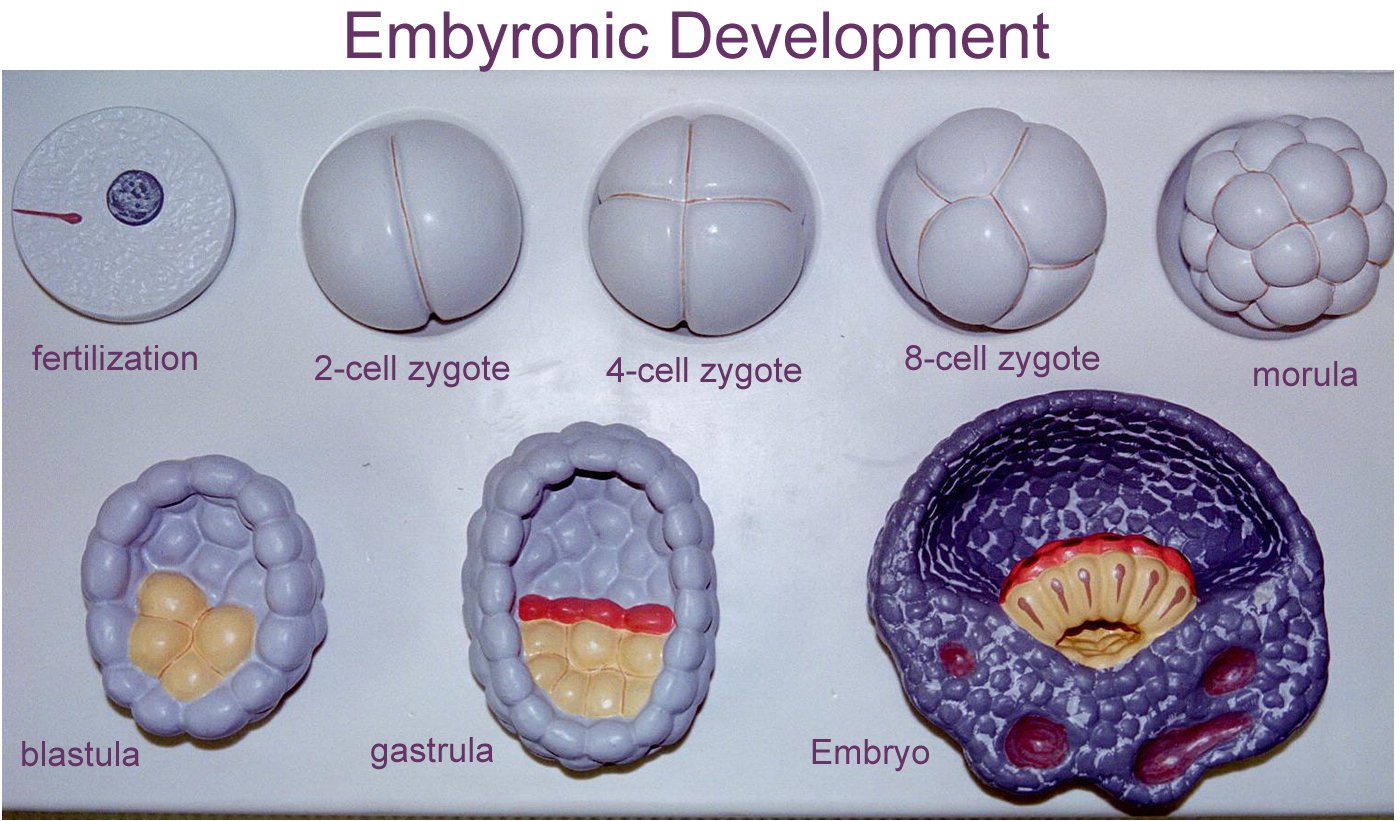
Embryonic Development
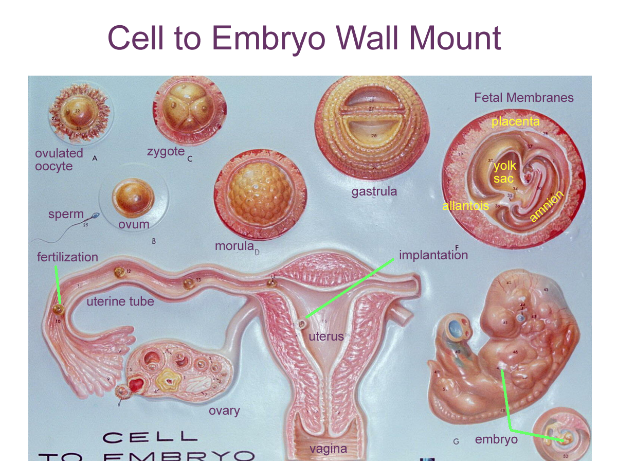
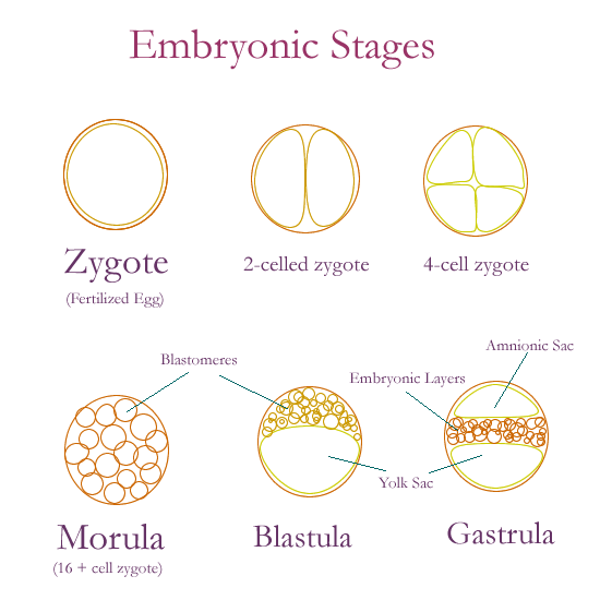
Fertilization occurs in the ampulla of the oviduct and involves the joining of the haploid pronuclei of the egg and sperm to create a single celled fertilized egg called the zygote. At this time the chromosome number is restored to the diploid (2n) number and the sex of the individual is determined. Within 24 hours the zygote begins to move down the Fallopian tube and divide by mitosis to produce daughter cells called blastomeres. The mitotic division of the zygote is called cleavage. From one cell, two cells, then four cells, and 8 cells by mitotic division. Within 3 days there are 16 blastomeres and the zygote is termed a morula. Cells continue to divide as the morula enters the uterine cavity. Fluid from the uterus enters the morula and creates a sphere with a fluid filled hollow ball of cells known as the blastula. The outer rim of cells is called the trophoblasts and the inner group of trophoblasts will produce a hormone that signals pregnancy to the ovary so that the corpus luteum is maintained and progesterone is secreted for the uterine lining.
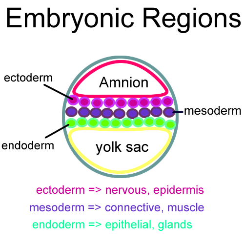
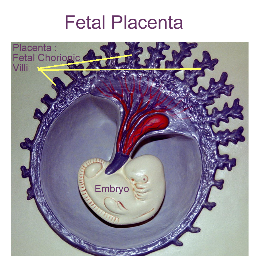
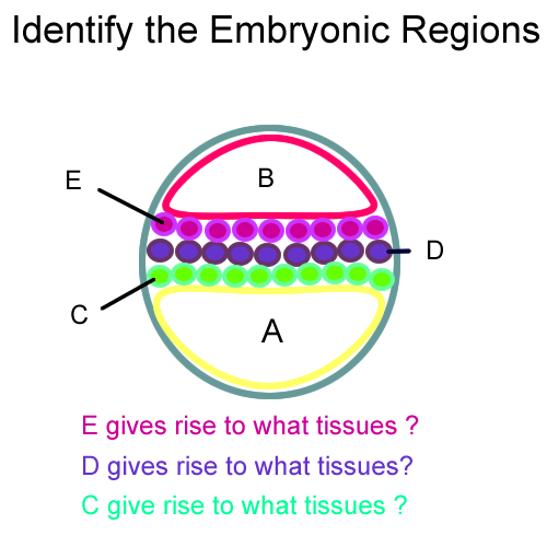
This hormone human chorionic gonadotropin (hCG) is the signal to the ovary and also forms the basis of the urine pregnancy tests. The outer group of trophoblasts forms chorionic villi that digest through the uterine lining during implantatin to help create the fetal portion of the placenta. The inner mass of cells becomes the embryo once the cells undergo gastrulation. The embryo after gasrulation consists of three germ layers: the ectoderm which forms the nervous system and the epidermis of the skin; the endoderm which forms the other epithelial linings, and the mesoderm which forms all the connective and muscle tissues.
Embryonic Models
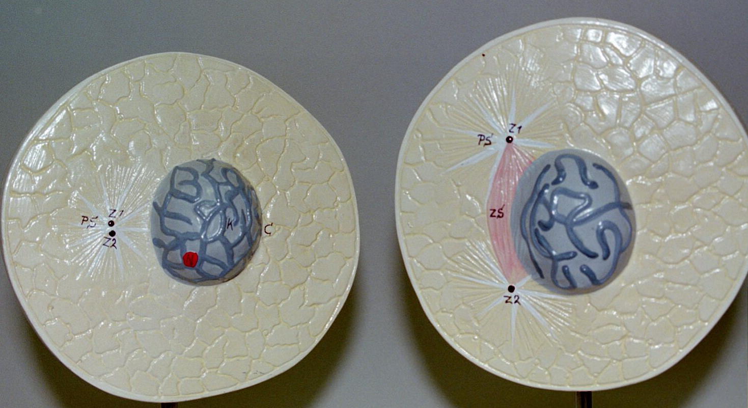
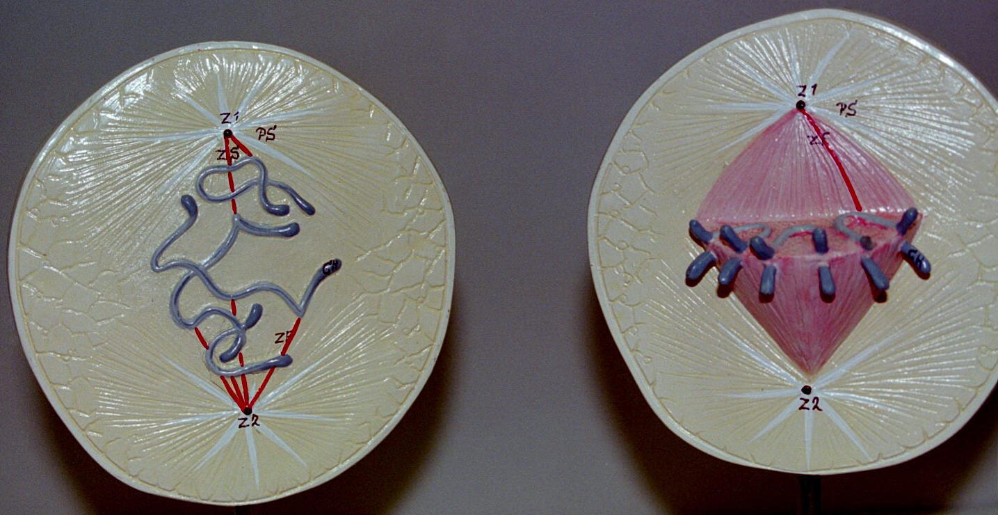
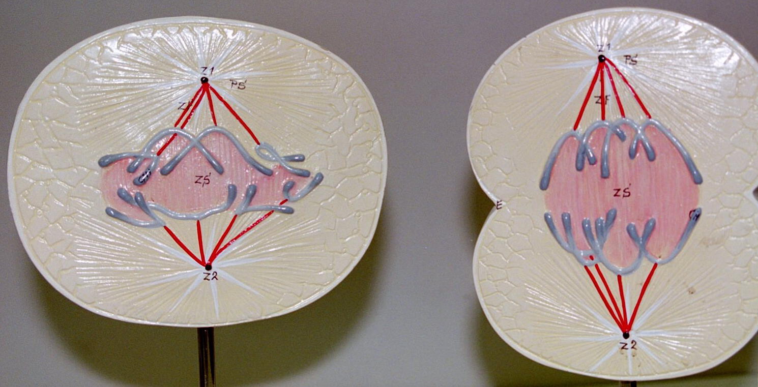
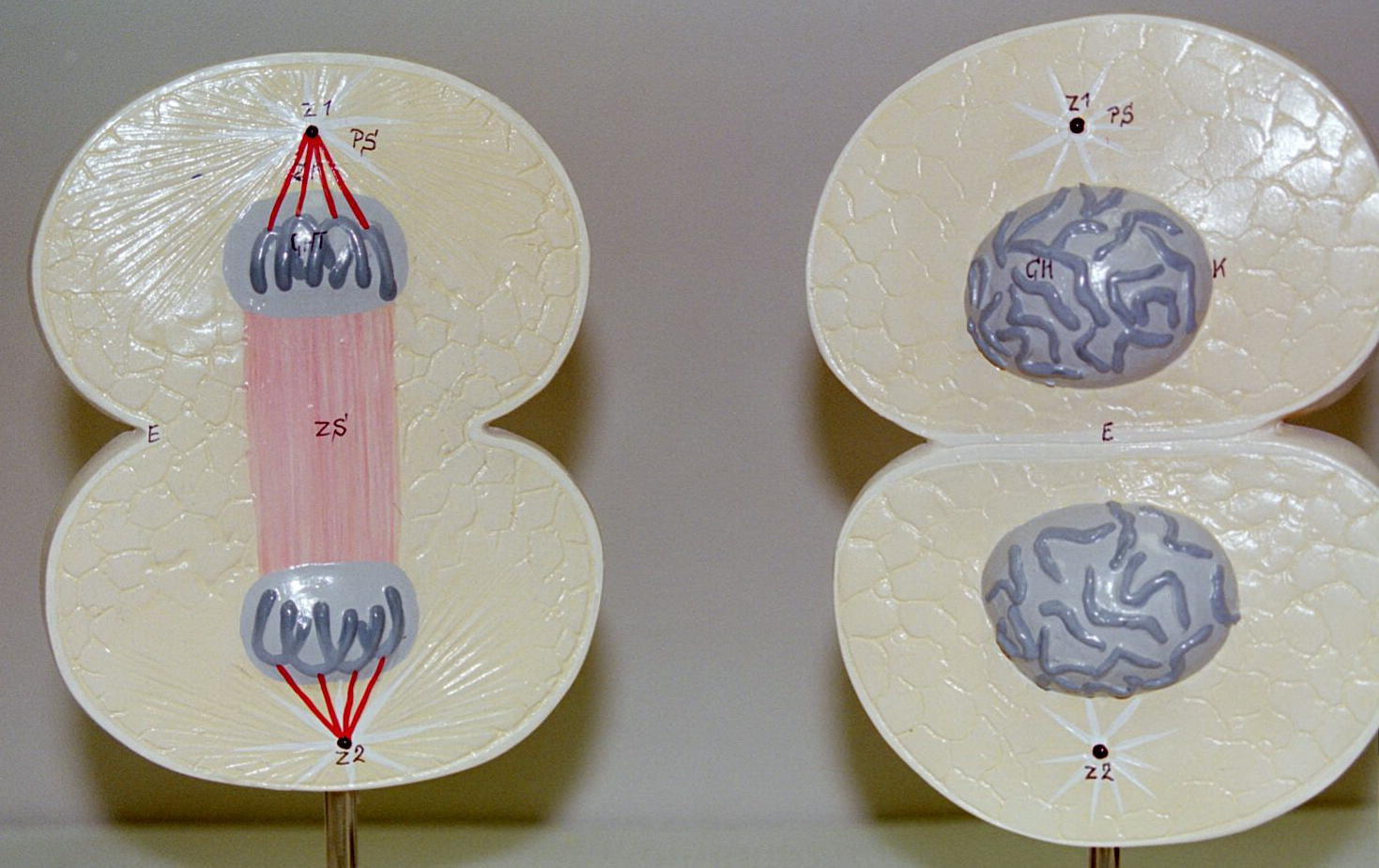
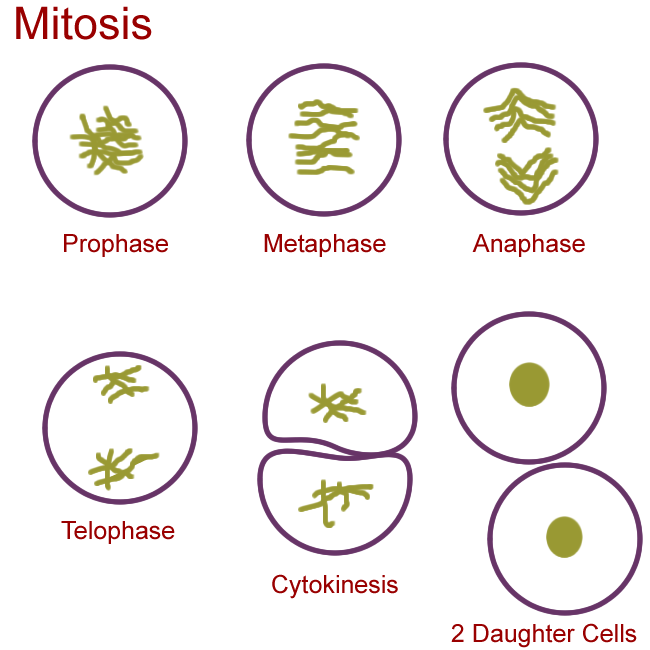
Mitosis models: Prophase, Metaphase, Anaphase, Telophase
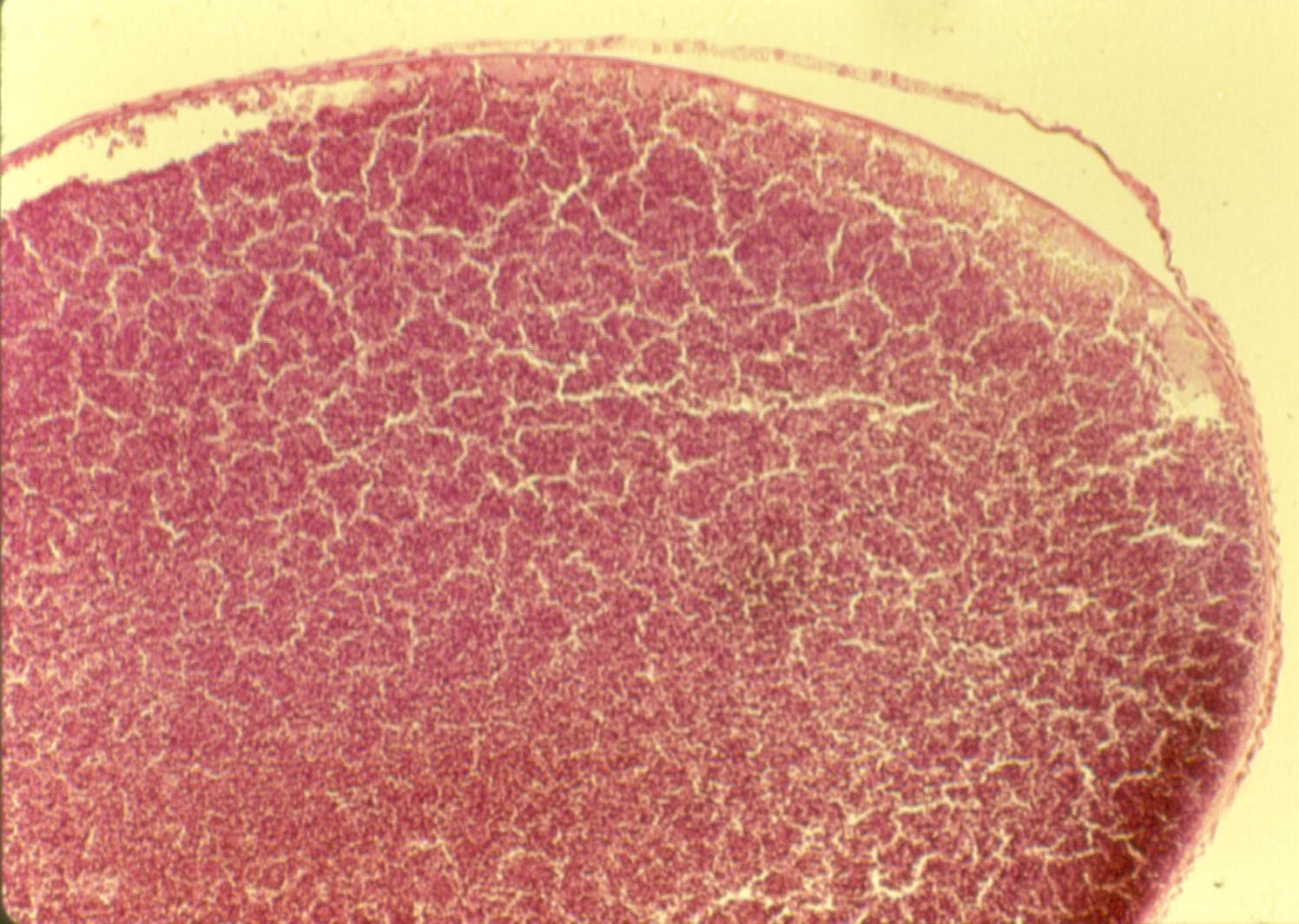
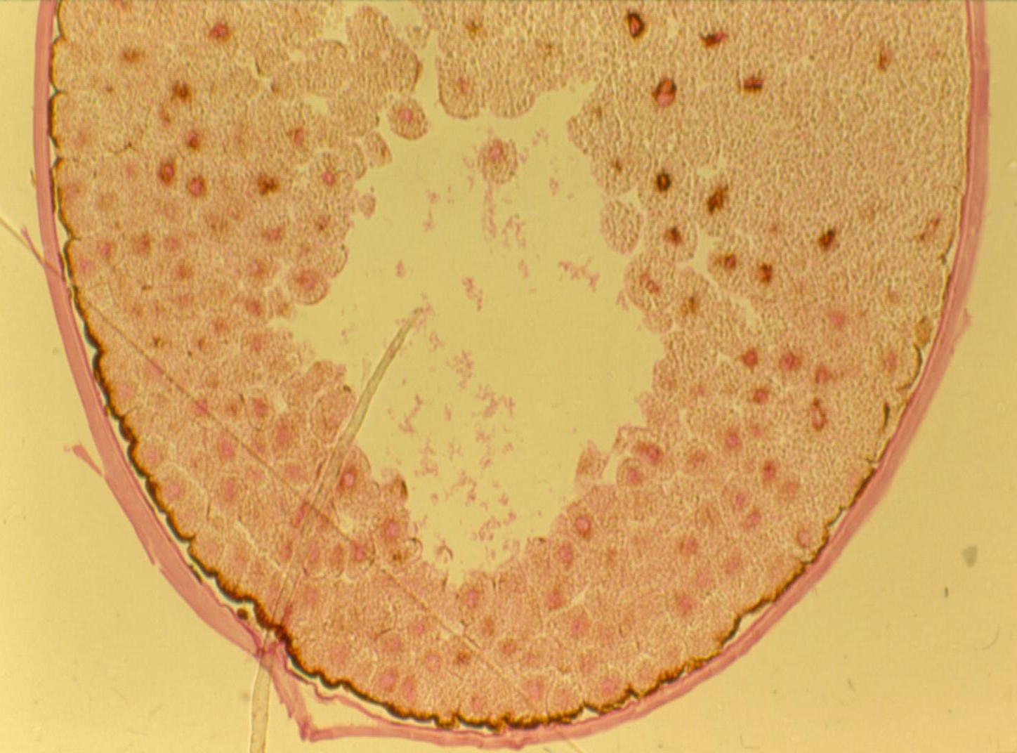
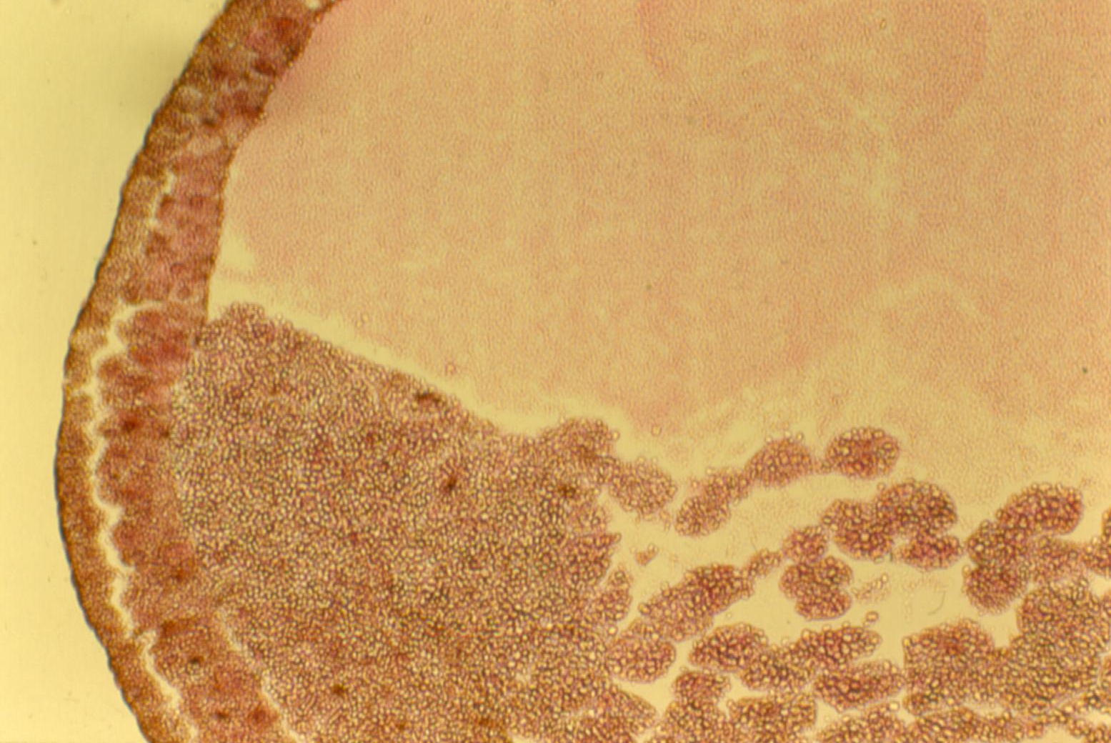
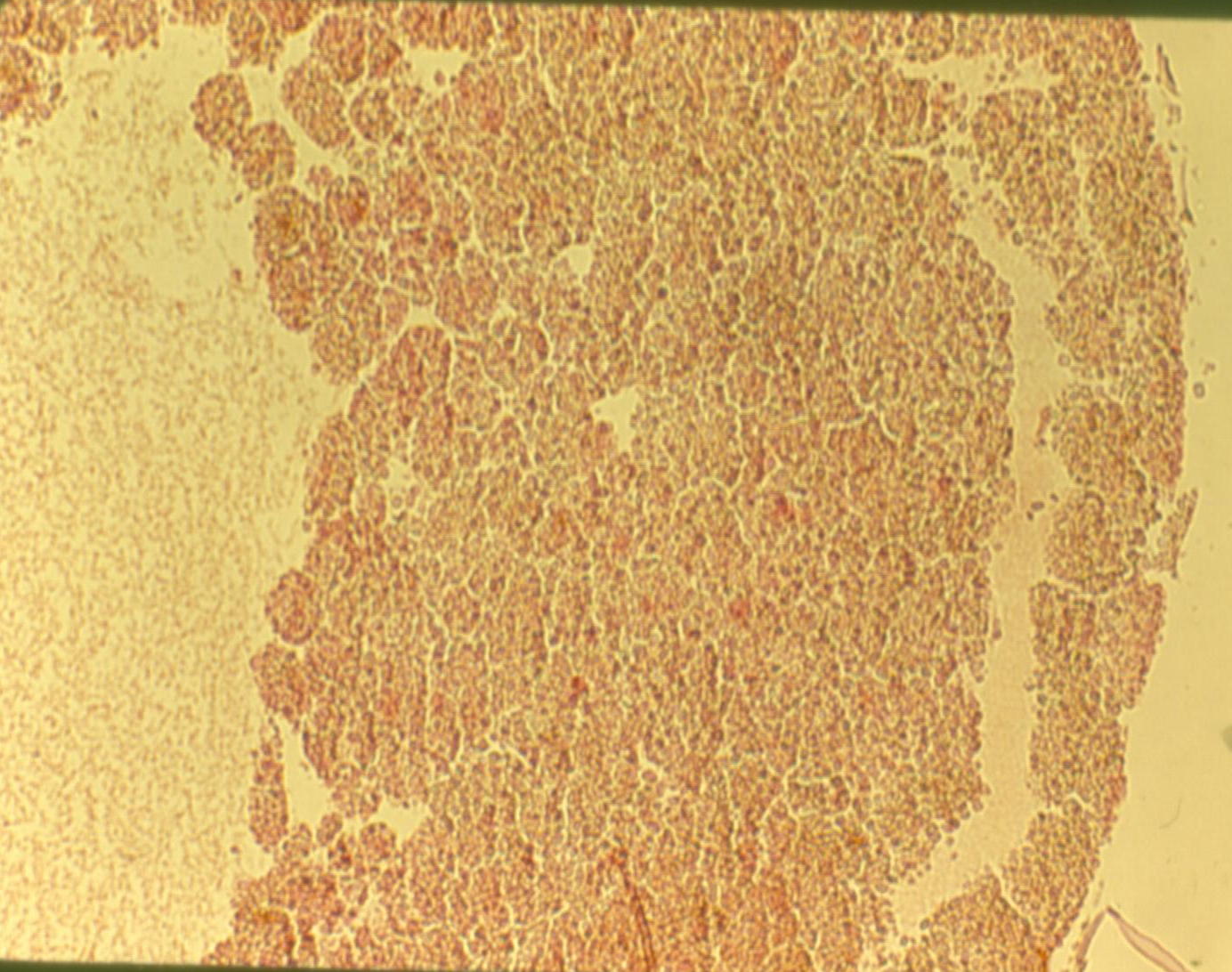
Zygote histology: Zygote, Early Cleavage, Late Cleavage
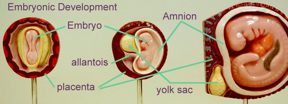
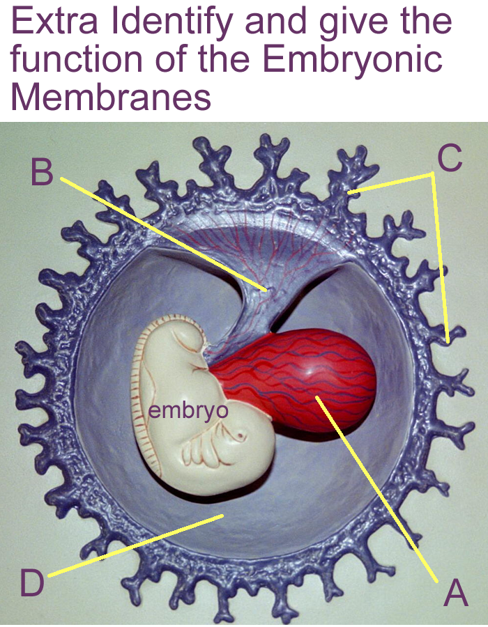
Extra Embryonic Tissues
In order to support the embryo, extra (external) embryonic membranes form at 20 days.
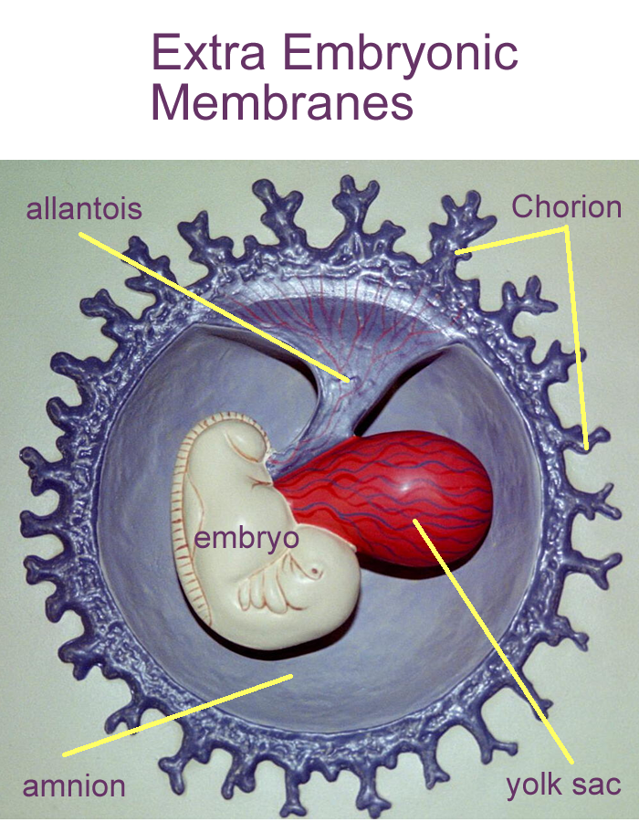
The extra embryonic membranes are:
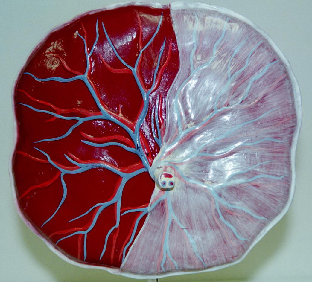
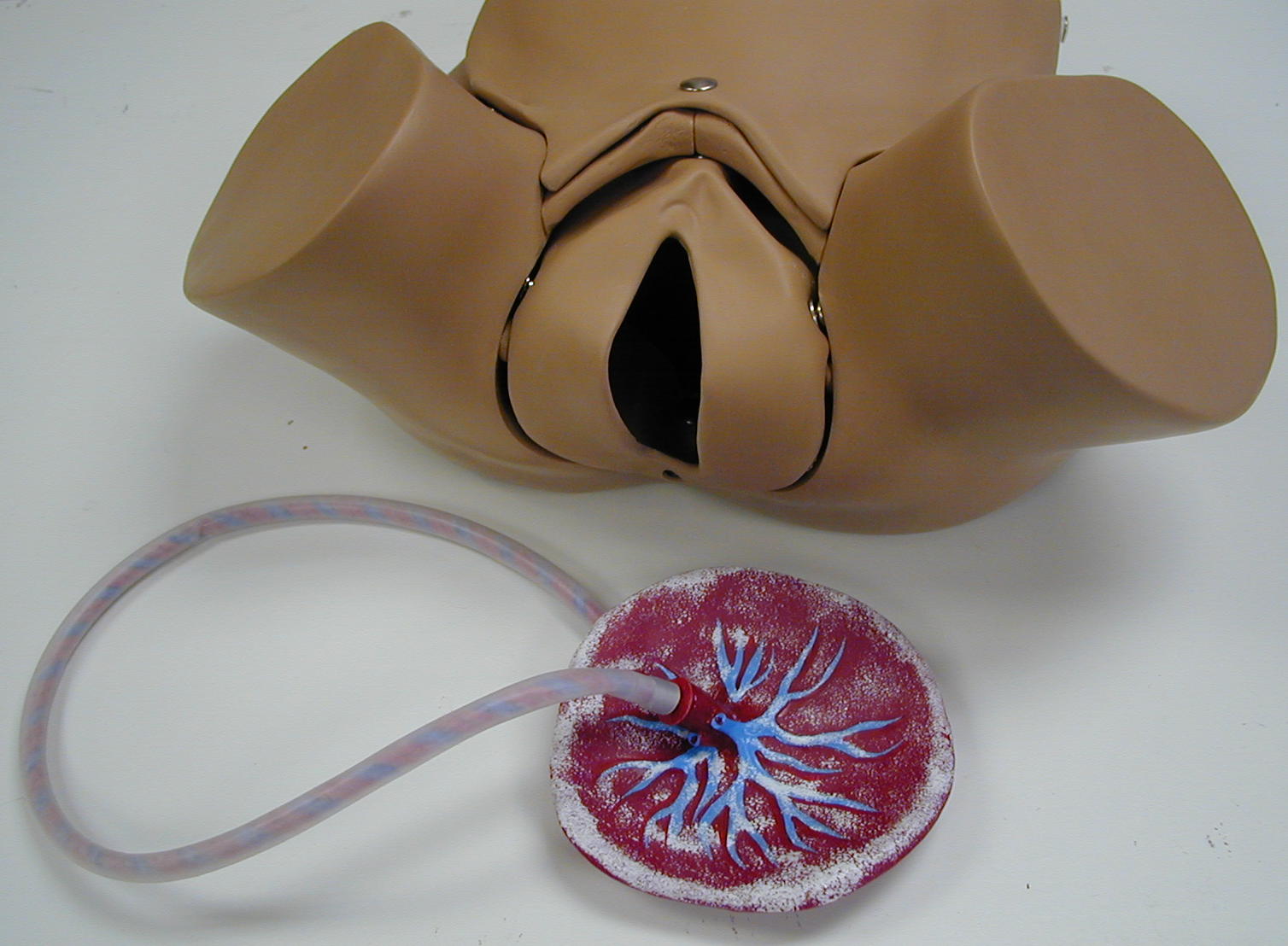
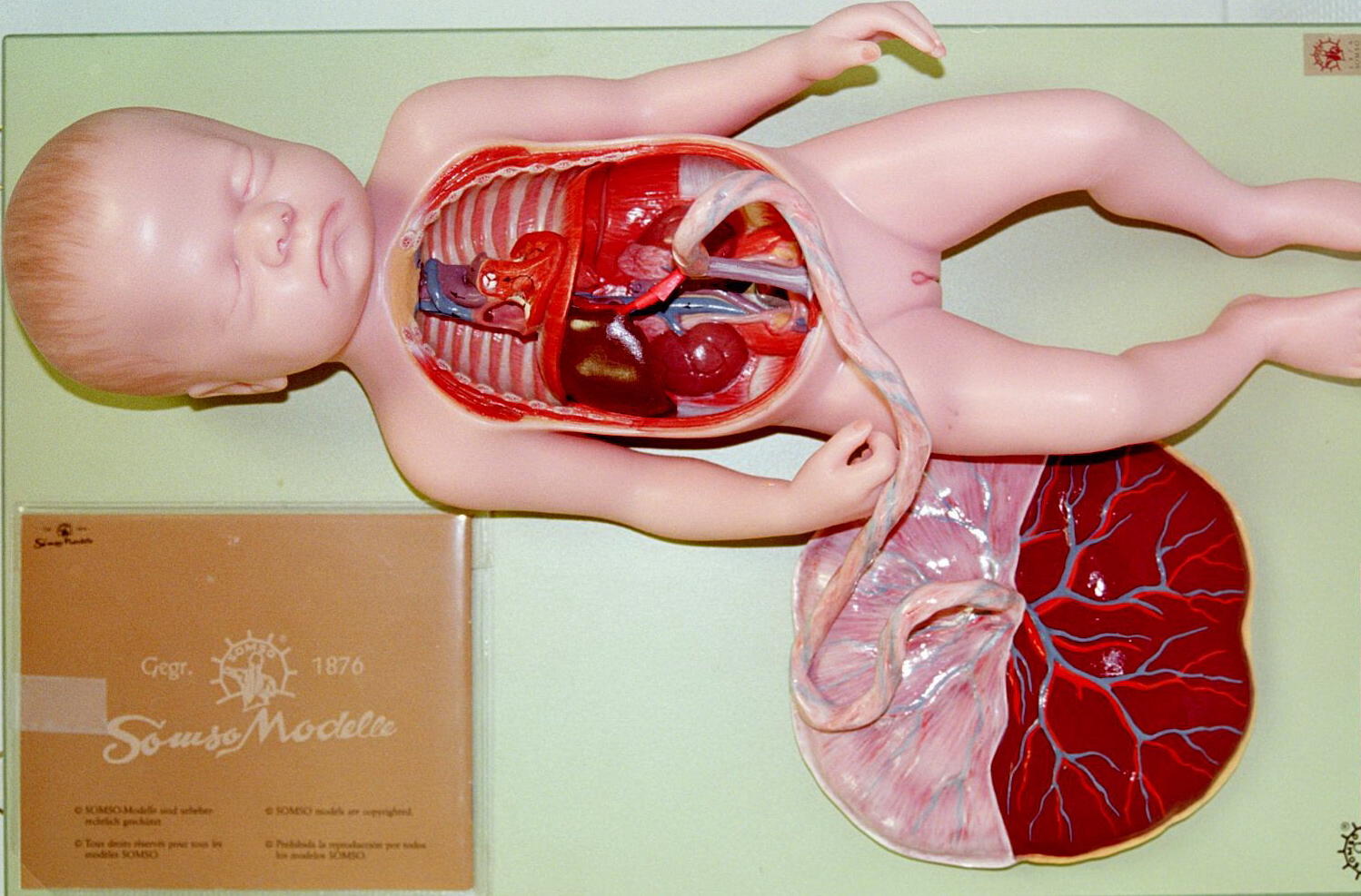
The placenta is a temporary endocrine structure composed of fetal chorionic villi and maternal blood supply. In addition to producing progestins, the placenta allows for nutrient and waste exchange between the fetus and mother.
The amnion is a thin membrane formed above the ectoderm. It contains amnionic fluid that helps provide a cushion and allows for embryonic development and movement.
The yolk sac is an outpocketing of the embryonic endoderm. It provides blood stem cells and the reproductive stem cells for the gonads. Eventually it is incorporated into the umbilical cord.
The allantois is also an outpockeing of the embryonic endoderm. It forms the umbilical cord and part of the lower digestive and urinary tracts.
Extra Embryonic Membrane Models
Placenta Models: Fetal Chorionic Villi, Afterbirth
Gestation
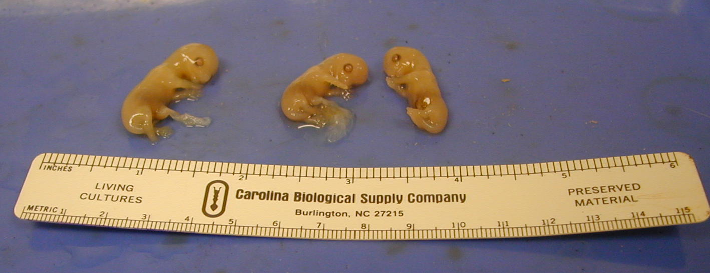
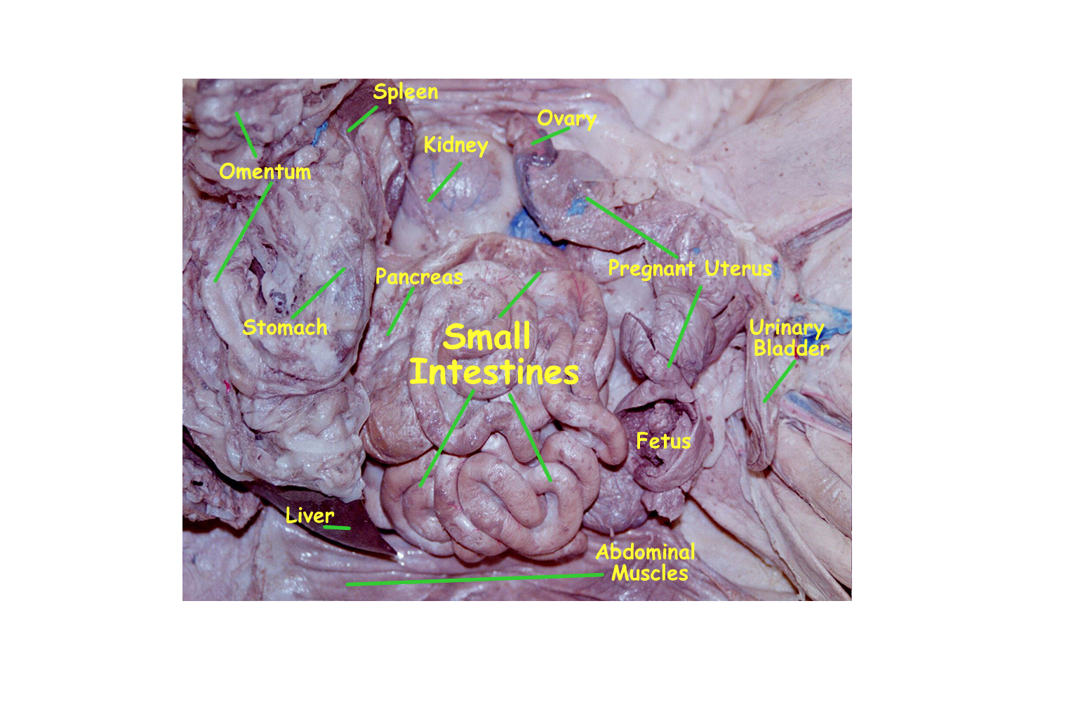
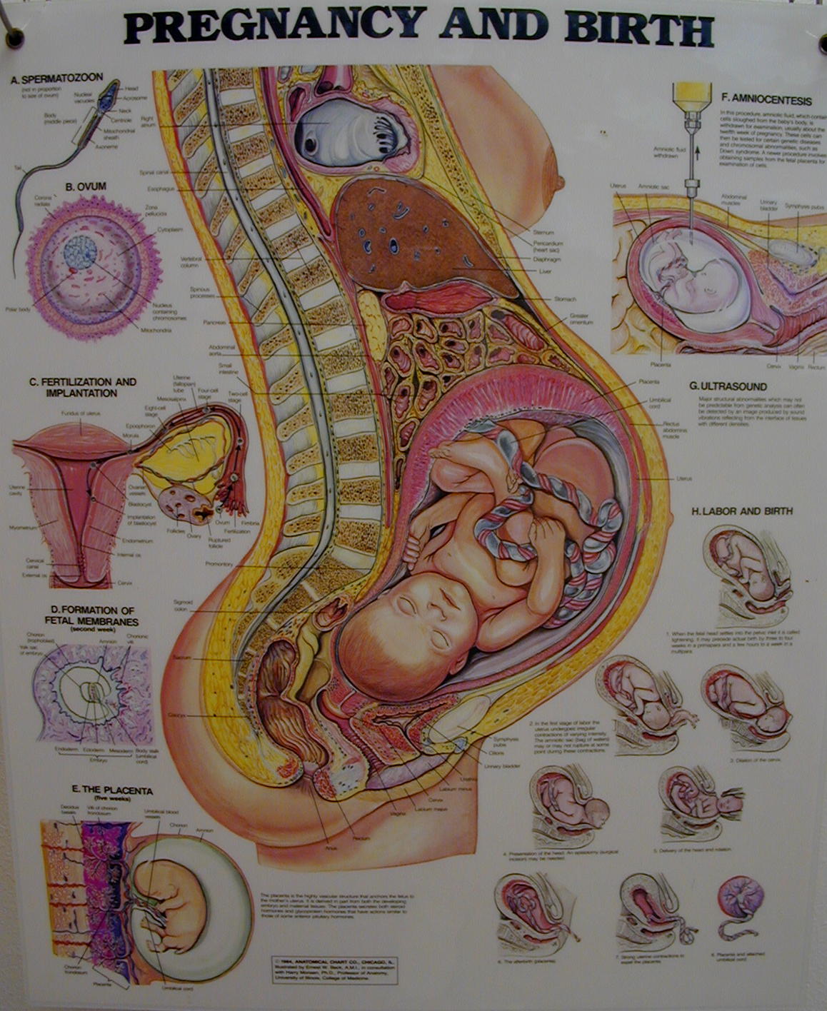
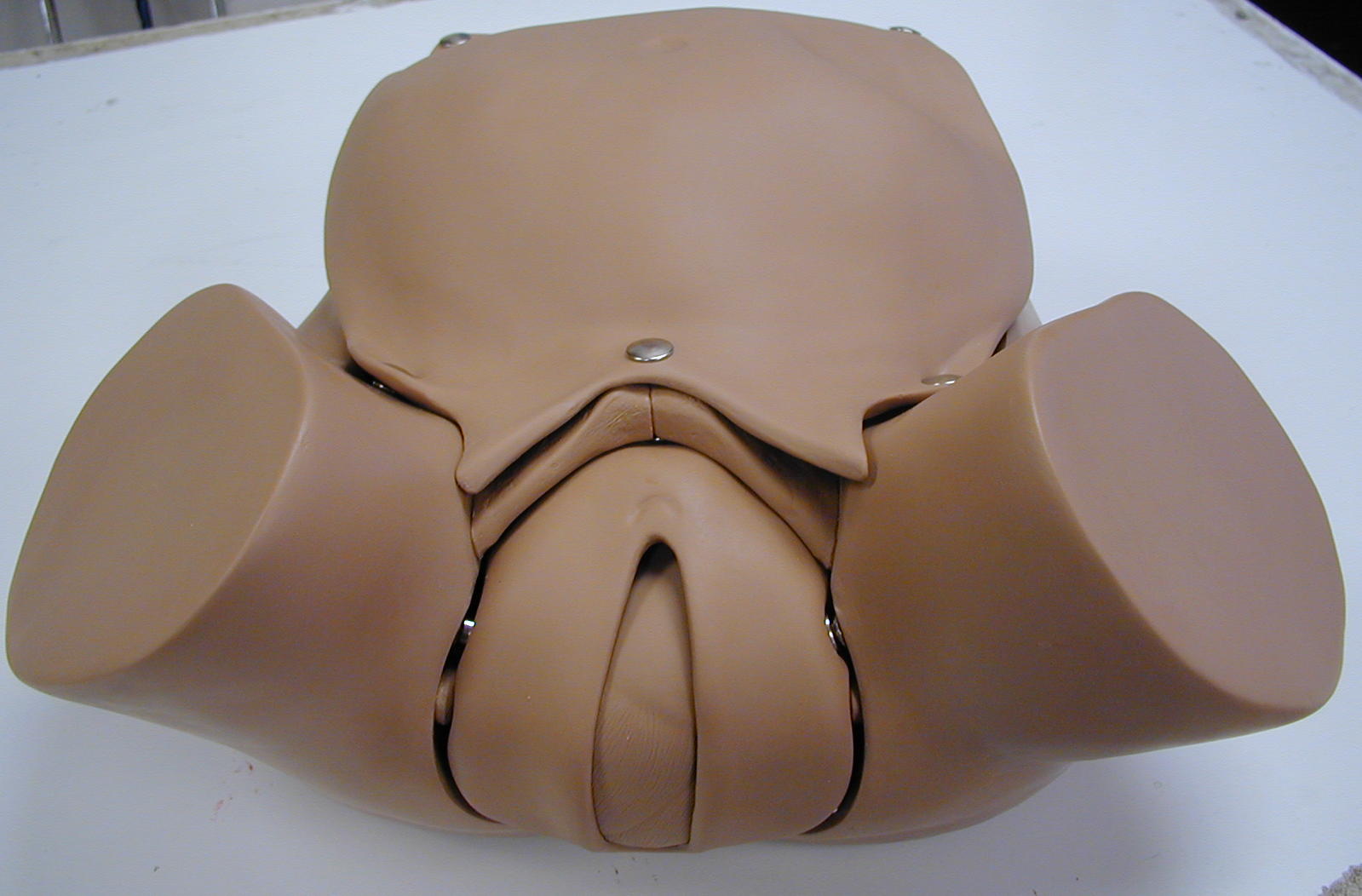
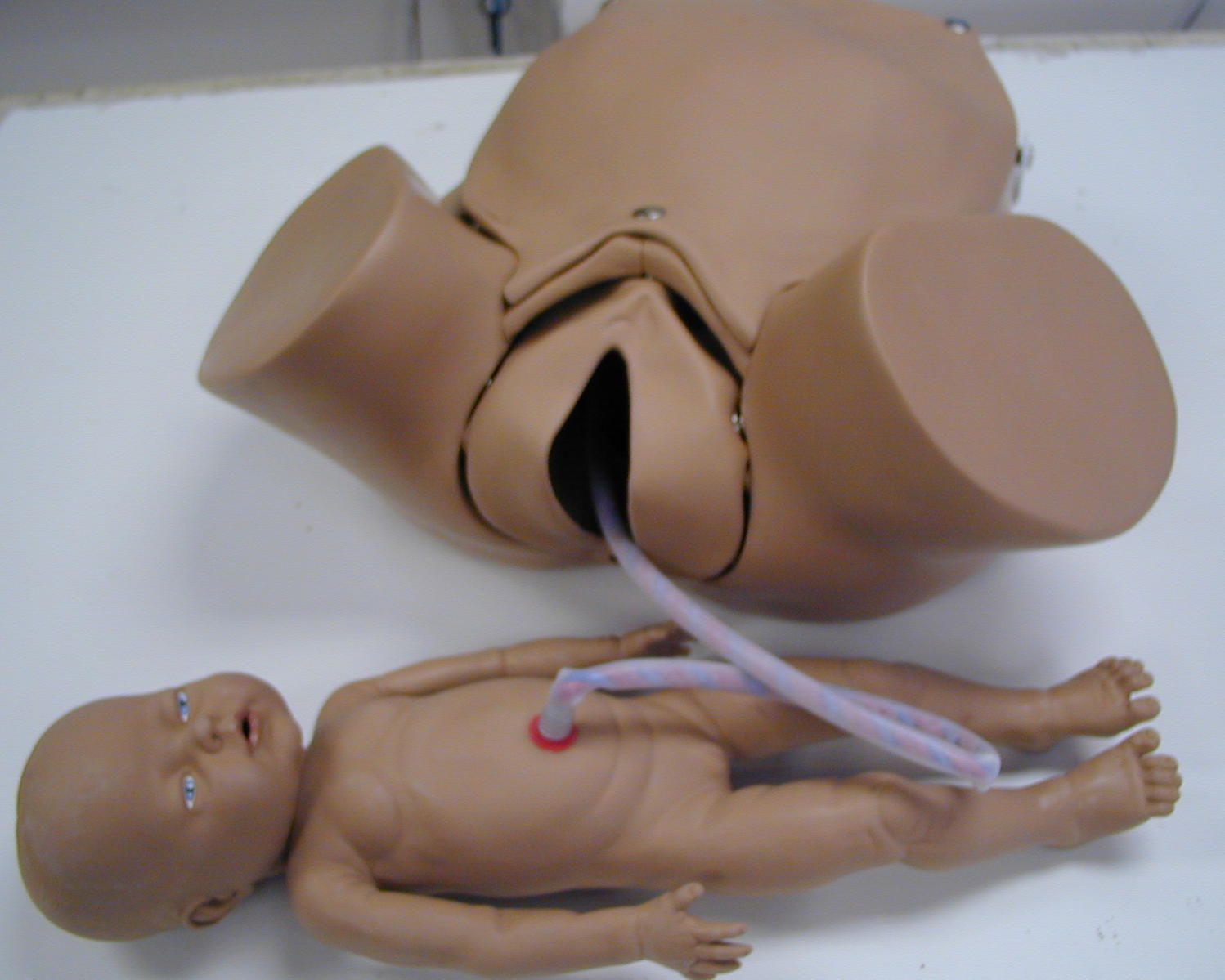
The period of the embryo occurs from fertilization through the second month or 8 weeks. During this time the tissues and organs are forming to create the body systems. After 8 weeks, the embryo is termed a fetus. During the fetal period, the organs formed during the first 8 weeks continue to grow and mature until the end of gestation at 9 months or the 40th week. As pregnancy progresses, the uterus enlarges to accommodate the fetus. At parturition or childbirth the fetus is expelled during labor through the vagina to become a neonate.
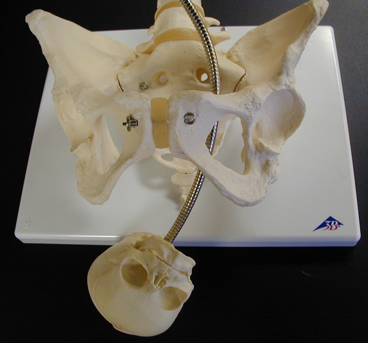
The three stages of labor are dilation of the cervix, fetal delivery, and placental detachment & expulsion. The primary hormone responsible for labor is oxytocin (OT) which comes from the posterior pituitary and is controlled through positive feedback mechanisms.
Labor Stages : Dilation/Crowning, Delivery / Expulsion, Placental
Fetal Circulation
Fetal circulation differences occur. In utero, there are shunts that allow for bypasses to the liver and lungs. In the heart the bypasses around the fluid filled lungs are the foramen ovale and the ductus arteriosus. In the liver, the bypass is the ductus venosus. Shortly after birth, the bypasses close and allow for blood flow through the lungs and liver.
Fetal Structure Adult Structure
Foramen ovale Fossa Ovalis
Ductus Arteriosus Ligamentum Arteriosum
Ductus Venosus Round ligament of the liver
pedia- child post- after
-sis condition meso- middle
troph/o- nutrition chori- fetal membrane
germ- bud gest- bear, carry
omphal/o- navel prim- first
pto- fall terat/o- malformed fetus
vita life viscer/a- organ
amni/o- amnion -arche beginning
-gravida pregnancy -para to bear (offspring)
-tocia childbirth, labor neo- new
I. ID development
A Drawings : Mitosis stages, Embryonic Regions
B Models: Identify Model A, Identify Model B
Concept Map: Make a concept map of the events of development starting with fertilization until birth. Include this map as part of your LAR lab report (if selected) as a document insert or as an addtional PDF document scan.
Premature Labor, Premature rupture of Membranes (PROM)
Abortion
Twins
Amniocentesis
Chorionic Villi Sampling
Fetal Alcohol Syndrome
Premature birth
Apgar score
Ectopic Pregnancy
Toxemias of Pregnancy: Preclampsia, Eclampsia
Placenta Previa
Abruptio Placentae
Cesarean birth (C-section)
Dilation and Curettage
Episiotomy
Tubal Ligation
Pap Smear
Contraceptives
Stem cell (blastomere) research
Congential Diseases
Nervous System: Cerebral Palsy, Spina Bifida, Hydrocephalus, Meningocele
Digestive System: pyloric stenosis, aganglionic megacolon
Cardiovascular System: heart defects, erythroblastosis fetalis
Musculoskeletal System: clubfoot (talipes), hip dysplasia
Genitourinary System: cryptorchidism, defects of ureter/bladder/urethra
Metabolic Disorders: cystic fibrosis, phenylketonuria (PKU)
Neonatologist
Pediatrics
http://www.nlm.nih.gov/medlineplus/healthtopics.html
http://www.lumen.luc.edu/lumen/meded/histo/frames/histo_frames.html
http://www.sciencedaily.com/news/health_medicine.htm
http://embryo.soad.umich.edu/carnStages/carnStages.html
http://www.track0.com/canteach/links/linkbodysystems.html
http://www.carr.lib.md.us/schs/science/anatomy/systems.html
http://www.nlm.nih.gov/medlineplus/pregnancyandreproduction.html
1. Define gamete and their sources
2. Compare and contrast meiosis and mitosis
3. Define fertilization
4. What is hCG and give its function
5. Name the extra embryonic membranes and their function
6. Define gastrulation
7. Name the three embryonic germ layers and the tissues they create
8. Define gestation and give the normal length of time for humans
9. Give the period of the embryo and the period of the fetus and describe the events that
occur in each period.
10. Name and describe the three stages of labor.
11. Name the fetal circulatory bypasses and what organ they are bypassing.
12. Name the adult structures that are formed from the closed fetal circulatory bypasses.