Biology 2404 A&P Basics Lab Exercise
7 Somatic & Special
| Objectives | Background | Medical Terms | Activities | Applications | Careers | WWW | Review Questions |
Students should be able to:
* give the receptor, location, function and cranial nerve(s) for the special senses
- Taste
- Smell
* Give the location, function, cranial nerve(s), organ anatomy, receptor anatomy
and a problem for the special senses
- Vision
- Hearing
- Balance and equilibrium
* Give the receptors, location, and function for somatic sensory
* Define related terms
* know the differences between somatic sensory and special sensory pathways,
how and where interpreted.
Read related material in textbook
Sensory receptors and pathways provide us information about our internal and external environment. The information provided allows us to analyze and appropriately respond to changing situations. The ability to react and respond helps in feedback mechanisms to maintain homeostasis.
Sensory pathways have specific components that provide organization and localization for their responses. The parts for sensory pathways are:
1) Receptors : dendrite regions of neurons, modified dendrites, or specialized cells that can be triggered by various stimuli. Such stimuli can be mechanical, chemical, or electrical.
2) Sensory Neuron : Part of the peripheral nervous system. Anatomically classified as either bipolar for special senses or pseudounipolar for somatic senses. Sensory neurons are also referred to as afferent, as the direction of the stimulus is brought (fer/o-) to (ad-) the Central Nervous System for processing, storage, and response.
3) Sensory Tracts : axons of sensory neurons that carry information to the CNS. These tracts are found in the white matter of the spinal cord or the brain. Sensory tracts can involve several neuronal synapses to direct conscious information to specific brain areas.
4) Sensory Areas : Specific areas within the brain lobes are responsible for receiving initial information. Once processed, other areas called association areas within the brain lobes are responsible for analyzing, storing, and sending assessments of the sensory input to the motor regions of the frontal lobe of the brain.
Other characteristics of sensory information include projection, phantom pain, intensity, contrast, adaptation, and after-image.
Projection and phantom pain are similar. Even though a particular sensory receptor picks up the signal, it is the brain that actually detects the sense, not the sensory receptor or neuron. When the impulses arrive, the brain creates a projection based on the type of sensory neuron, and the strength and duration of the initial stimulus.
Intensity is the weakness or strength of a sensory signal. Weaker stimuli activate only a small number of receptors. The more receptors that are activated, the more impulses are sent, and the more intense interpretation is given. Timing is important in helping with the interpretation of intensity. How fast an impulse travels is affected by size and myelination of the neuron.
The slower, smaller fibers will create a duller, diffuse interpretation to the sensation. The larger, myelated fibers will create an interpretation of very sharp, pin point localization.
Contrast is the ability to differentiate between two different sensory stimui, either from a previous or simultaneous sensation. When the brain compares the newer sensation to the other, it may be diminished or exaggerated. An example would be when you walk into an air conditioned room from an outside environment that is hot and humid. The air conditioned room feels much cooler than the actual thermostat reading.
Adaptation is a decreased response to a continuous stimulus. The receptors generate fewer impulses when a stimulus continues at the same intensity. An example of this is when someone who is wearing perfume or aftershave walks into the room. You detect the smell at first, but as they remain in the room, you become unaware of the olfactory sensation.
After image is the sensation that remains even after the stimulus has stopped. You may have experienced this when a bright light is shone into your eyes and the image is still seen on the retina. A bright light source could be a flashbulb from a camera, or the pictures taken of your eye during a retinal (fundic) exam.
Recall that the PNS is the peripheral nervous system and functions to send information to the CNS that allows for delivery of information to effector cells. The information that travels to the CNS does so along sensory afferent neurons. Motor efferent neurons carry information from the CNS for an action response by effector cells such as muscle or glands.
The sensory portion of the PNS division can be divided into two parts:
A) General (Somatic) Sensory
B) Special Sensory
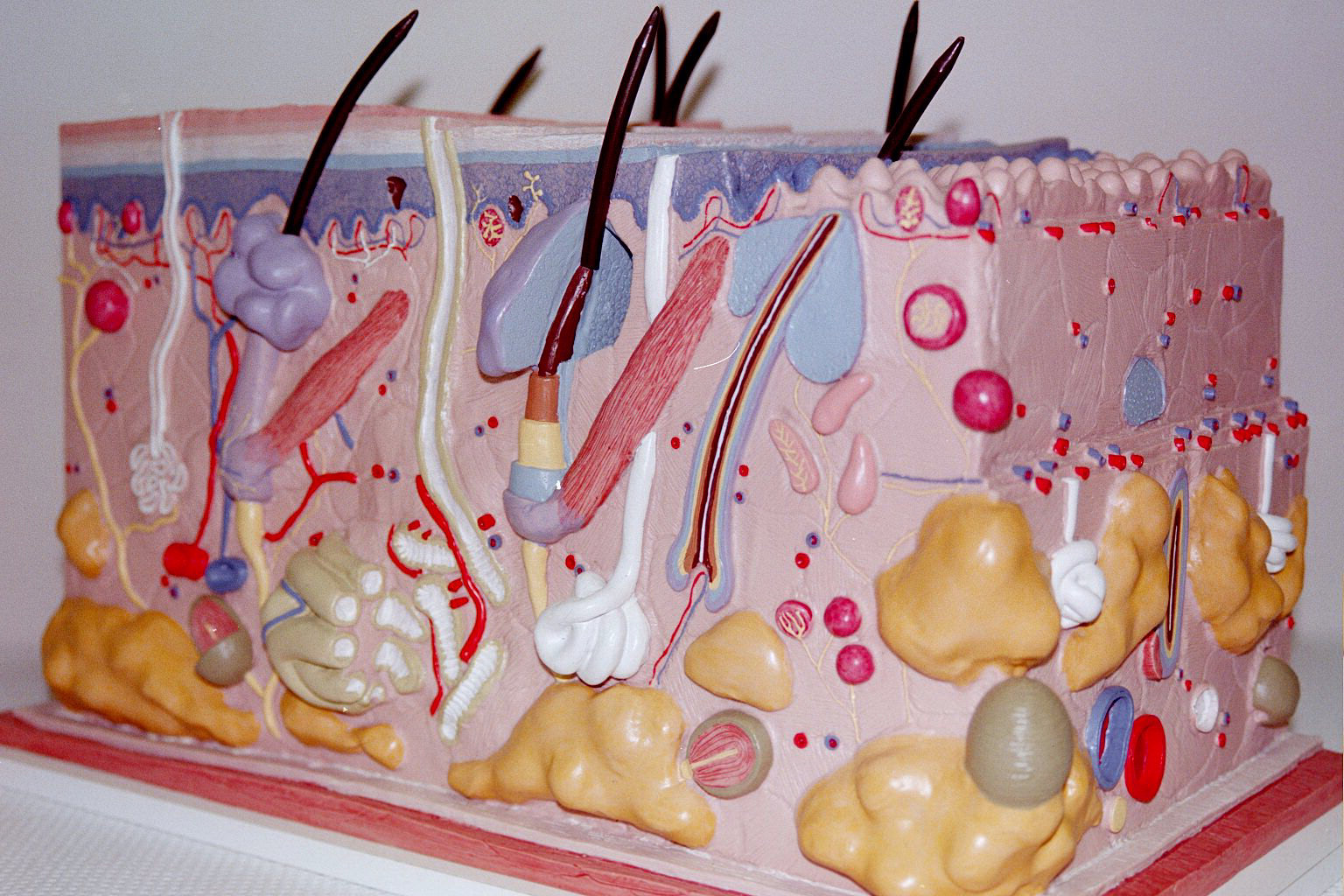
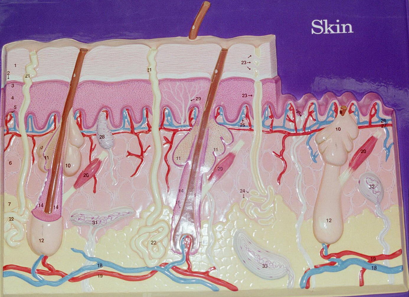
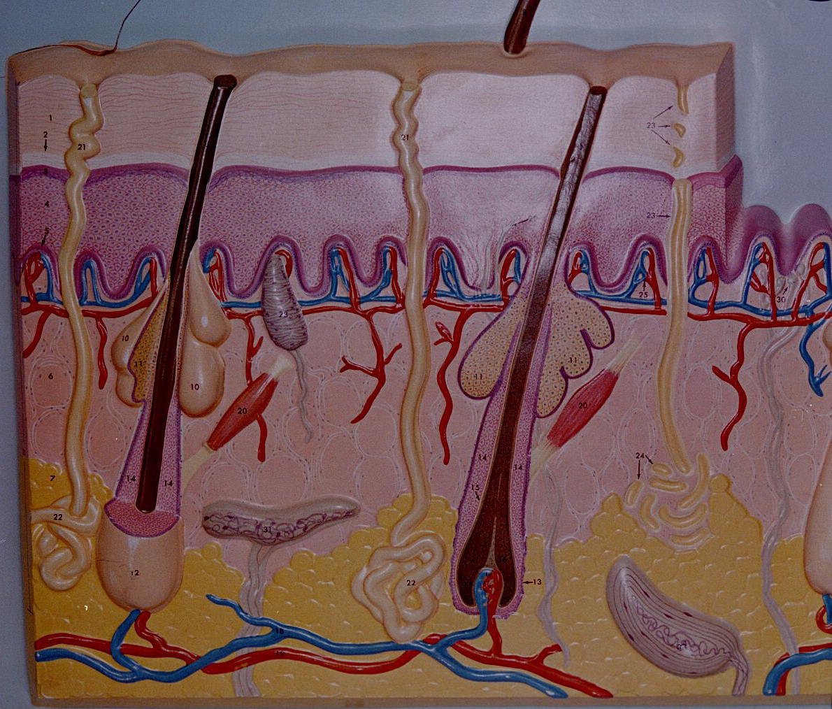
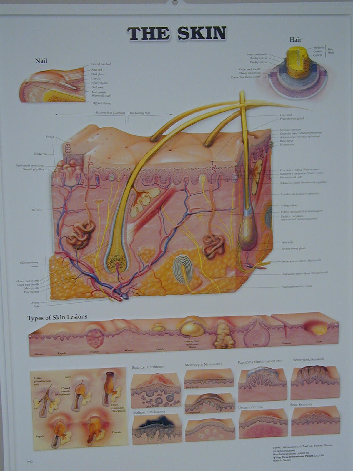
General or somatic (body) senses deal with receptors located in the skin, tendons, and muscles (skeletal, cardiac, and smooth). These sensory receptors respond to a specific stimulus and impulses are carried by the PNS, spinal nerves or cranial nerves, to the CNS. General or somatic sensory neurons are pseudounipolar in structure.
The impulses detected by the general senses are pain, temperature, touch, pressure, and position. These impulses follow specific pathways to the brain where they are analyzed, potentially stored for further comparisons, and usually acted on by returning a motor response command from the brain to the body.
The intensity of a sensation depends on its frequency. The more frequently an impulse is received, the stronger the sensation is interpreted. The frequency of the impulse is determined by the type and location of the sensory receptor, as well as its size and myelinatin properties. The general body sensory receptors are as follows:
Thermoreceptors
Thermoreceptors are responsible for transmitting the sense of temperature.
The two types of temperature receptors are cold and hot and both are located in the skin. Thermoreceptor anatomy consists of free nerve endings located within various regions of the skin.
Cold receptors are located in the upper regions of the dermis. They are more numerous than heat receptors. Cold receptors are most sensitive to temperatures below 20° C or 68 ° F. Temperatures below 10 °C or 50° F stimulate both cold and pain receptors.
Heat receptors are located deep in the dermis. They are also anatomically free nerve endings and are sensitive to temperatures above 25° C (77 °F).
Temperatures above 45° C (113° F) stimulate both heat and pain receptors.
Mechanoreceptors
Mechanoreceptors are responsible for transmitting information regarding mechanical displacement of tissues. The two types of mechanoreceptors are designed for touch and pressure. Two types of touch are pressure are light, superficial and heavy, deep.
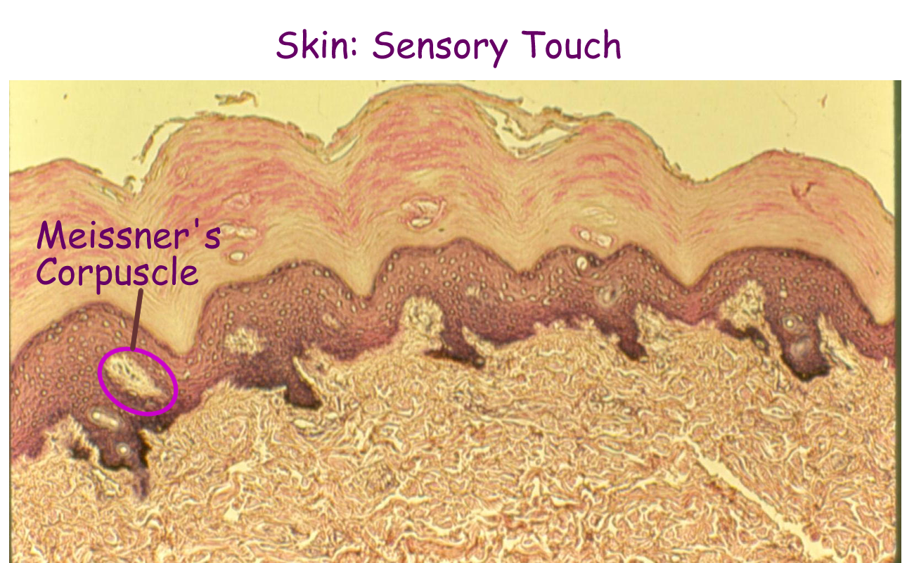
Touch receptors are the Merkel disc, Meissner corpuscles, and Krause end bulb. These receptors have a light connective tissue wrapping around the dendrite end of the neurons. Free nerve endings can also be involved with touch and are seen wrapped around the base of hair follicles. The merkel disc and meissner corpuscles are located in the papillary layer of the dermis.
They are responsible for light touch or very light stimuli. Meissner’s corpuscles have specific body locations, such as the genitalia, mouth, and hands. Krause end bulbs are in the mid dermis.
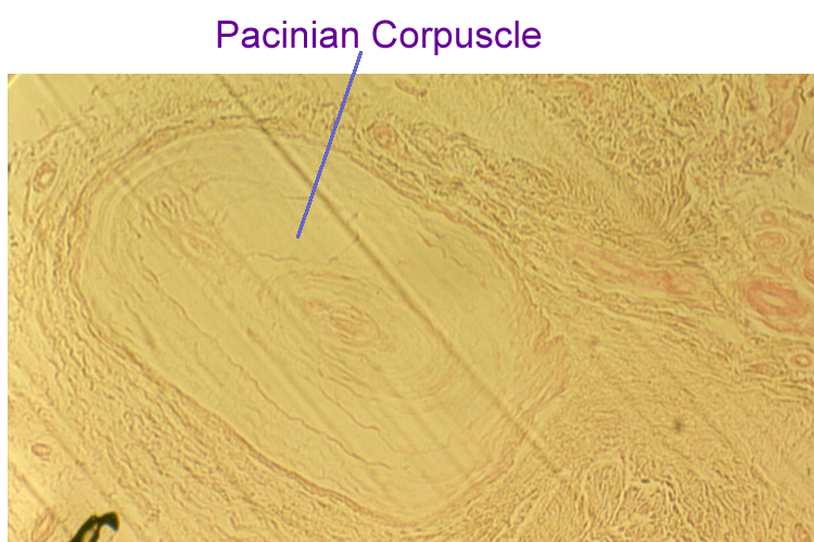
Pressure receptors are the Pacinian corpuscle and the Ruffini end bulbs. Both have more extensive layers of connective tissue covering the dendrite portion of the neuron and both are located mid to deep in the dermis. Pacinian corpuscles are also found in ligaments and tendons of joints. Pressure receptors respond to heavy touch, deep pressure, and vibration.
Nocireceptors
Nocireceptors are responsible for transmitting the sensory impulse of pain. The receptor anatomically is a free nerve ending and is located in the epidermis and upper dermal layers of the skin. Recall that any skin temperature changes above or below the thermoreceptor threshold can trigger a pain sensation.
Another pain phenomenon involves the autonomic nervous system (ANS).
The visceral organs have stretch receptors, pressure receptors, and chemical receptors. When one of these receptors is stimulated, the signal is sent along the same pathways as pain from the skin. Therefore, all sensory information from the ANS is then interpreted as pain from the skin, since it follows the somatic pain pathways to the brain. This is called referred pain. The back pain that a patient may feel could be muscle pain, skeletal pain, or kidney pain. The internal body organ’s signal is referred to a regional site and then sent to the CNS (thalamus then the parietal lobe) by somatic pain pathways. Another example of referred pain is the classic “heart attack” pain that most human males feel such as a pain that radiates down the left arm and into the chest. Recent studies indicate that women have different regions of pain such as the shoulders or neck that can be associated with “heart attacks.”
Pain nerve fibers come in two types. Ones that are small and myelinated give the sense of a very sharp, localized, and acute (sudden) pain sensation. Impulses are conducted more quickly and can stop quickly once the pain source is removed. Those nerve fibers that are larger and nonmyelinated give the sense of dull, diffuse, and chronic (long term) pain sensation. Impulses travel more slowly to the CNS and may linger once the original stimulus is removed.
The midbrain and diencephalon can regulate pain impulses to the brain and can respond quickly by signaling motor responses from appropriate CN or spinal nerves. Also, the brainstem and diencephalon areas can release pain inhibiting substances such as enkephalins and endorphins.
Think of when you may have stepped on something sharp. You reacted immediately before you had a chance to think. We are consciously aware of the pain stimulus and the parietal lobe of the cerebral cortex still receives the pain impulse in order to determine location and intensity so that an appropriate motor response can be maintained or initiated.
Proprioceptors
Proprioceptors are responsible for transmitting the sensory impulse of position. This primarily involves the position and movement of body joints, the proper tension in tendons and ligaments, and the amount of muscle length and contraction state. The purpose of Proprioceptors is to prevent connective tissue strain or muscle sprain.
An example of a proprioceptor is a muscle spindle, which is a modified skeletal muscle fiber with a sensory nerve wrapped around it. Muscle spindles are responsible for information relating to stretch and are sometimes called stretch receptors.
Several pathways allow for these impulses (position, stretch, contraction, tension, etc.) to reach the cerebral cortex so that we are aware of where our body is in a particular location. Another pathway sends this proprioceptive information to the cerebellum where it is integrated with signals from the inner ear to help maintain balance and equilibrium. This also allows the cerebellum to coordinate and integrate cortical signals for any muscle activity.
Other terms used for proprioception is muscle sense or kinesthetic sense.
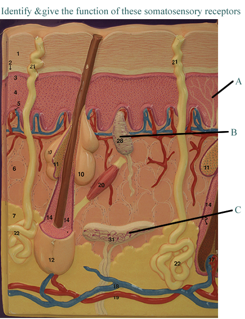
Skin Wall Mount with Somatic Sensory
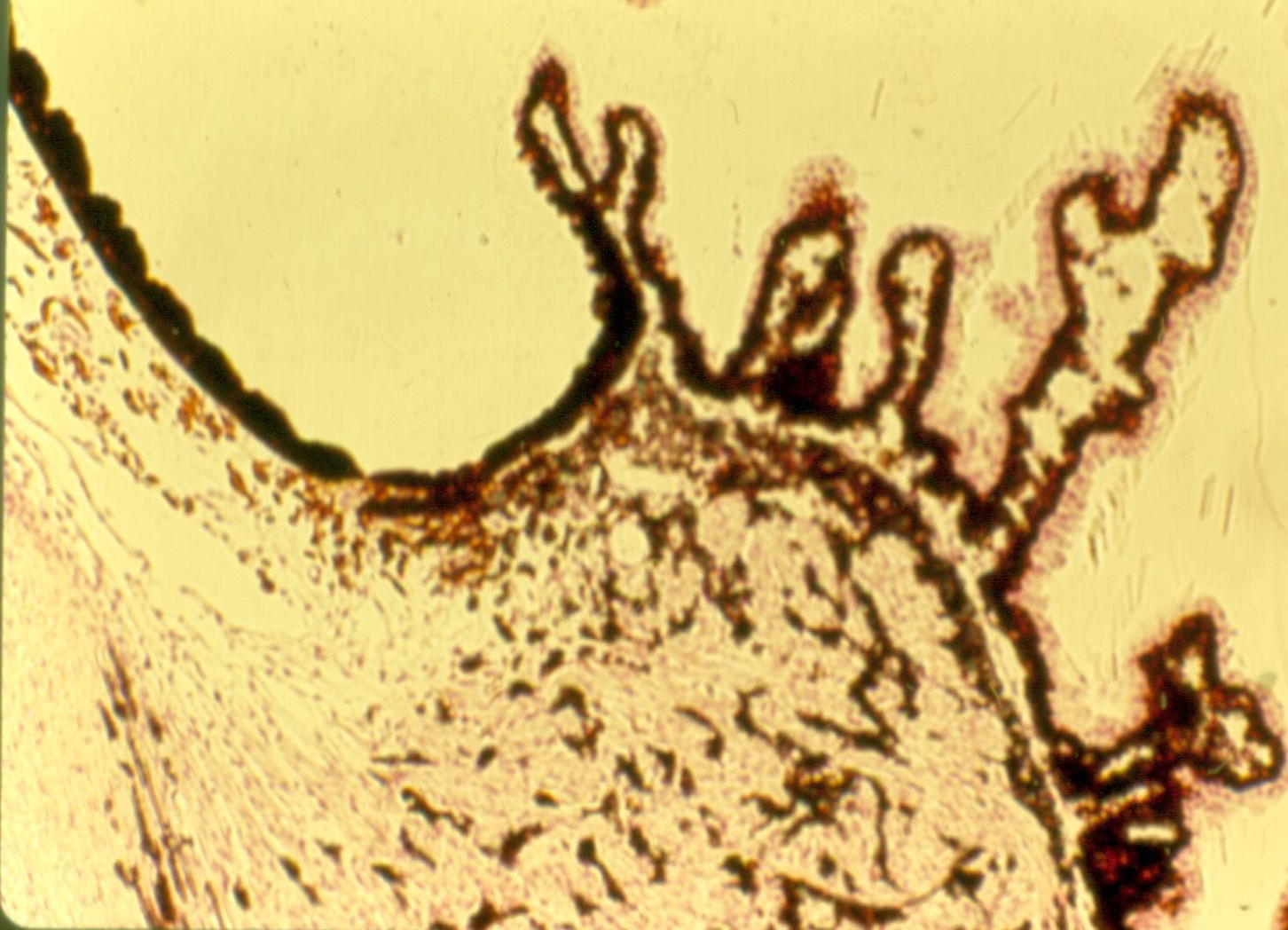
Skin Sensory Histology: Meissner Corpuscle, Pacinian Corpuscle
SPECIAL SENSES:
The five special senses are taste, smell, hearing, vision, and balance/equilibrium.
Special senses have a bipolar neuron involved in the anatomy, either as a receptor or specialized cells that are tied to the receptor. They are located in protected areas by bones of the skull. Sensory information is directed along a cranial nerve to one of the primary sensory lobes of the brain and then assessed and possibly stored in the association areas of the brain.
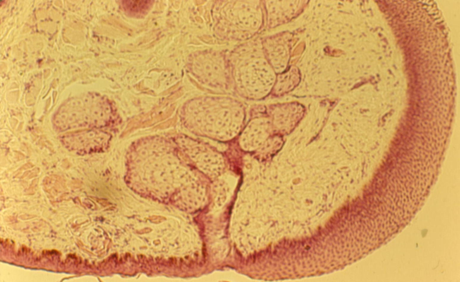
Chemical Senses of Taste and Smell
Both of these senses require that the chemicals be dissolved for sensory stimulation.
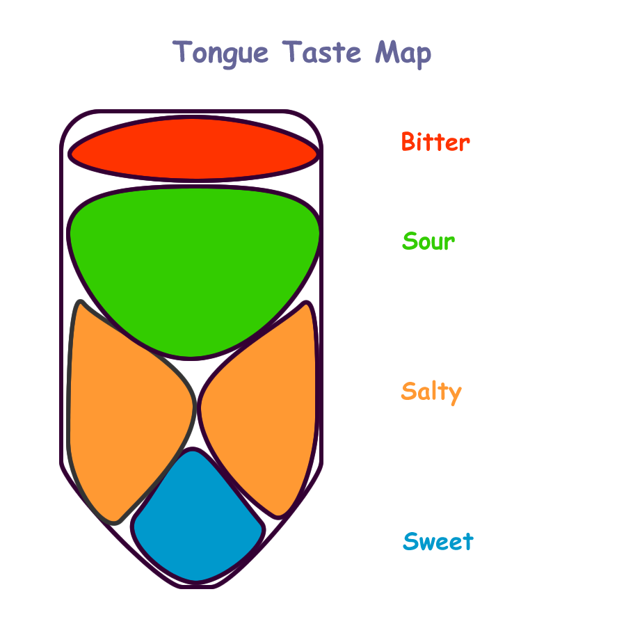
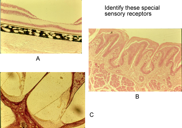
Taste or gustation occurs in the tongue and pharynx. Chemicals must be dissolved in saliva in order to stimulate the taste buds located on the tongue projections (papilla) and in the lining of the pharynx or throat. Taste buds have modified hair cells called a taste cell whose ‘hair’ protrudes from an opening called the taste pore. Taste cells are located in the sides of the papilla and are protected by supporting cells. When a chemical is dissolved in the saliva produced by the salivary glands, it covers the sides of the papilla and enters the taste pore. This contact with the hairs of the taste cell will stimulate the taste cell to trigger the cranial nerves to send a signal to the brain. The cranial nerves involved with taste are CN VII (facial), CN IX (glossopharyngeal), and CN X (vagus). The sensations interpreted in the brain are sweet, sour, salty, and bitter. Smell also plays a role in how we interpret smells as evidenced by a cold or stuffy nose, when nothing seems to taste right. The sense of taste becomes less acute as we grow older or if salivary secretions diminish.
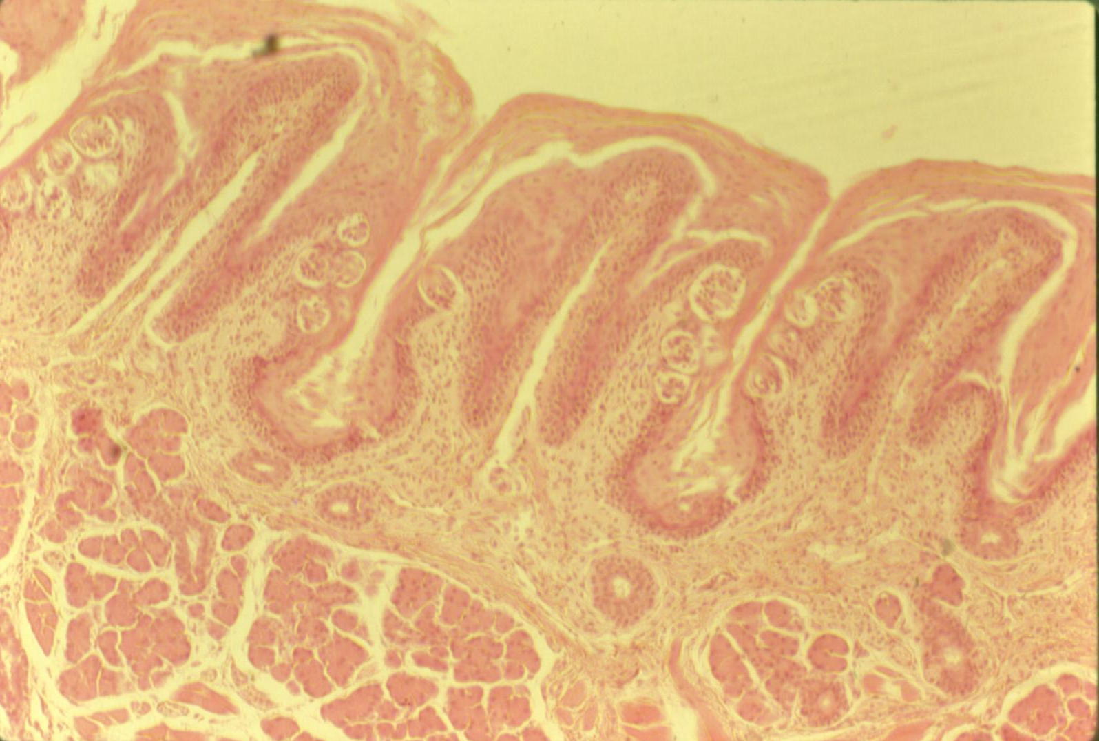
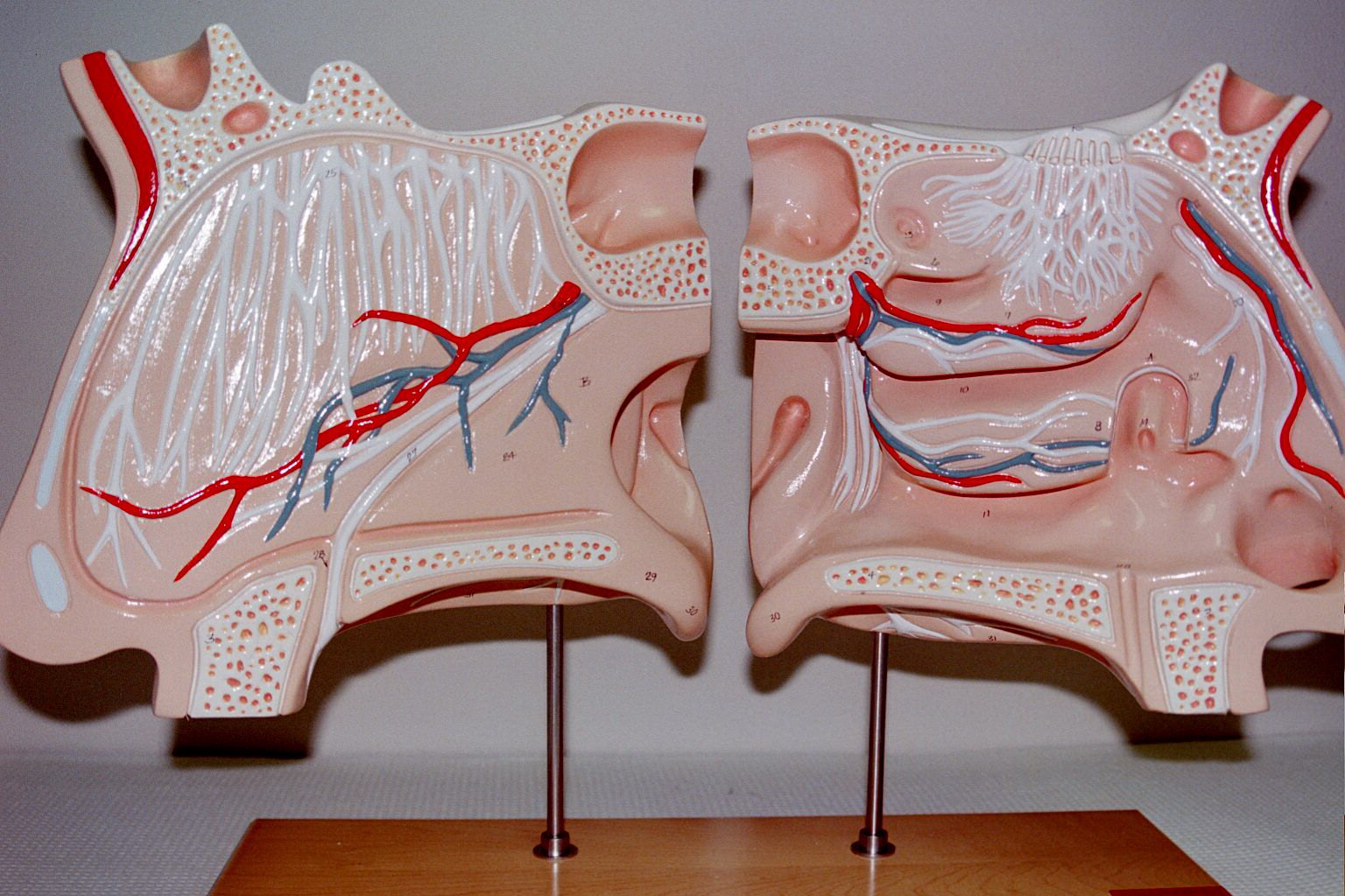
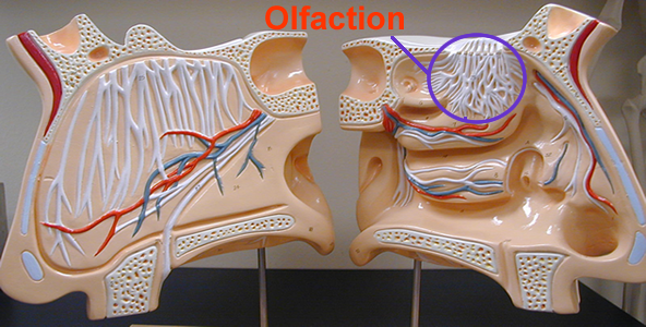
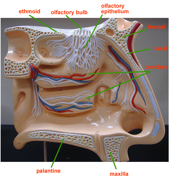
Smell or olfaction occurs in the upper regions of the nasal cavity. Chemicals must be dissolved in air in order to stimulate the olfactory receptors in the superior nasal conchae region. The olfactory receptors are the dendritic ends of neurons that hang down in the spaces above the conchae. These dendrites have modified receptors known as olfactory hairs and are protected by supporting cells. The other structures important to smell are the goblet cells that produce mucus. Mucus allows the dissolved chemicals to be trapped and concentrated in order to stimulate the olfactory hairs. The stimulus travels through foramina (holes) in the cribiform plate of the ethmoid bone and synapse on the olfactory bulb. This olfactory bulb continues as CN I, the olfactory nerve. The axonal pathways of CN I continue to the base of the brain and tie into a functional system for emotions known as the limbic system.Storage of smells occurs in the association areas of the temporal lobe.
The sense of smell is poor in humans when compared to other animals. Several
smell sensations are interpreted and can trigger an emotional response. There
are thousands of odors, some examples include putrid, burnt,
Ol
factory
receptor cells easily adapt and are important in aiding in the sense of taste.
Olfactory epithelium histology
Vision
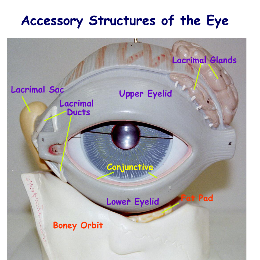
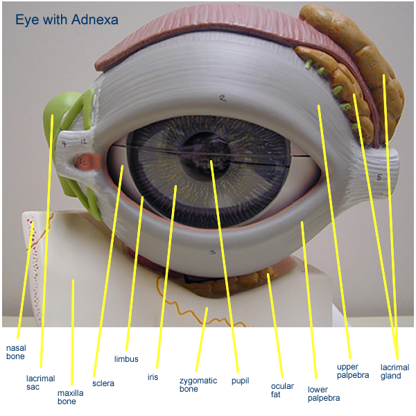
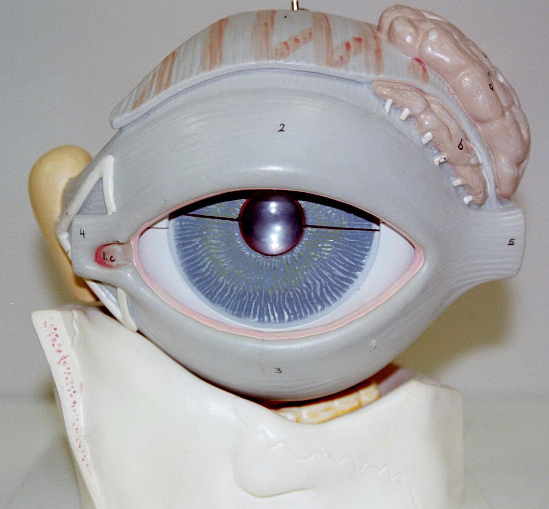
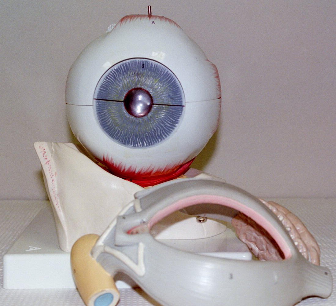
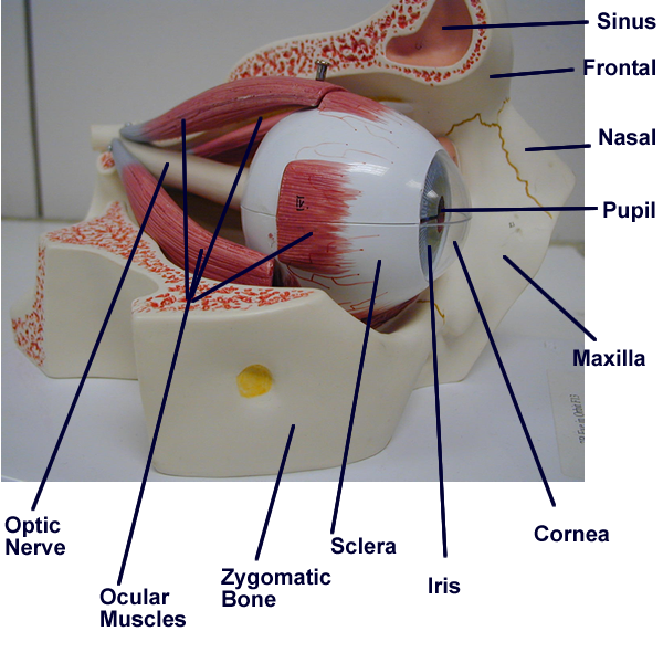
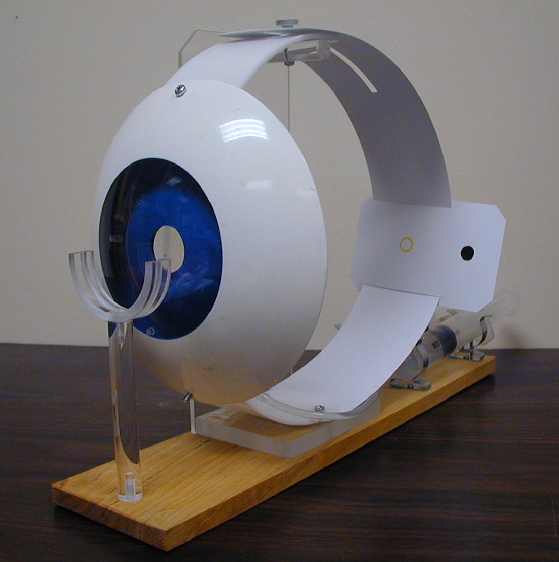
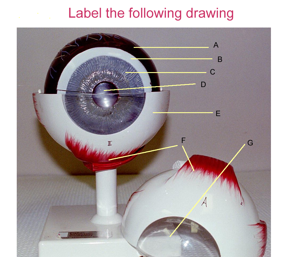
The eyes contain the receptors for sight and are protected and aided by accessory structures.
The accessory structures of the eyeball or globe are:
Eye brows and eye lashes: protection
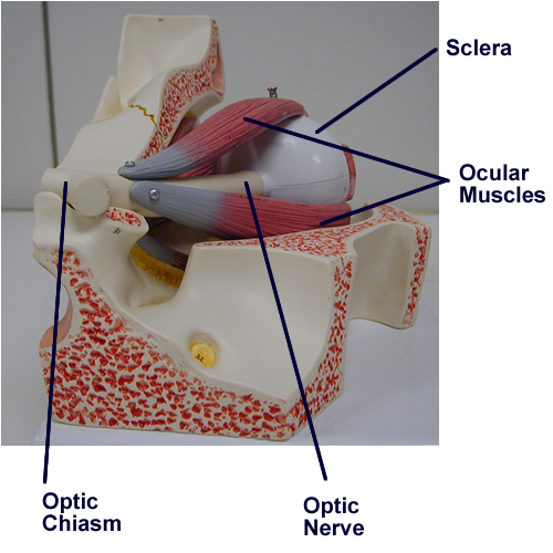
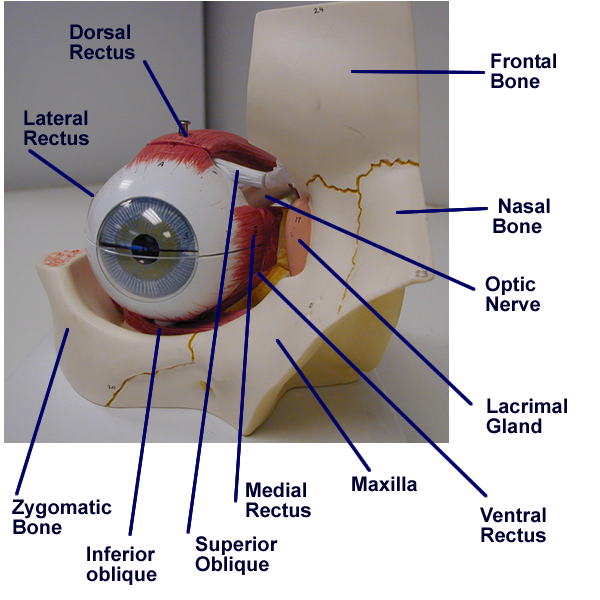
Extrinsic ocular muscles: movement
Lacrimal Apparatus: tear production and elimination
Conjunctiva: lining for immune defense
Orbital fat pad: cushion
Boney orbit: protection
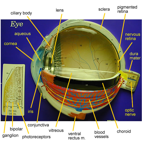
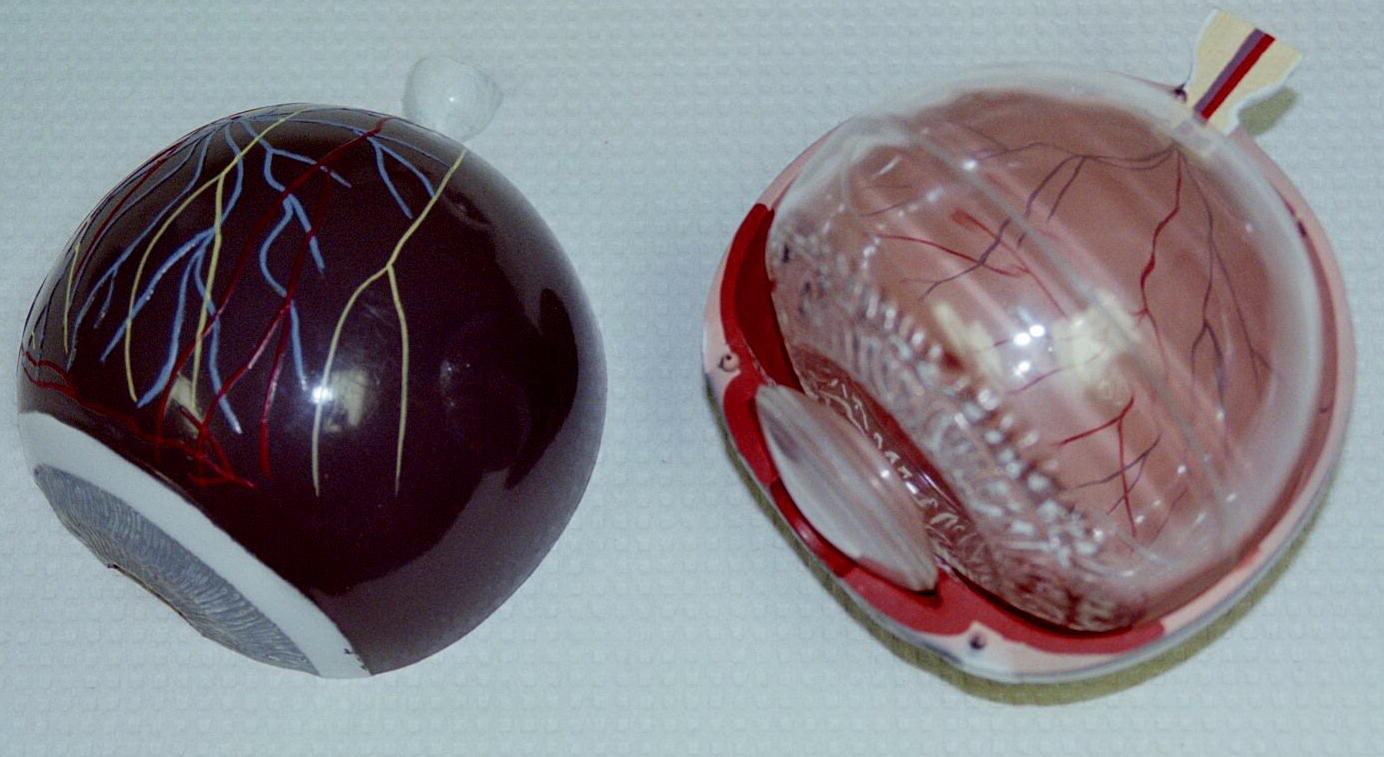
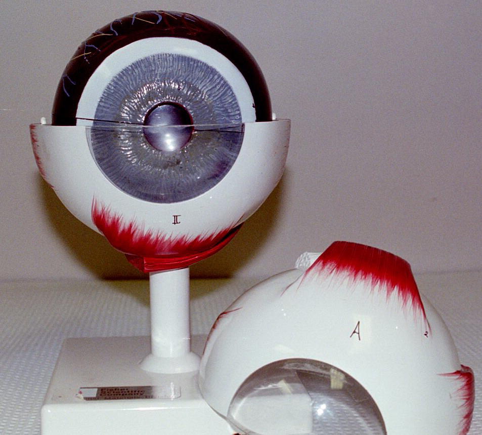
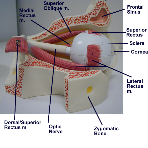
Eye model with accessory structures
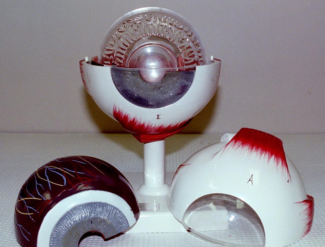
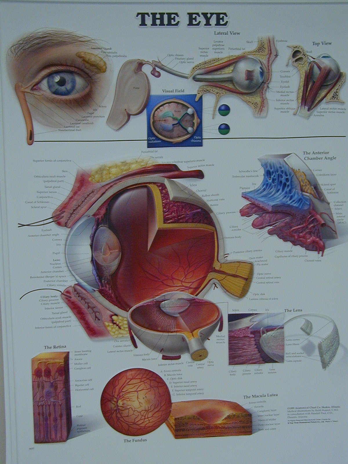
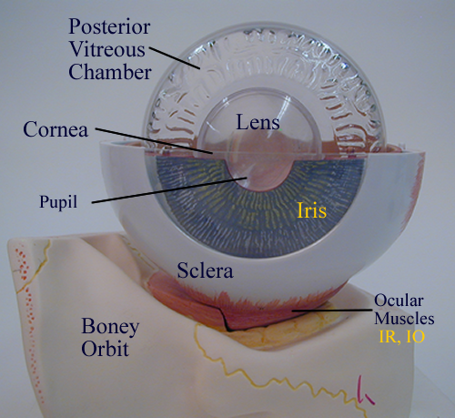
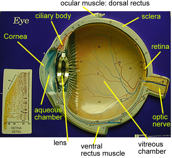
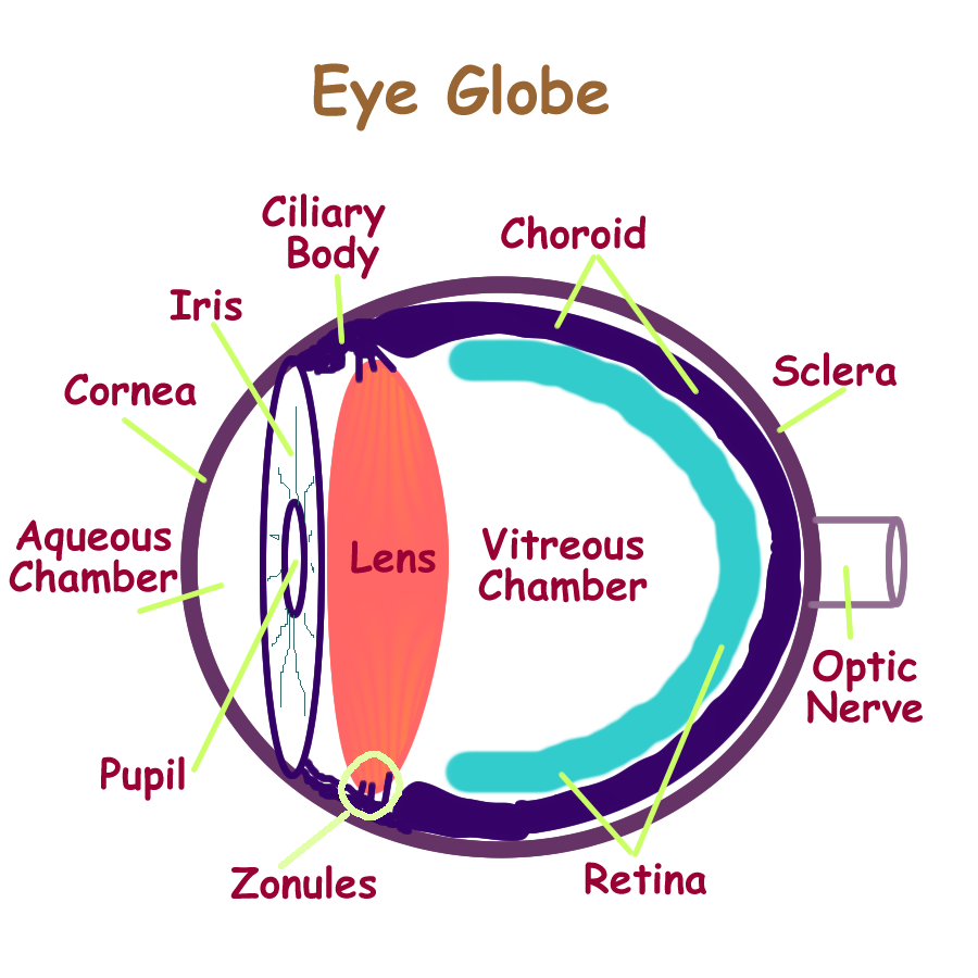
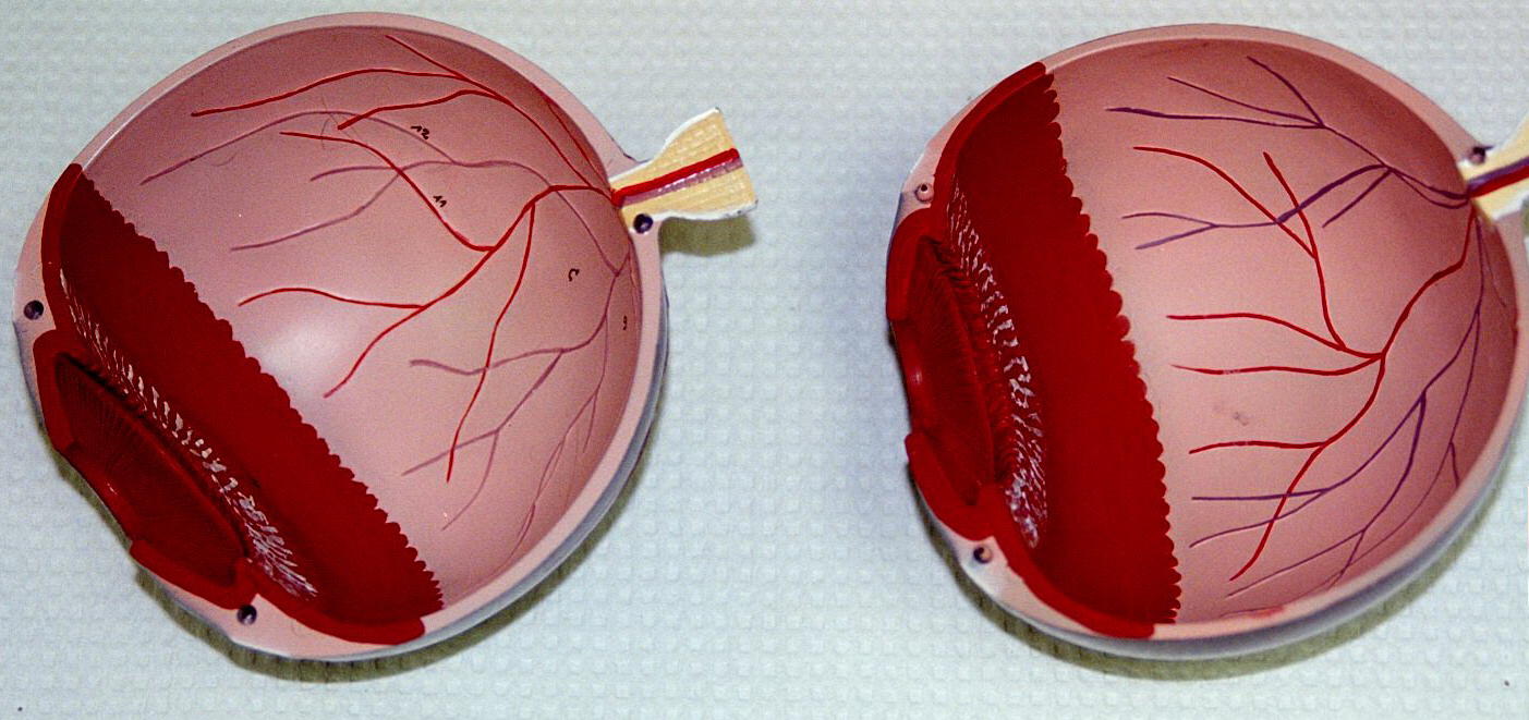
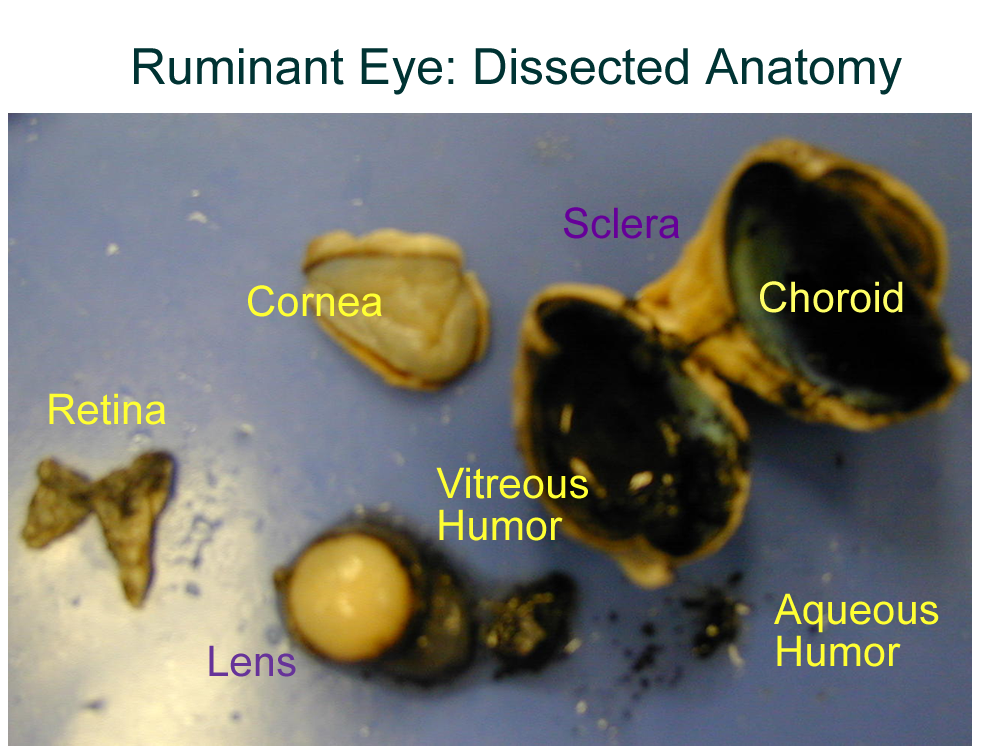
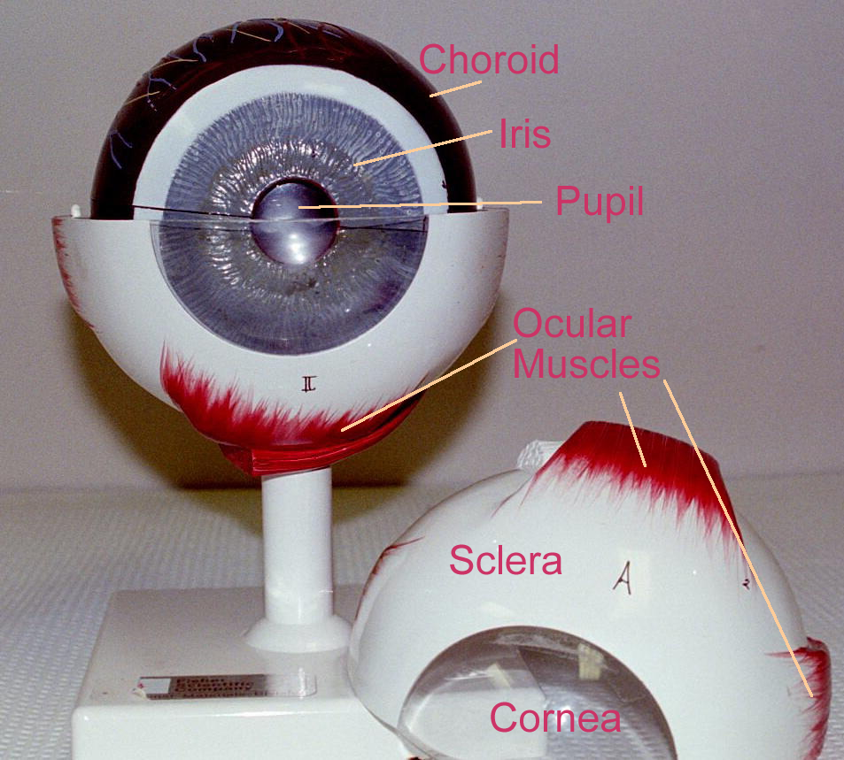
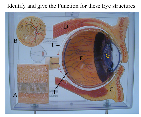
The eyeball is a globe that consists of three outer layers with inner fluid chambers.
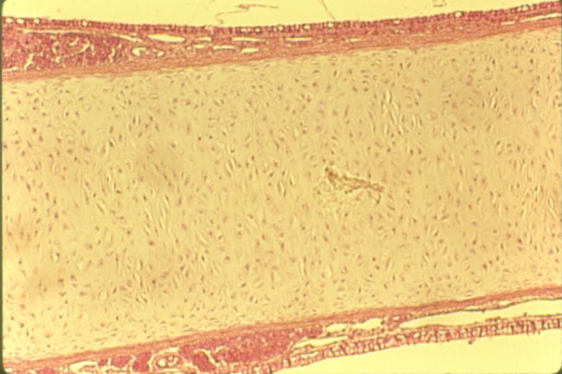
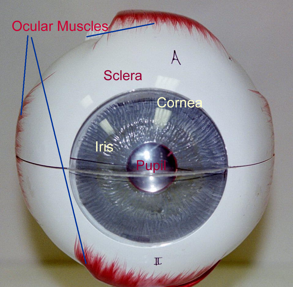
The outer layer or tunic is called the fibrous tunic that is divided into the sclera and the cornea. The sclera is tough, dense connective tissue that helps maintain eyeball shape and allows for muscle attachment for movement. The cornea is primarily connective tissue with two epithelial linings. The transparency of the cornea is due the collagen fiber arrangement that creates the stroma and a sodium ion pump in the inner lining that moves water out of the stroma. This transparency allows light to enter the eye and encounter inner chamber structures.
The second layer of the eye is the pigmented, vascular tunic, also known clinically as the uvea. This tunic is divided into three areas: the choroid, the ciliary body, and the iris. This tunic has extensive blood vessels for blood supply and melanocytes for pigment. The choroid follows the sclera at the back of the eye and the pigment of this layer helps to absorb light to prevent glare. The choriod continues anterior to become the ciliary body. The ciliary body occurs behind the junction of the cornea and sclera called the limbus. The ciliary body’s blood supply produces the fluid found in the front chamber of the eye. The iris is the visible portion of this tunic and can be seen when looking through the cornea. This creates the eye “color” and muscles in this tunic control the pupil opening. Light entering the eye at the cornea will travel through the aqueous fluid created by the ciliary body then through the pupil of the iris.
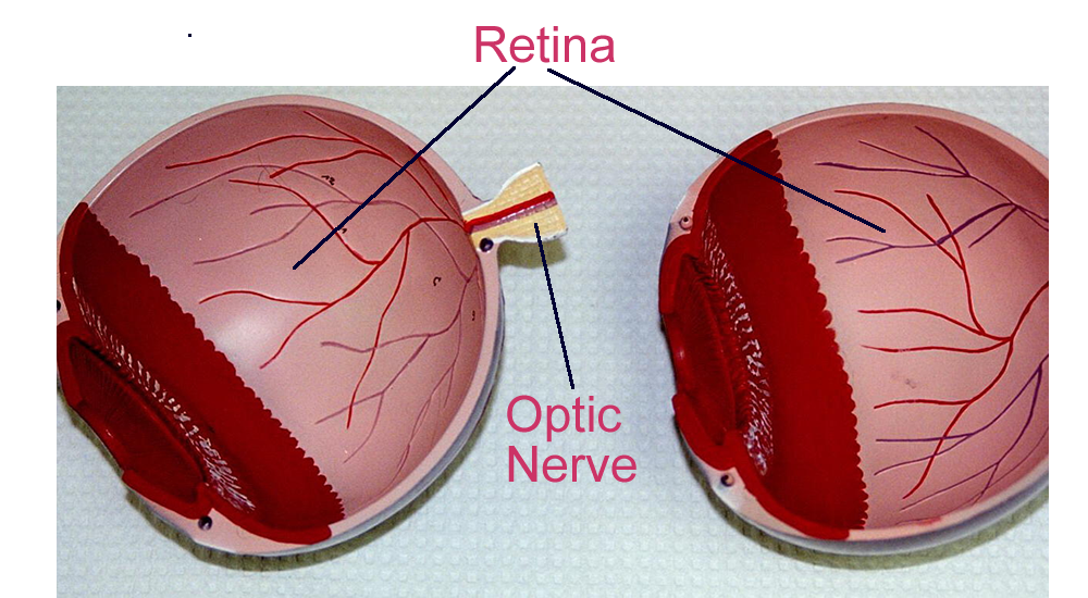
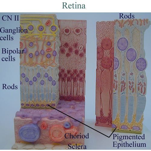
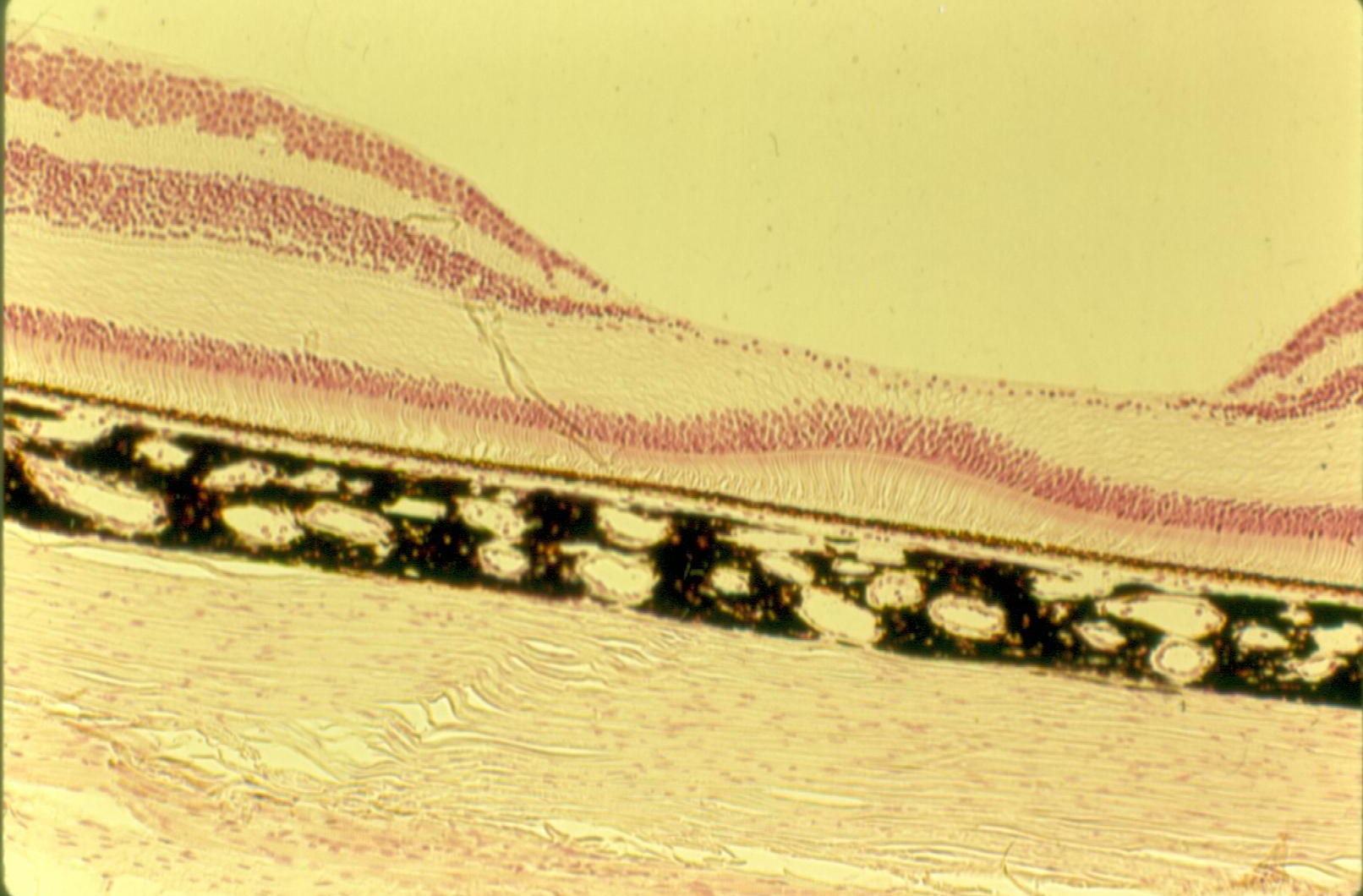
The third, innermost layer is the nervous tunic, also known as the retina. The retina consists of two parts: a pigmented epithelial layer and several nervous layers. The pigmented epithelial layer absorbs light, stores vitamin A, and helps in photoreceptor repair or regeneration.
The nervous layers of the retina begin with the specialized receptors for light.
These photoreceptors are named for the shape of their modified dendrites. Rods have elongated regions and detect low energy light level, diffuse, gray tones. Cones have triangular regions and detect high energy light levels, sharp, color. Rods are located throughout the retina whereas cones are primarily concentrated in one area known as the macula lutea or yellow spot.
There is a depression in this macula where there is only one layer of neurons, which consists of cones. This depression is called the fovea centralis.
Rods and cones will synapse on a second neuron layer formed by the bipolar neurons. The final layer of the retina consists of ganglion cells. The axons of ganglion cells form the optic nerve that continues as CN II. As these axons leave the globe, there are no dendrites for stimuli. This creates the optic disc or blind spot.
Visual sensory information follows optic tracts. Part of this information crosses in front of the pituitary gland to create the optic chiasm. The tracts continue and tie in with brainstem and the diencephalon regions for reflexes. Final processing of visual information occurs in the occipital lobes.
There are other neurons involved with the retina that control communication between the neurons that make up the three major retinal layers. They are the amacrine and horizontal cells.
The inner fluid chambers of the eye are divided into two major areas and contain several structures. The lens consists of specialized proteins contained in an elastic capsule. Due to the arrangement of the protein fibers, the lens is also transparent and allows for light passage to the retina. The lens is suspended by connective tissue fibers that extend from the ciliary body of the uvea. These suspensory ligaments or zonules can change the shape of the lens by pulling on the elastic capsule.
Changing the lens shape helps to fine focus the light to a point on the retina. The lens also helps divide the inner chambers of the eye into the anterior and posterior chambers.
The posterior chamber consists of a gelatinous protein fluid called the vitreous humor.
The anterior chamber has a watery fluid called aqueous humor. Recall that the aqueous humor is produced by the ciliary body. It helps to maintain the shape of the cornea, allows for light passage, as well as provides nutrients to the corneal stroma and inner lining. The anterior chamber of the eye can be subdivided into the posterior and anterior segments. The iris is the reference for this subdivision.
The anterior segment of the anterior chamber is between the cornea and iris. The posterior segment of the anterior chamber is between the iris and the lens. Both fluid humors allow for light passage through the eye to the retina.
Eye model with accessory structures
Eye Wall Mount:
Eye Parts : Boney Orbit, Choriod, Uvea, Chambers, Retina
Eye in situ: Lateral, Posterior, General
Ocular Muscles: Lateral
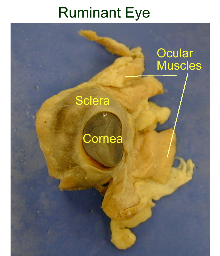
Ruminant Eye dissection : Outer Eye, Inner Contents
Physiology of Vision
Light is part of the visible spectrum of electromagnetic energy. Light travels
in wave packets called photons. The light waves can be referenced as the colors
we see. The light spectrum of color is ROYGBIV = red, orange, yellow, green,
blue, indigo, and violet. The presence of all light waves or colors creates
white. The absence of light waves or colors is black. Other colors that we
are familiar with are a combination of the various light waves.
In order for light to stimulate the retina, it must be focused to a point. The bending of light rays as it enters different density mediums (air to water) is called refraction. The surfaces of the eye that help to bend light to a point are concave surfaces. The cornea and lens provide the concave surfaces for refraction. They must remain transparent in order for light to pass through. The cornea is the strongest refractive surface and does not normally change shape. The lens can change shape due to the pull of the suspensory ligaments on its elastic capsule which helps to focus light on the retina when looking at closely held objects. The eyes also rotate medially and the pupil opening constricts to limit the amount of light. All these changes to observe a close object are called convergence.
When light is focused to a point on the retina, the photoreceptors detect the stimuli.
Photoreceptors have chemicals that are sensitive to light striking the retina. These chemicals are made up of a modified vitamin A molecule and a photopigment. In rods, the photopigment is rhodopsin.
In cones, there are three photopigments that correspond to the light waves that represent the colors red, blue, and green. The light stimulus detected by the photopigments in the photoreceptors is converted to a chemical signal that is transmitted to the bipolar and then the ganglion layer to the optic nerve, CN II. As discussed earlier, the stimulus travels along the optic tracts and ends at the cerebral cortex of the occipital lobes to form a real, inverted image. Since both lobes are involved, the brain integrates the two images to create binocular vision and allows us to interpret the images right side up.
Eye model parts: Accessory, Globe, Tunics, Retina
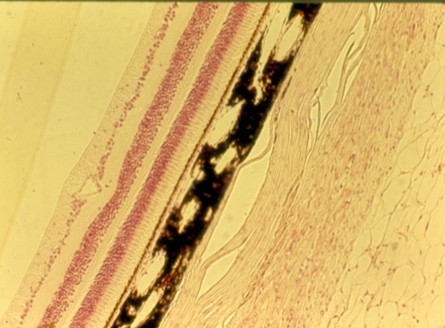
Histology
Cornea
Retina at the Fovea Centralis
Hearing, Balance, and Equilibrium
Anatomy
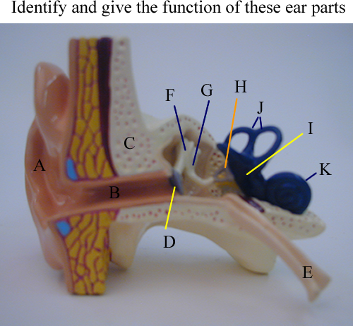
The ear contains the receptors contain the sensory receptor for hearing, balance and equilibrium.
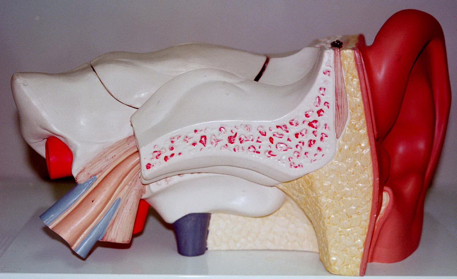
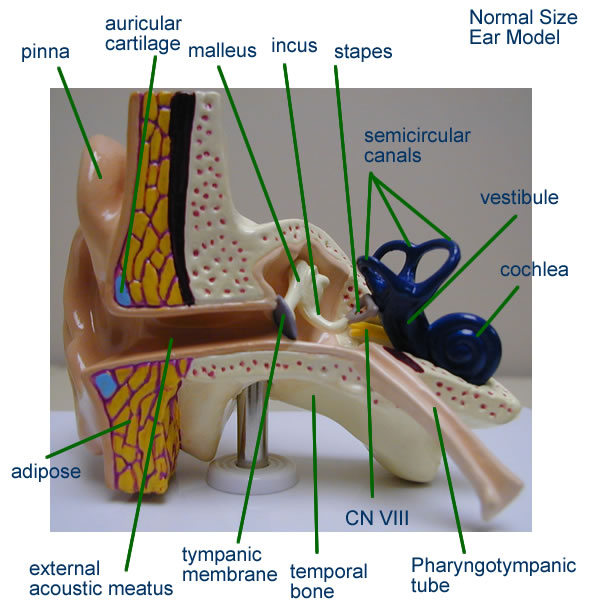
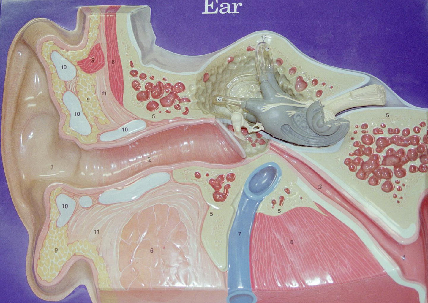
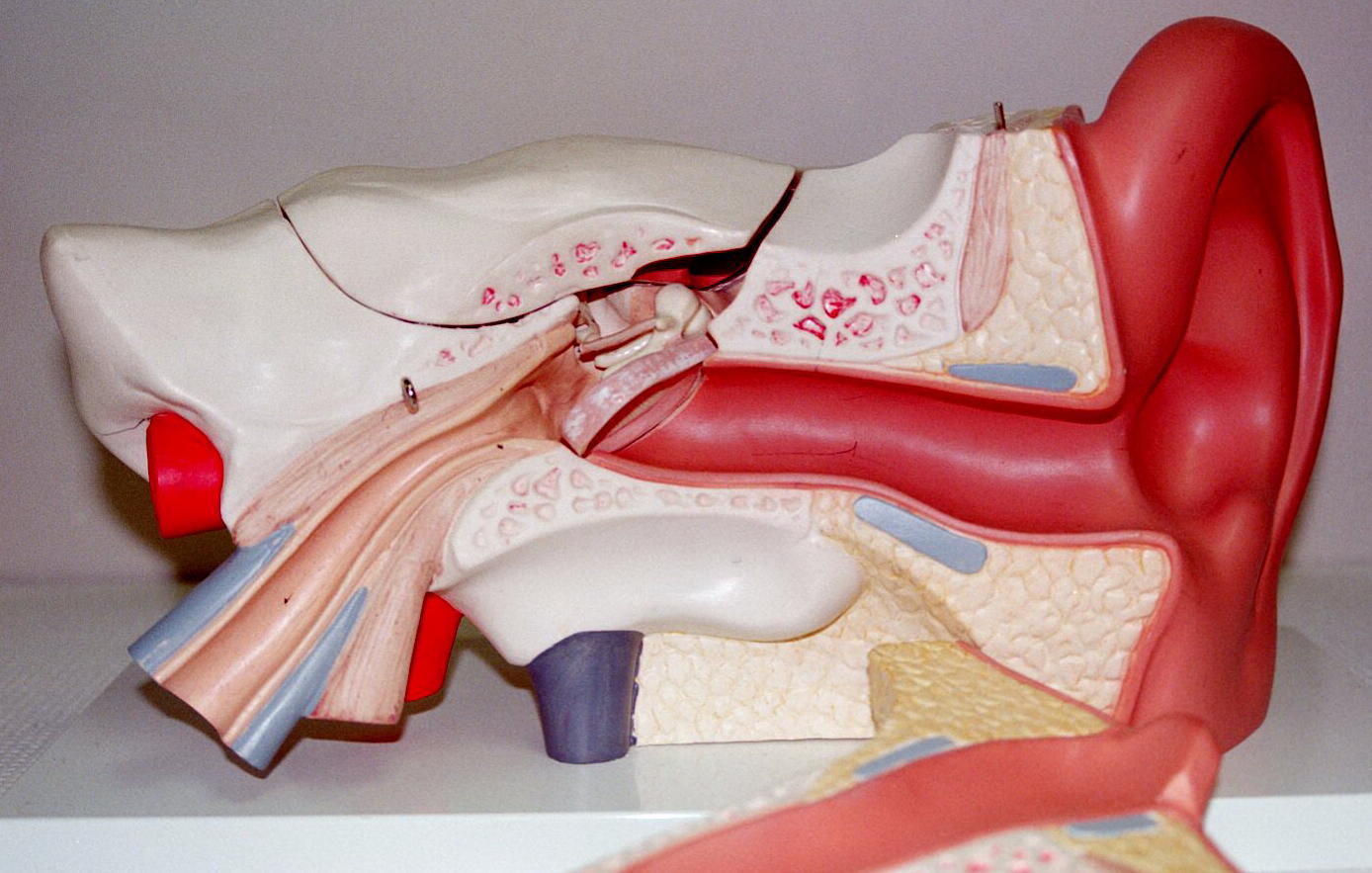
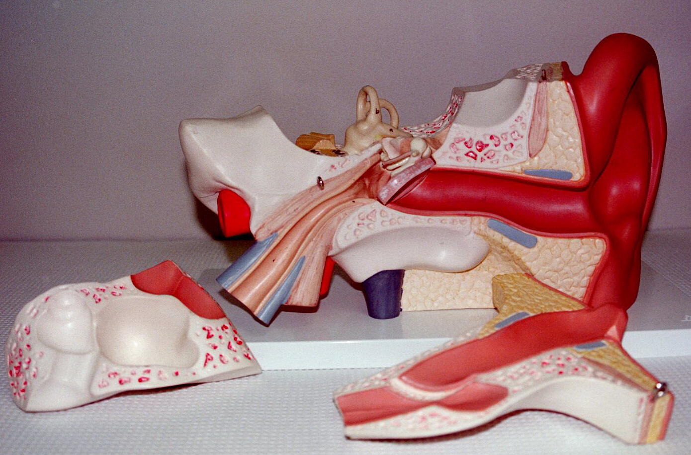
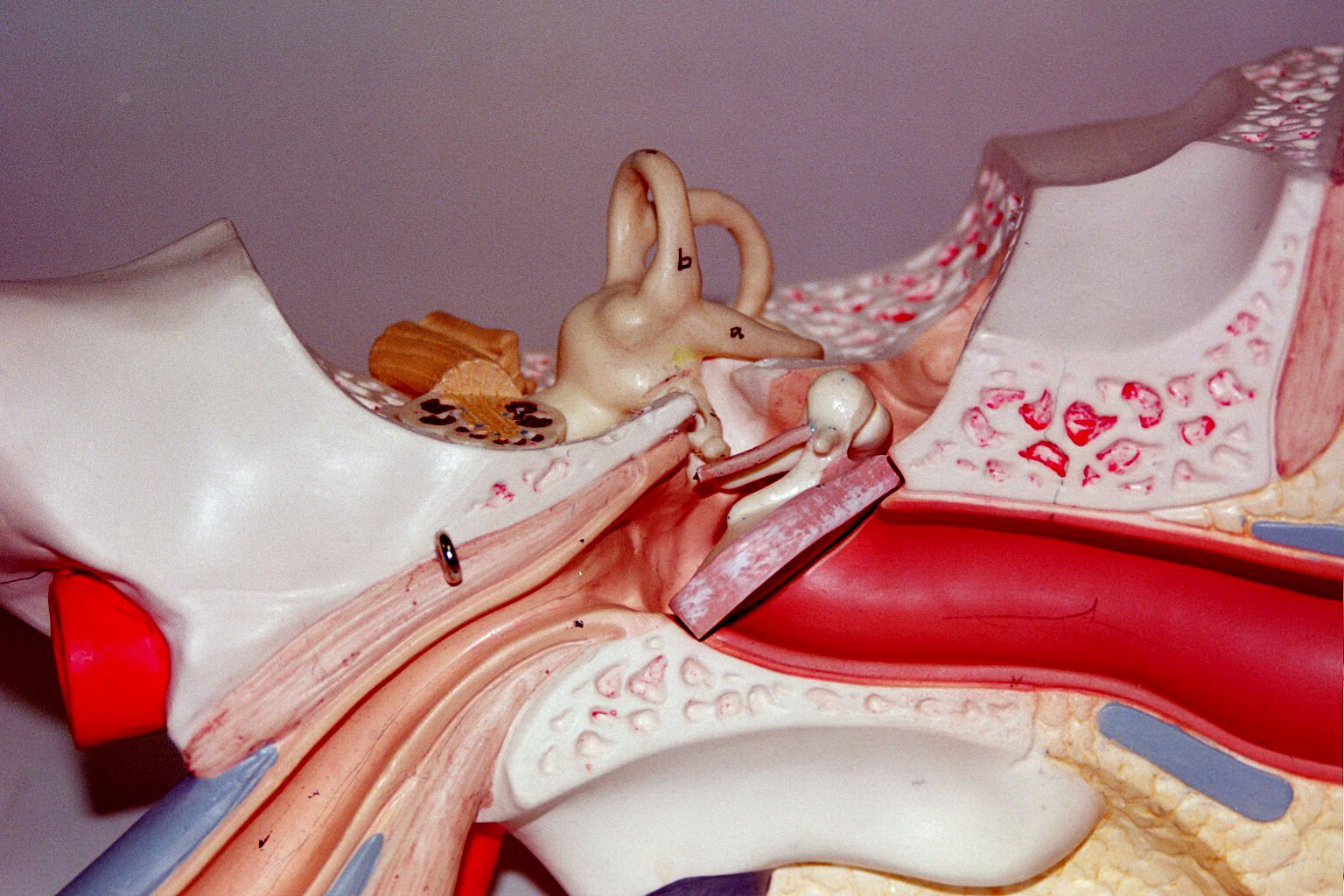
The three parts of the ear are external, middle, and internal.
The external ear is a boney and cartilaginous structure that consists of the auricle (pinna), ear canal (auditory meatus), and ear drum. The function is to conduct sound waves to the ear drum or tympanic membrane. The external ear canal also has Ceruminous glands produce cerumen (ear wax) that helps protect and keep the tympanic membrane clear.
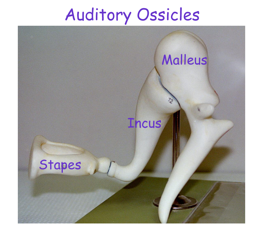
The middle ear is an air filled chamber that contains the tiny ear bones called ossicles. The three bones that make up the ossicles are the malleus, incus, and stapes. The malleus is attached to the tympanic membrane and vibrates in response to sound waves striking the membrane. The ossicles then convert sound energy to mechanical energy. The stapes connects to inner ear structures and helps set fluid in motion. The other structure associated with the middle ear is the Eustachian tube or pharyngotympanic tube. This muscular tube allows for air pressure equalization across the tympanic membrane. You may have experienced your ears “popping” when there is an altitude pressure change such as felt when flying or scuba diving. Swallowing or yawning helps to open the tube and allow for pressures to equalize across the ear drum.
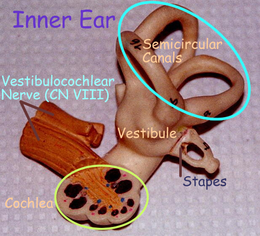
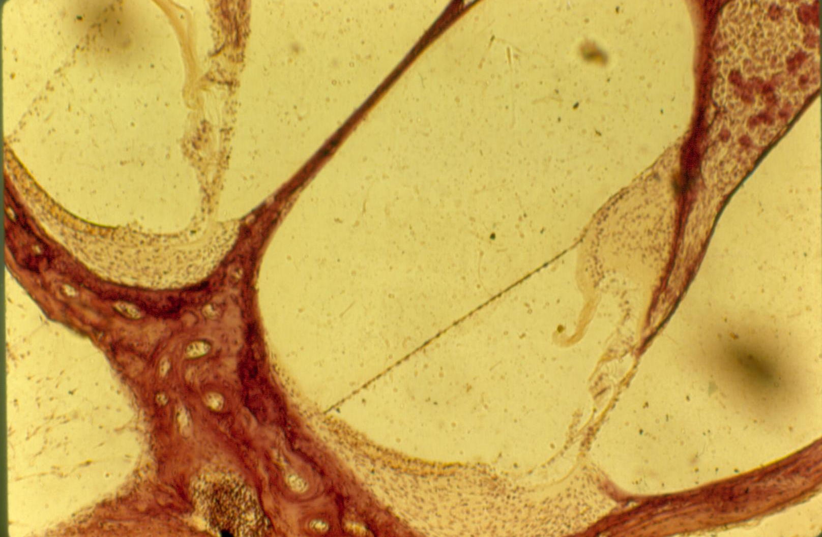
The inner ear is a fluid filled chamber contained within the temporal bone. The chamber is subdivided by connective tissue sacs to create three regions. Within the fluid filled connective tissue sacs are receptors for hearing or balance and equilibrium. The fluid inside all these sacs is called endolymph while any fluid outside the three connective tissue sacs is called perilymph.
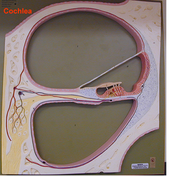
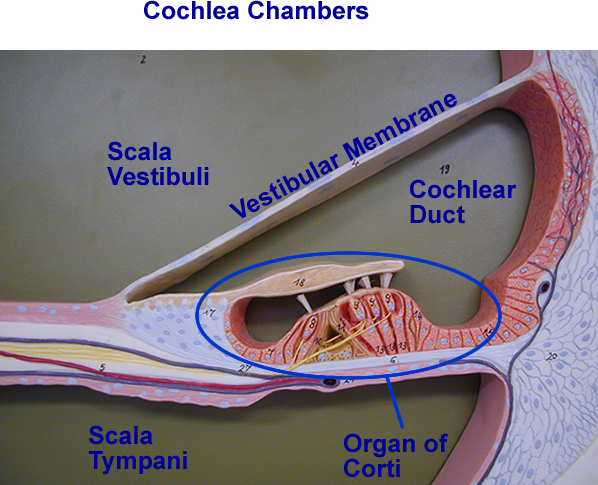
The boney chamber regions are called the cochlea, vestibule, and semi-circular canals.
Remember that each region has its own fluid filled sac with a specialized receptor.
Physiology of Hearing
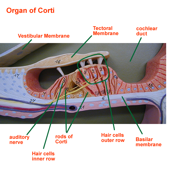
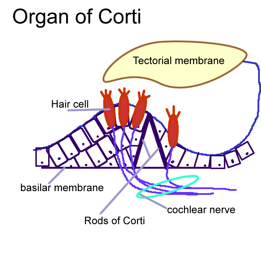
Sound waves are collected by the pinna, sent down the auditory tube, and cause vibrations at the tympanic membrane. The motion at the ear drum sets the ossicles in motion. This mechanical motion creates perilymph fluid motion around the cochlear duct. The endolymph in the cochlear duct moves the basilar membrane of the organ of Corti. The basilar membrane supports the hair cells and allows the cells come in contact with the tectoral membrane. The region of the cochlea that is affect is dependent on the energy and frequency of the sound waves entering the ear.
The cochlea is a snail shell shape chamber that contains the fluid filled sac called the cochlear duct. Within the cochlear duct is the receptor called the spiral organ of Corti.
Specialized hair cells connected to neurons are stimulated by fluid movement. The hair portions of these cells come in contact with a gelatinous membrane called the tectoral membrane. When the hair cells bend it then signals their neurons to generate impulses. The neuron involved with transmitting the electrical signals is the cochlear branch of cranial nerve eight (CN VIII). Signals are sent to the temporal lobes ford analysis, storage, and interpretation for possible motor response.
Physiology of Balance and Equilibrium
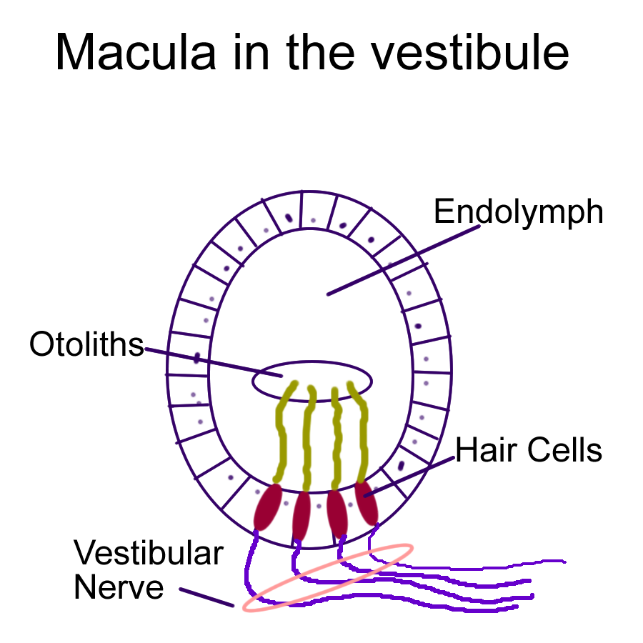
The vestibule and semi-circular canals form the vestibular apparatus. The vestibule contains the fluid filled sacs called the saccule and utricle. Within these sacs is the receptor called the macula. It too has hair cells that respond to fluid changes. Another important structure of this receptor is the crystals or otoliths that sit on top of the cells. Fluid changes occur in response to straight line or static changes of gravity. The gravitational forces pull on the otoliths which bend the hair cells. The stimulus that is generated travels along the vestibular branch of cranial nerve eight (CN VIII). Information then goes to the cerebellum, brain stem, and temporal lobes of the cortex to help maintain balance and equilibrium.
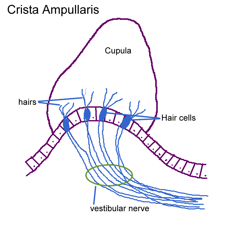
The semicircular canals are narrow half circle tubes that contain the fluid filled sac called the semi-circular sacs. At the ends of the sacs is an expanded region called the ampula. In the ampulla region is the receptor called the crista ampullaris. The crista contains hair cells that are covered by a tall gelatinous material called the cupula. Fluid in motion pulls the cupula which in turn moves the hair cells. These receptors detect dynamic, circular or rotational changes to help maintain balance and equilibrium when spinning or turning.
Ear Model: Outer ear, Middle Ear, Inner Ear
Cochlea Model
Drawings of Receptors
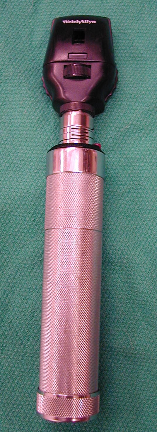
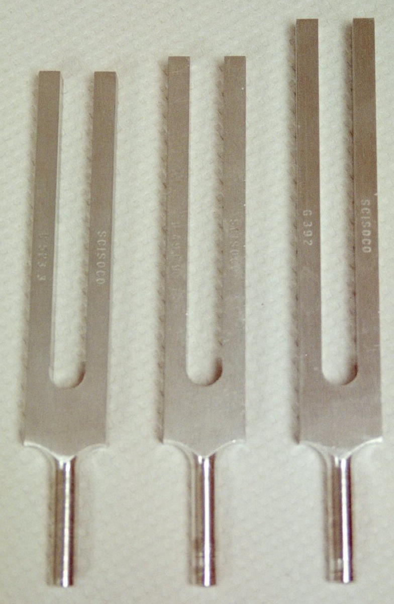
Instruments : Otoscope, Tuning Forks
Cribr- sieve dipl/o- double
Emmetr/o- normal measure lacr/i- tear
Mio- less, smaller ocul/o- eye
-opia eye ot/o- ear
-phasia speech rhin/o- nose
sclera/o- hard -scopy view, see
trop/o- turning acou- hearing
aur- ear blephar- eyelid
cili- eyelid -flect bend
glott- tongue irid- colored circle
palbebr/o- eyelid phac/o- lens
phon/o- sound ophtalm/o- eye
presby- old kerat/o- horn, cornea
phthi decay, waste dacry- tear
Somatosensory:
Identify and Function of Eye
Hearing and Balance Experiments:
Concept Map: Make a concept map of the sensory system (somatic and special senses) using the receptors, functional type, location, nerve pathway, structure and final body effect. Include this concept map in your LAR lab report (if selected) as a document insert or as an addtional document of a PDF scan of the map.
Case Study
Referred pain Hyposmia
Cataracts Glaucoma
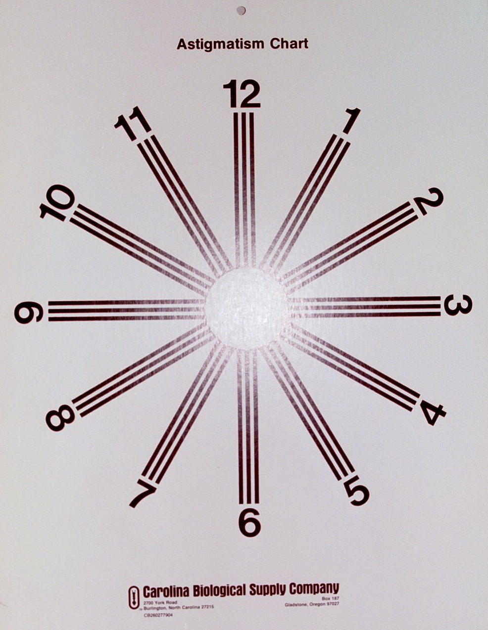
Floaters Astigmatism
Myopia (nearsightedness) Hyperopia (farsightedness)
Presbyopia Nystagmus
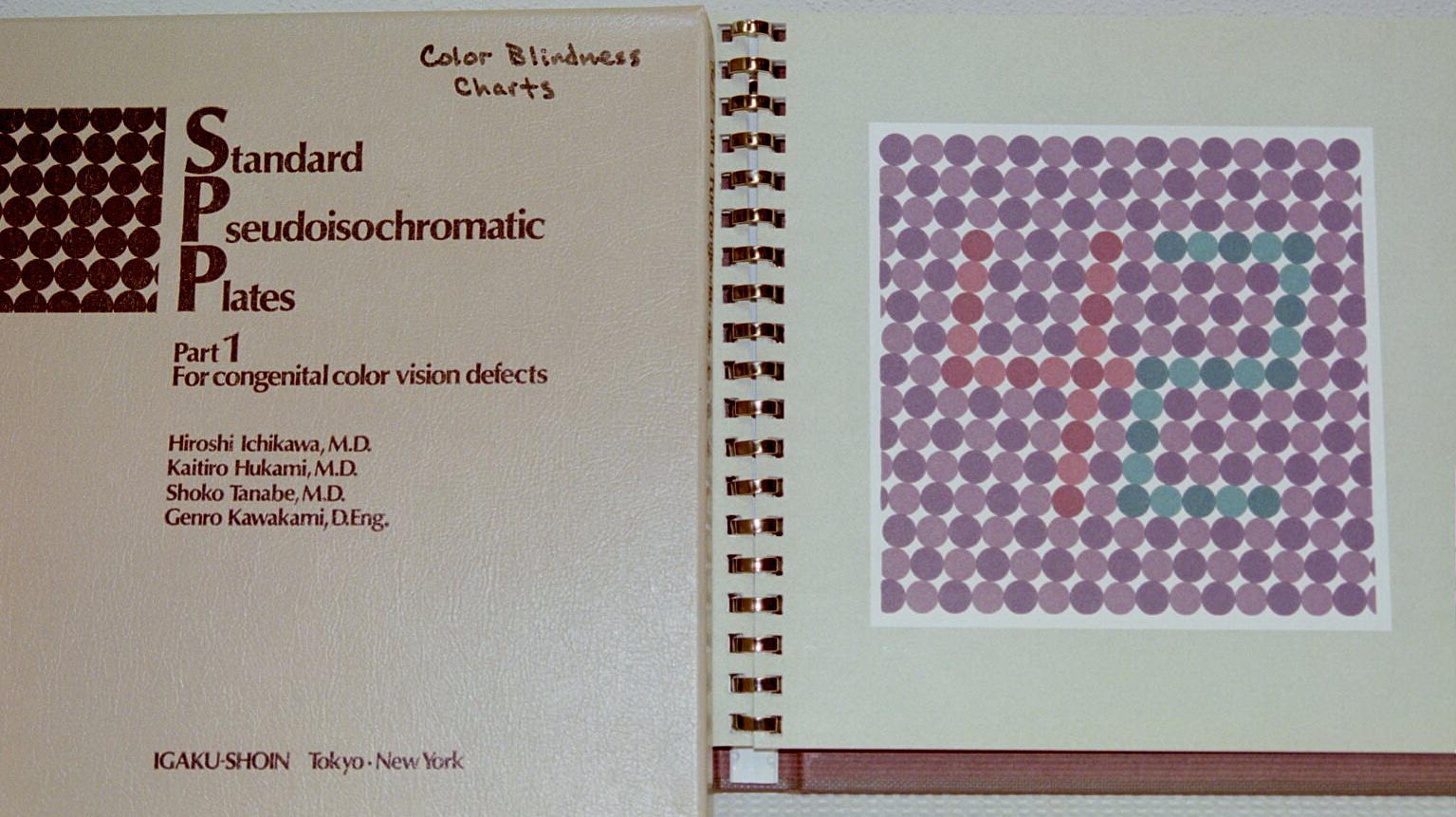
Night blindness Color blindness
Retinal Detachment Macular Degeneration
Conjuctivitis Uveitis
Blepharitis Strabismus
Conduction deafness Nerve deafness
Central deafness Motion sickness
Otitis Externa Otitis media
Otosclerosis Meniere’s Disease
Ophthalmologist
Optometrist
EENT
Audiologist
http://www.slider.com/Health/Senses/Hearing/Anatomy-Physiology.htm
http://www.slider.com/Health/Conditions_and_Diseases/Ear_Nose_and_Throat.htm
http://www.slider.com/Health/Conditions_and_Diseases/Eye_Disorders.htm
http://www.slider.com/Health/Senses/Touch_and_Sensation/Pain.htm
http://www.slider.com/Health/Senses/Touch_and_Sensation/Pruritus_-_Itching.htm
http://www.growing.com/doxys/eent.html
http://www.nlm.nih.gov/medlineplus/anatomy.html
http://www.nlm.nih.gov/medlineplus/earnoseandthroat.html
http://www.nlm.nih.gov/medlineplus/eyesandvision.html
http://www.nidcd.nih.gov/health/kids/teachers/hear_s.htm
http://www.medem.com/MedLB/article_detaillb.cfm?article_ID=ZZZYXNW46JC&sub_cat=198
http://access.parkercc.edu/Academics/Clinical/Clinical_Diagnosis_I/Ear_Nose_Throat.htm
http://www.bcm.tmc.edu/oto/otologyprimer/vertigo/vestdisperipheral.html
http://www.nei.nih.gov/health/eyediagram/index.htm
Study Guide and Review Questions
1. Name the cranial nerves involved with:
a) Vision
b) Hearing
c) Balance and Equilibrium
d) Olfaction
e) Gustation
2. Somatic senses detect what types of stimuli?
3. Define adaptation
4. Define referred pain
5. Name the brain lobes involved with sensory information.
6. Describe the neuron anatomy of somatic sensory vs. special sensory
7. Give the specific receptor location for the following:
a) Rods
b) Cones
c) Organ of Corti
d) Crista Ampullaris
e) Macula
f) Nocireceptors
g) Thermoreceptors
h) Touch / Pressure
i) Taste buds
j) Olfactory hair cells
k) Proprioceptors
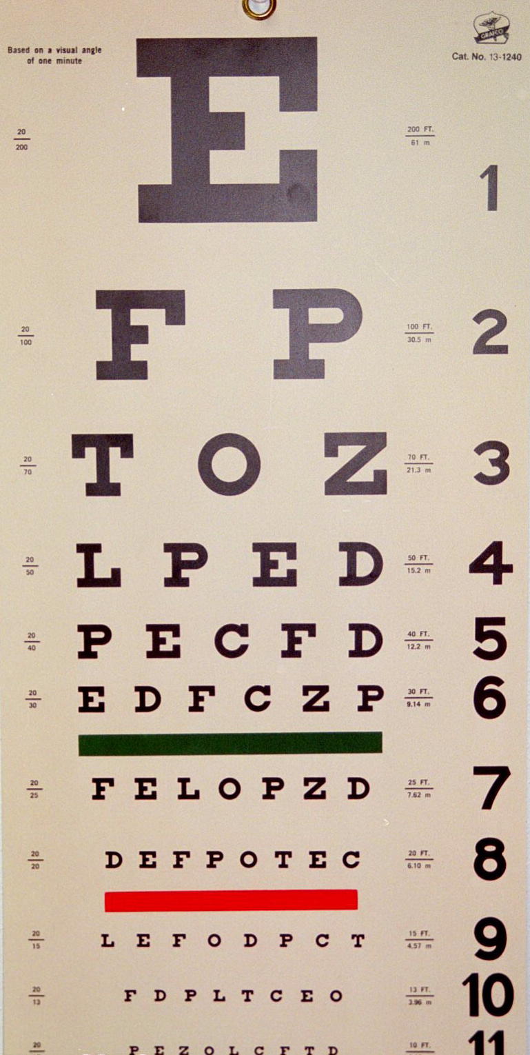
8. Name the instruments used to detect and test sensory responses for:
a) pain
b) vision
c) hearing