Biology 2404 A&P Basics Lab Exercise 22: Male Reproductive System Dr. Weis
| Objectives | Background | Medical Terms | Activities | Applications | Careers | WWW | Review Questions |
Students should be able to:
* Name, identify, and give the function for the organs related to the male reproductive system:
Primary Sex organ
Duct or tubular system
External Genitalia
Accessory glands
* Name the source and feedback control for the male hormones
* Describe anatomy of the male gamete
* Define spermatogenesis and spermiogenesis
* Define related terms
Read related material in textbook
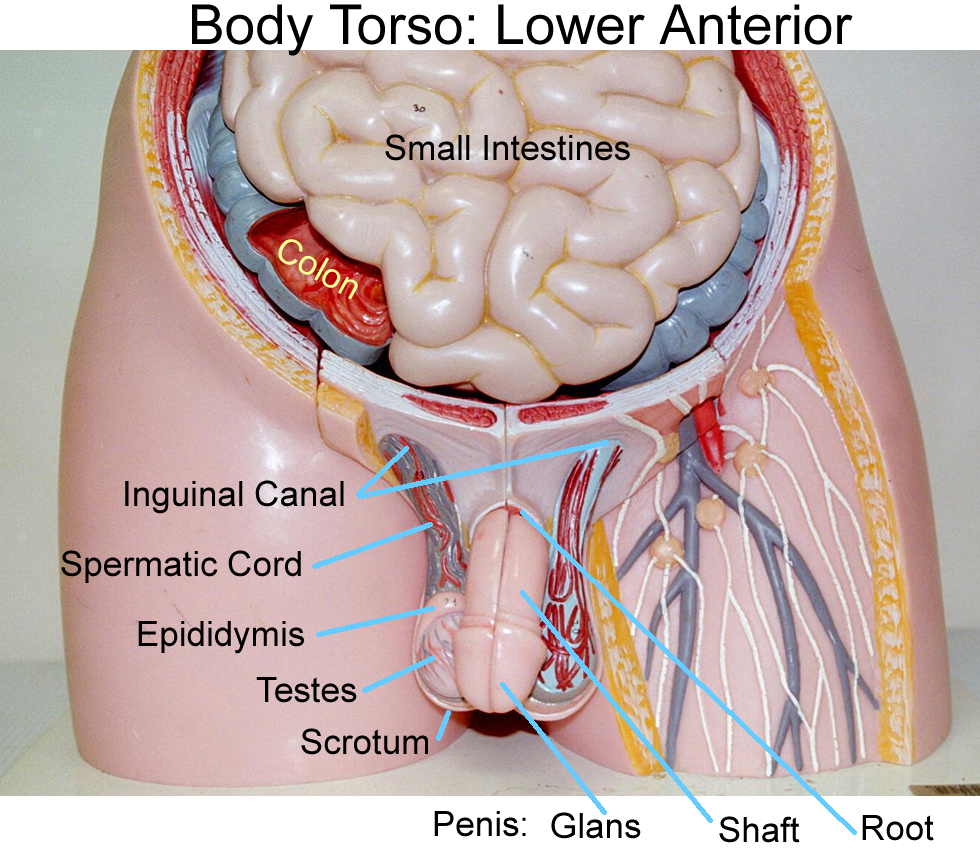
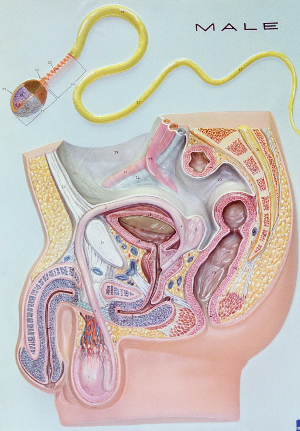
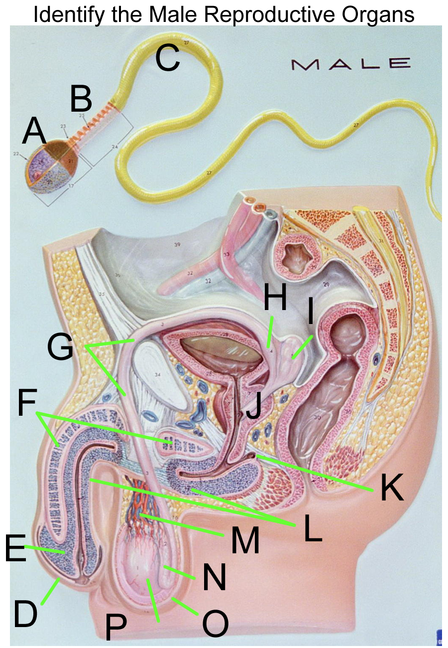
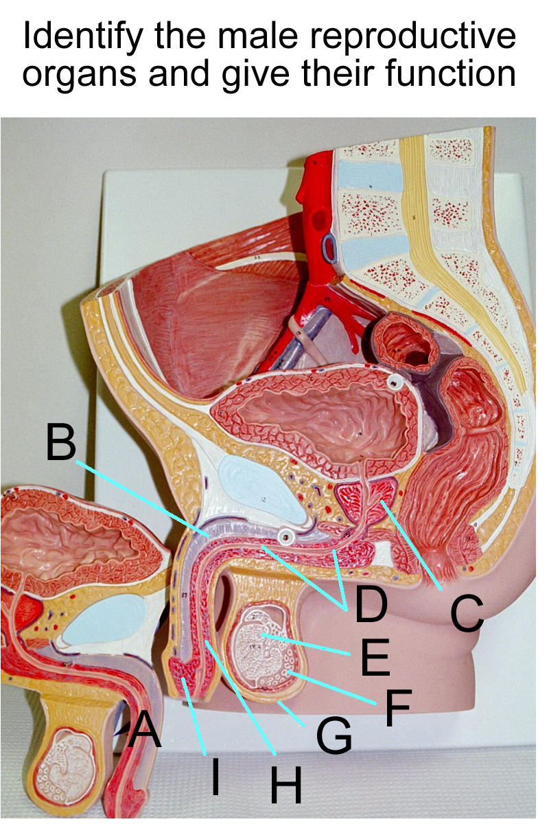
The male reproductive system is composed of:
Primary sex organ testis
Duct or tubular system: epididymis, vas deferens, ejaculatory duct, urethra
External genitalia scrotum, penis
Accessory Glands seminal vesicles, prostate, bulbourethral glands
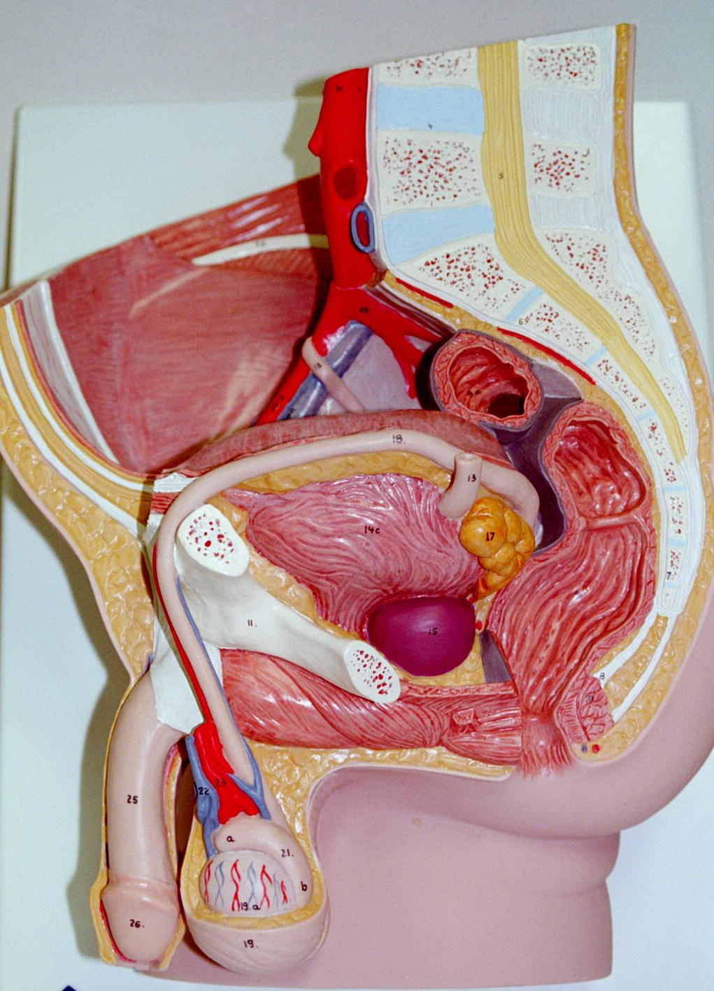
Testes
The male gonads, the testes, are paired oval glands that sit externally in the scrotum of most mammals. They are covered by fibrous connective tissues, the tunica externa and the tunica albuginea. The tunica albuginea divides the testes internally into compartments called lobules which house the seminiferous tubules. One to three seminiferous tubules are contained within a lobule division.
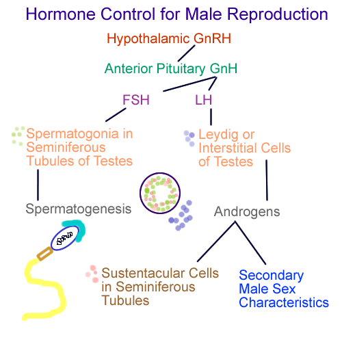
The process of spermatogenesis occurs in the seminiferous tubules and relies on proper body temperature and hormone levels. Spermatogonia within the seminiferous tubules divide by mitosis into two daughter populations.
One of the daughter groups, the primary spermatogonia, then begins the process of meiosis. Primary spermatogonia divide to produce secondary spermatogonia, then continue to develop into immature sperm called spermatids. Meiotic division creates 4 haploid cells in a two division cycle. These haploid spermatid cells then leave the testes to mature into spermatozoa or sperm. GnRH from the Hypothalamus signals FSH from the AP to trigger spermatogenesis in the testes, given proper temperature and hormone levels.
Other cells in the seminiferous tubules are the sustentacular cells. The sustentacular cells function in spermatogenesis to bind testosterone in the testes to allow for adequate hormone levels. They also provide the blood-testes barrier to prevent immune cells from attacking the developing sperm.
Surrounding the outside of the seminiferous tubules are clusters of cells known as Sertoli, Leydig, or Interstitial cells. These cuboidal cells are responsible for androgen production under the direction of GnRH from the Hypothalamus and LH from the AP.
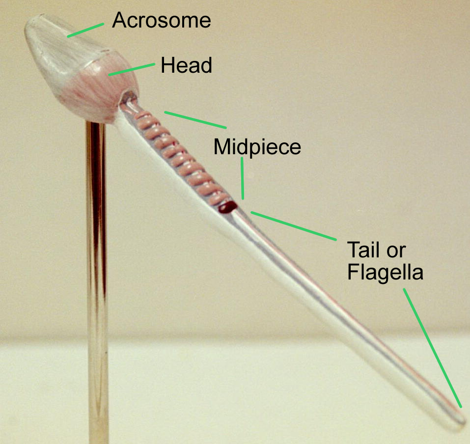
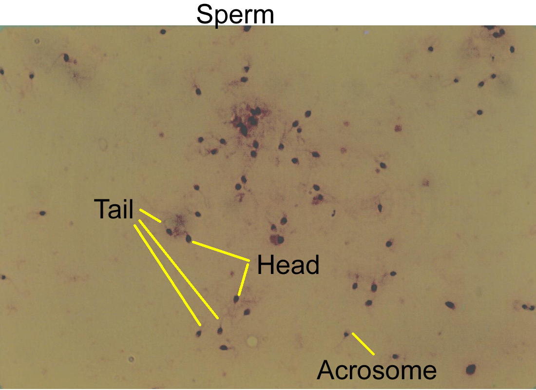
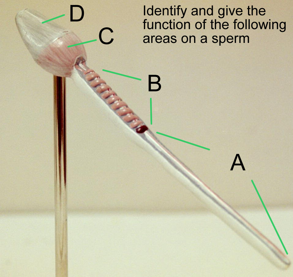
Once the spermatids form in the seminiferous tubules, they are transported to the epididymis for maturation and storage. 300 million sperm are produced daily and once they leave the male reproductive system, have a life expectancy of about 48 hours in the female reproductive system. Mature sperm or spermatozoa have a head, midpiece, and tail. The head contains half of the chromosomes for the individual and is covered by a connective tissue sac called the acrosome. The acrosome contains enzymes that aid in digestion through the surrounding structures of the ovulated oocyte. The midpiece contains mitochondria that produce energy in the form of ATP for the tail or flagella.
Once activated by accessory gland secretions, the flagellum propels the sperm through the female reproductive tract. Male reproductive fertility assessment is based on number of sperm, number of alive verses dead, normal anatomy, and normal forward movement.
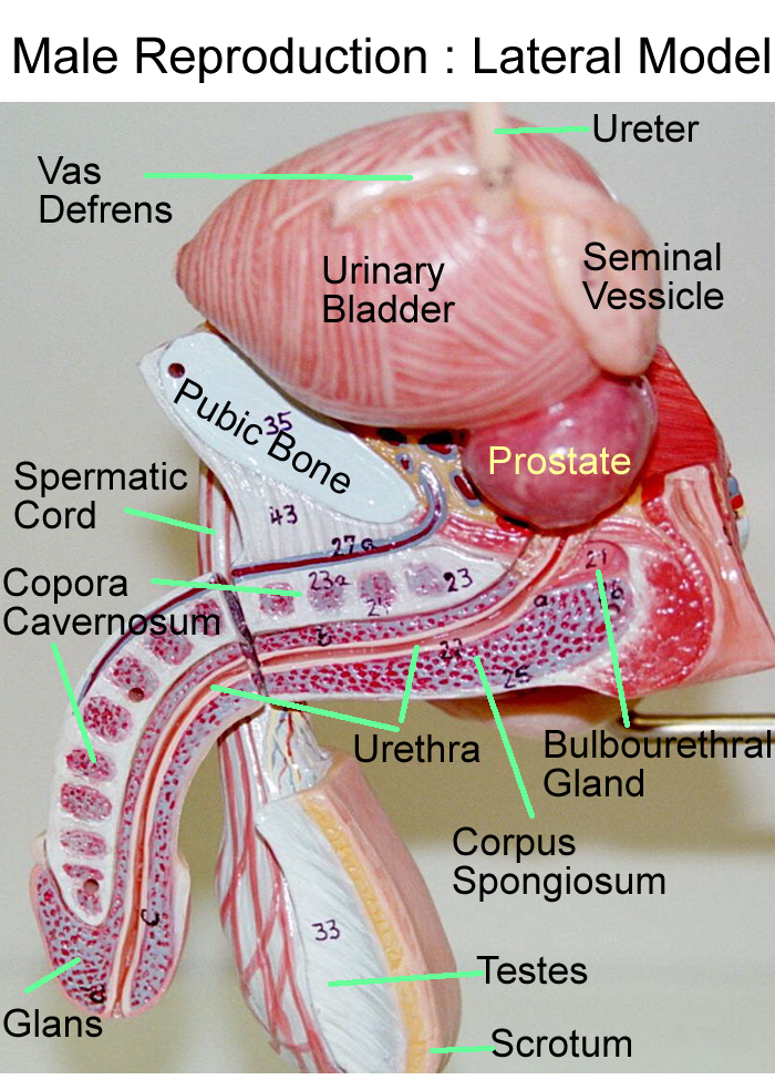
Male repro wall mount with sperm
Hormonal Control of the Testes
Duct System
Besides spermatogenesis, testosterone and its precursor DHEA, contribute to secondary sex characteristics. Bone growth, muscle development, body hair patterns, laryngeal changes, and behavioral changes are seen with secretion of these androgens.
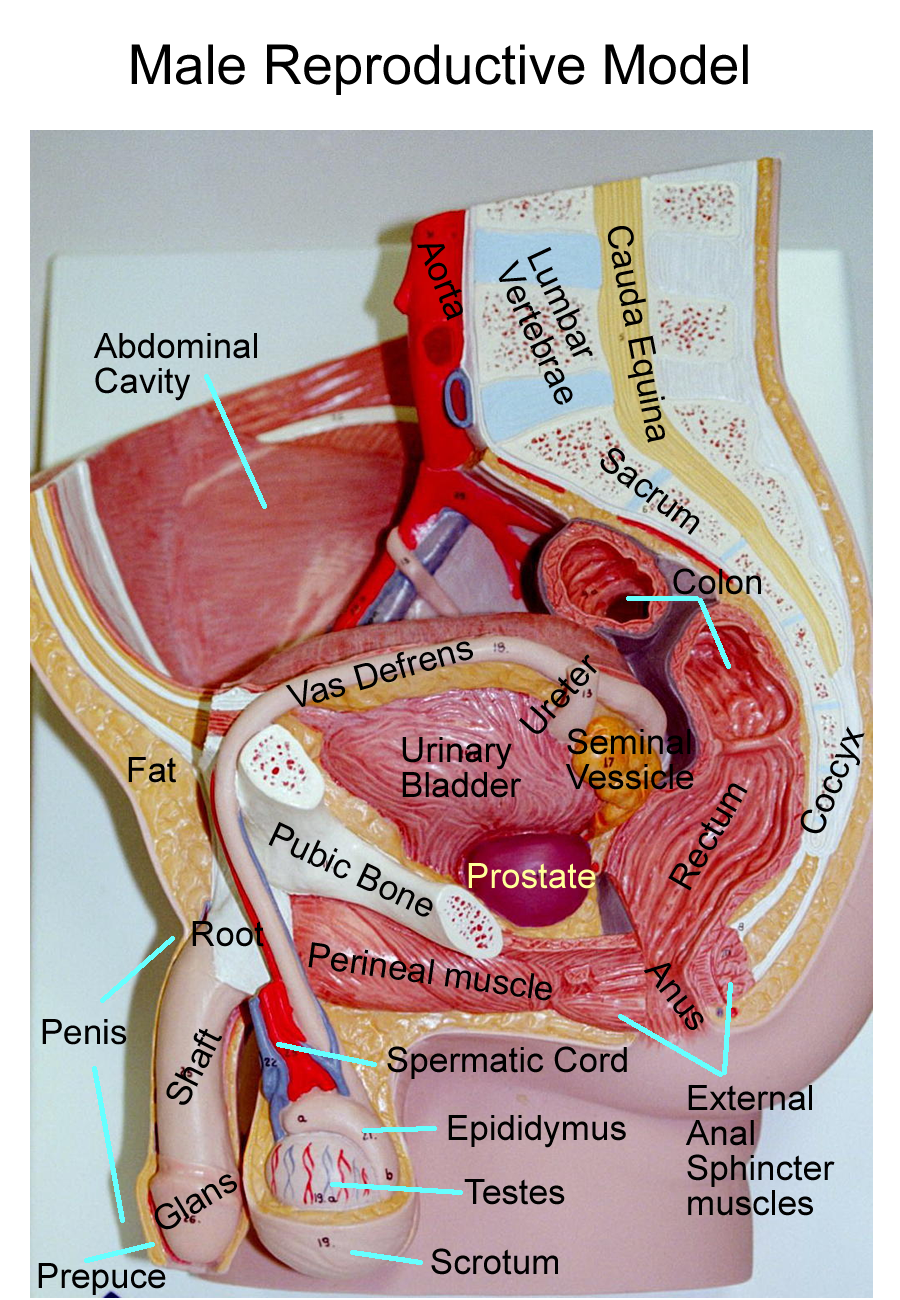
The sperm are stored and mature in the tail of the epididymis. During the male climactic, the smooth muscle movements within the ducts will help propel the sperm outward. From the epididymis, the sperm travel through the vas (ductus) deferens which is part of the spermatic cord. The spermatic cord has a connective tissue covering that contains vessels, nerves, and ducts that aid and supply the male reproductive organs.
The vas travels over the urinary bladder and around the ureters to the urethra.
As the vas joins with duct openings from accessory glands, the tube expands to form the ejaculatory duct. The ejaculatory duct continues as the urethra. In the male the urethra functions for sperm transport and urine transport, although not at the same time.
The male urethra is 8 inches long and divided into three regions: the prostatic urethra that travels through the prostate, the membranous or penile urethra that travels through the body of the penis, and the spongy urethra that is surrounded by the spongy tissue of the glans penis. The urethra has a dilated portion, the ampulla, before it opens at the external urethral meatus.
External Genitalia
The external genitalia of the male are comprised of the scrotum and penis. The scrotum is a two compartment, connective tissue sac covered by skin and hair. Each sac of the scrotum contains and supports one of the testes. The scrotum allows the testes to be external from the abdominal cavity to allow for proper temperature maintenance, which is 3 degrees below normal body temperature. The scrotum helps to regulate this temperature by two muscles in its wall. The cremaster muscle helps to raise or lower the testes and the dartos muscle changes the surface area of the skin for heat exchange depending on external environmental temperatures.
The penis is primarily connective tissue with blood supply. The gross regions are the root, shaft (body), and the glans. The root and body are formed by three erectile tissues:
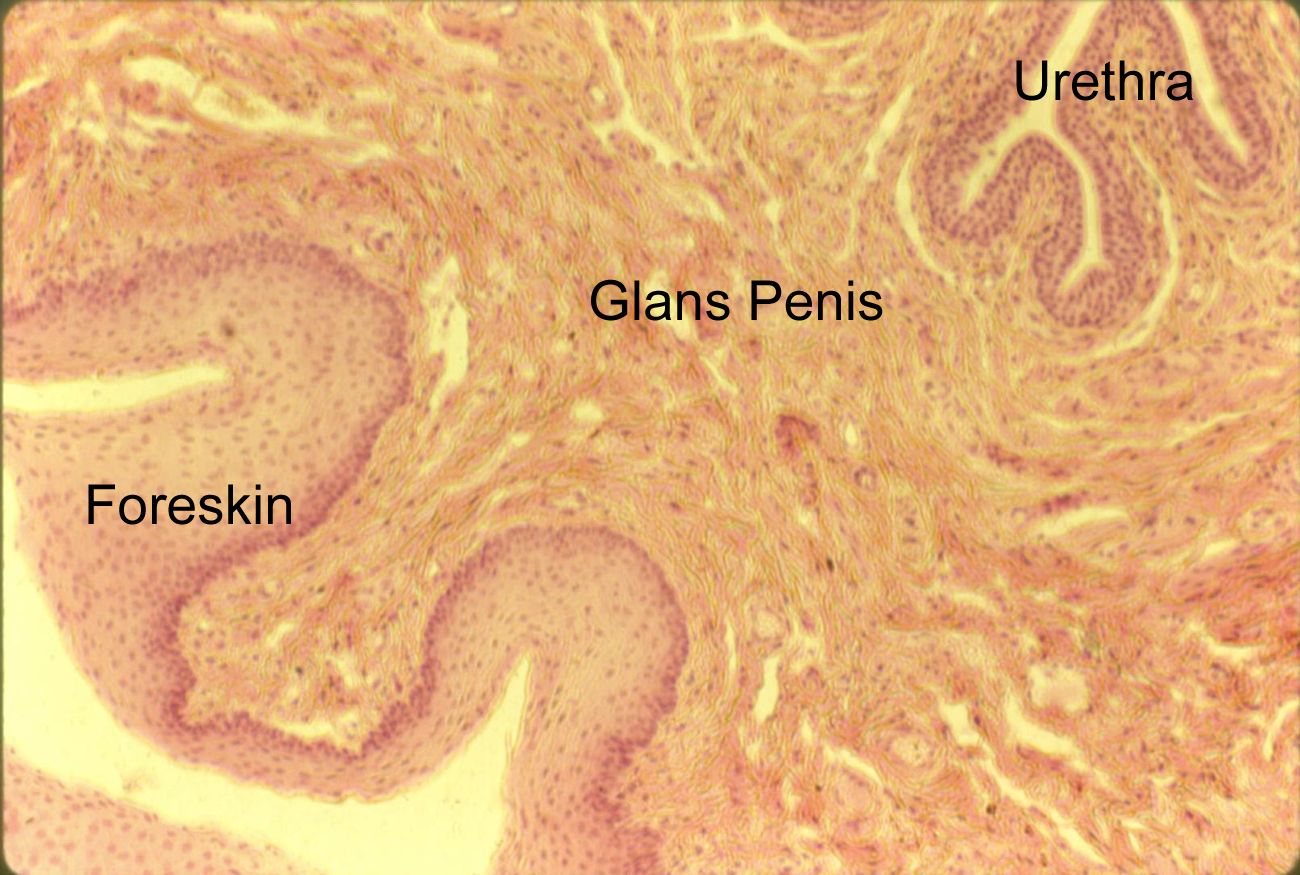
2 corpora cavernosa and 1 corpora spongiosum. The corpora spongiosum houses the urethra as it passes through the penis. At the glans penis, only corpora spongiosum is present. In the male climactic, the blood supply in the penis changes. Arteries dilate, while veins constrict. This allows the connective tissue sinuses to fill with blood to create an erection to allow the penis to penetrate the female vagina and deliver semen into the female reproductive tract. The glans penis is surrounded proximally by an extra fold of skin called the prepuce or foreskin.
Sagittal, Transverse cut of penis
Accessory Glands
The accessory glands of the male are the seminal vesicles, the prostate, and the bulbourethral (Cowper’s) glands. These glands secrete most of the liquid portion of the semen with the seminal vesicles producing 60% of the volume, the prostate producing about 20 % of the volume and the Cowper’s glands producing 10% of the semen volume.
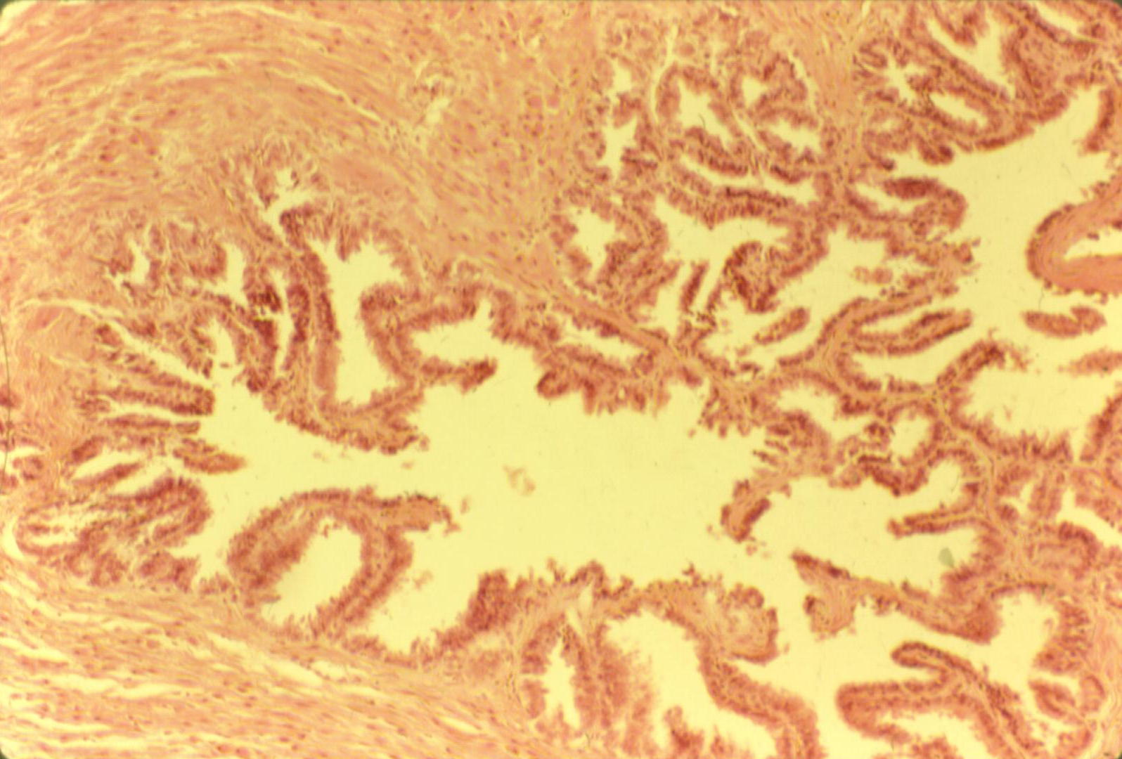
The seminal vesicles are paired structures that sit posterior to the urinary bladder and they join with the vas deferens to form the ejaculatory duct. The function of the seminal vesicles is to produce an antibody and nutrient rich fluid containing sugars such as fructose. IgA antibody helps defend the sperm and the sugar fructose is converted to ATP for energy needed to move the flagellum of the sperm. The single prostate gland sits at the base of the urinary bladder and encloses the prostatic urethra. Their secretions aid in sperm motility. The paired bulbourethral (Cowper’s) glands lie at the bulb or root of the penis. Their alkaline secretions help neutralize any acidic substances in the urethra, such as urine. All accessory glands secrete an alkaline rich fluid to help neutralize the acidic pH of the female reproductive tract.
Male repro models : Large, Small
Histology: prostate, seminal vesicles
coel, -coel cavity orchid/o- testicle
crypt- hidden andr/o- male
balan/o- glans penis epididym/o epididymis
prostat/o- prostate spermat/o- sperm
vas/o- vas deferens, duct vesicul/o- seminal vesicle
jact- throw rete- net
I. ID repro organs
A Drawing
B Models :
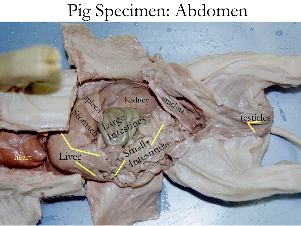
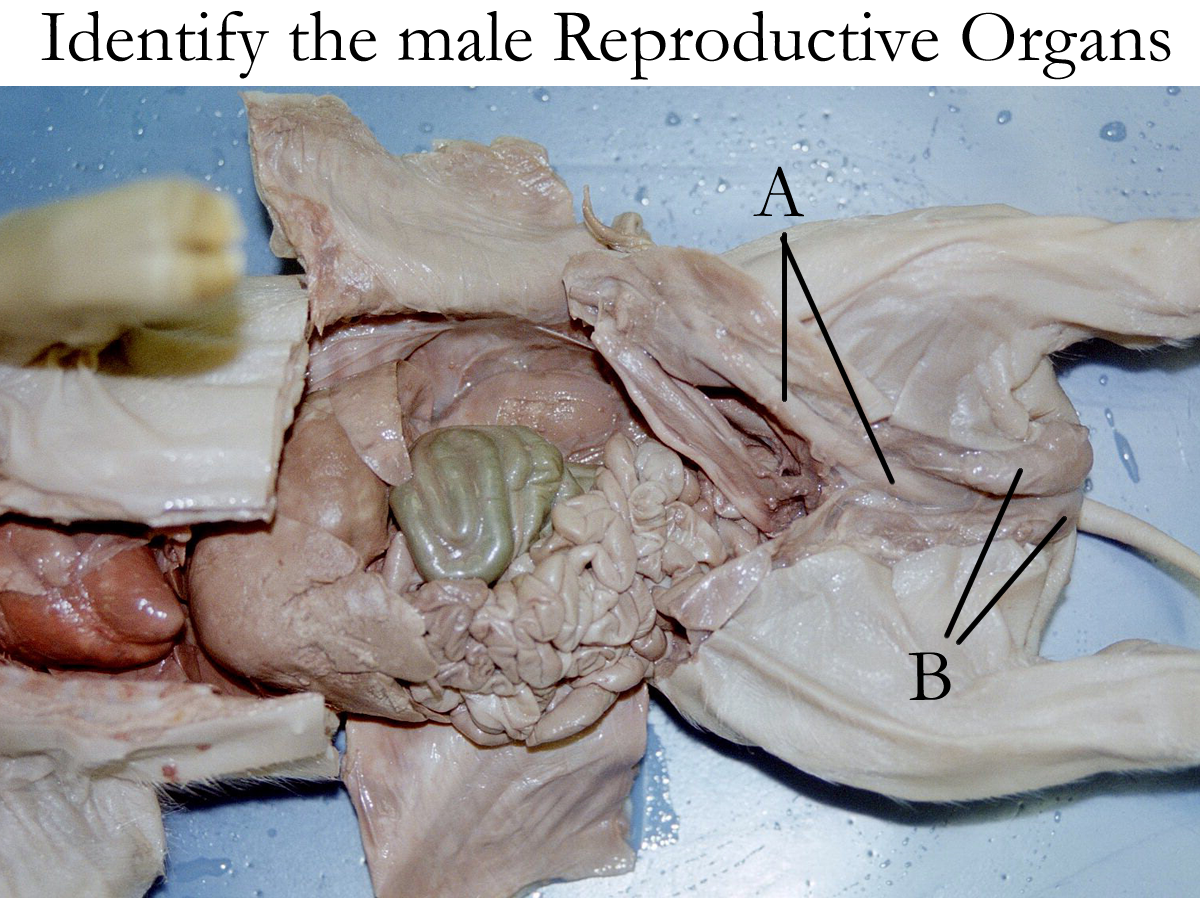
C Specimen
Concept Map: Make a concept map of the male reproductive system (gross and histo) anatomy, location, and physiological function. Include hormones, sources, targets, and effects. This map should be part of your LAR lab report (if selected) as a document insert or as an additional PDF document scan.
Benign Prostatic Hypertrophy(BPH)
Prostatic Cancer
Prostatitis
TURP
Epididymitis
Orchitis
Erectile Dysfunction (impotence)
Premature ejaculation
Cryptorchidism
Balano Prostatitis
Testicular Cancer
Sertoli Cell tumor
Infertility
Vasectomy
Circumcision
Aspermia, Oligospermia
Urologist
http://www.nlm.nih.gov/medlineplus/healthtopics.html
http://www.lumen.luc.edu/lumen/meded/histo/frames/histo_frames.html
http://www.gen.umn.edu/faculty_staff/jensen/1135/webanatomy/
http://calloso.med.mun.ca/%7Etscott/second.htm
http://www.track0.com/canteach/links/linkbodysystems.html
http://www.carr.lib.md.us/schs/science/anatomy/systems.html
http://www.leeds.ac.uk/chb/humbmods.html
http://www.med.virginia.edu/med-ed/phys/practice_board.html
1. Describe the histology of the testes and give its functions.
2. Name the function of the male reproductive ducts:
a) epididymis
b) vas deferens
c) ejaculatory duct
d) urethra
3. Name the three accessory organs for the male reproductive system.
Give their location and function.
4. Give the function of the scrotum
5. Describe the anatomy and function of the penis.
6. Describe the anatomy of a spermatozoon
7. Define mitosis
8. Define meiosis
9. Define spermatogenesis
10. Define spermiogenesis
11. Describe the endocrine control for the testes
12. Name and define the two parts of the male climactic.